The nerve conduction study came back normal. So did the EMG. Your reflexes are fine, your strength is fine, and the neurologist has written “no evidence of peripheral neuropathy” in the letter that went back to your GP.
Meanwhile your feet burn every night, your hands go strange in the cold, standing up in the shower makes the room swim, and the fatigue that got you the ME/CFS diagnosis in the first place has not moved an inch.
There is a specific reason those two things can both be true, and it is not that you are imagining the burning. It is that the standard nerve test does not look at the nerves that are most likely involved.
Over the past decade a body of research has been accumulating around a finding that keeps surprising people: a substantial share of patients carrying a chronic fatigue syndrome or fibromyalgia label have measurable damage to their smallest nerve fibers. Not all. Not even most. But enough that the question is now worth asking out loud in a clinic room.
Why a Normal EMG Does Not Mean Your Nerves Are Fine
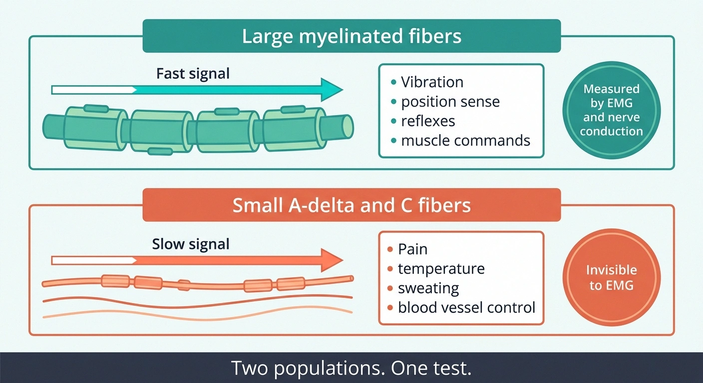
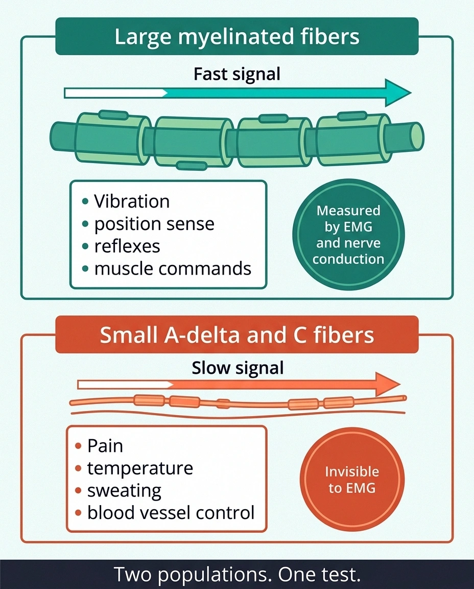
Nerve conduction studies and electromyography measure electrical signals travelling along large, myelinated nerve fibers. Those are the fibers that carry vibration sense, position sense, and the motor commands that move your muscles. They are thick, they are fast, and they are the only fibers those tests can see.
Your smallest nerve fibers are a different population entirely. Thinly myelinated A-delta fibers and unmyelinated C fibers are too slender and too slow to register on standard electrodiagnostics. They are also the fibers that carry pain, temperature, itch, and every autonomic signal your body sends to blood vessels, sweat glands and gut.
So a normal EMG rules out large-fiber problems. It says nothing whatsoever about the small ones. This is not a controversial point or a fringe position; it is a basic property of the test. It is simply not communicated well, and thousands of people walk out of neurology appointments believing they have been cleared when they have only been partially examined.
If your symptoms are burning, temperature weirdness, patchy numbness, and autonomic trouble, and your EMG was clean, you have been tested for the wrong thing.
The Fibers Nobody Tested
Small fiber neuropathy is damage to those thin fibers. When they degrade, the endings that reach into your skin thin out and retract, and the signals they carry get scrambled.
The symptom set is distinctive. Burning that is worse at rest and worse at night. Skin that hurts when the bedsheet touches it. Temperature that reads wrong, so a room feels freezing while your feet feel like they are in an oven. Pins and needles that come and go without pattern. And, because these same fibers run the autonomic system, symptoms that most people would never file under “nerves” at all: lightheadedness on standing, blotchy or purple feet, sweating too much or not at all, an unreliable gut.
Our full overview of small fiber neuropathy covers the condition on its own terms. What matters here is the overlap, and the overlap is large.
What the Skin Biopsies Actually Found
The evidence base starts with fibromyalgia, because that is where the first careful study was done.
In 2013, Anne Louise Oaklander and colleagues published work in the journal Pain examining people who had been given a fibromyalgia diagnosis by an independent physician. They took 3mm skin punch biopsies and counted the nerve fiber endings in the epidermis. In 41% of those patients, the biopsy was consistent with small fiber neuropathy, meaning the fiber density fell below the 5th percentile of normal. In matched controls, the figure was 3%.
That study has been replicated and extended repeatedly since. The current picture across the literature is that somewhere in the range of 40% to 60% of people carrying a fibromyalgia label show small fiber abnormalities on skin biopsy.
The ME/CFS numbers landed in similar territory. In one studied cohort, 38% of ME/CFS patients had small fiber polyneuropathy confirmed on skin biopsy. The detail that made that finding interesting was what those patients had in common: 93% of them also had POTS or orthostatic intolerance.
More recently, work published in the European Journal of Neurology in 2025 looked at small fiber neuropathy in both post-COVID condition and ME/CFS together. Both groups showed sensory small fiber involvement, identified through impaired heat detection thresholds and through increased tortuosity of the small fibers in the central corneal subbasal plexus, which is imaged with a non-invasive eye scan rather than a biopsy.
A word about what these numbers mean and do not mean. They do not mean fibromyalgia “is” small fiber neuropathy, or that ME/CFS is a nerve disease with a fatigue label stuck on it. They mean that a large minority of people inside those diagnostic categories have a findable physical abnormality that nobody looked for, and that finding it changes what can be offered.
One Fiber Population, Two Jobs

Here is the piece that makes the whole overlap make sense, and it rarely gets said plainly.
What the skin biopsies found, by group
Share of each group whose biopsy met criteria for small fiber neuropathy.
41%
Fibromyalgia-diagnosed patients, Oaklander 2013
38%
ME/CFS patients in a studied cohort
93%
Of those ME/CFS cases who also had POTS or orthostatic intolerance
3%
Matched healthy controls, same study
A large minority, not a majority. Which is exactly why the question has to be asked case by case rather than assumed in either direction.
The small unmyelinated fibers do two completely different jobs with the same cells. One job is sensory: they report pain and temperature from your skin. The other job is autonomic: they tell your blood vessels when to constrict, your sweat glands when to fire, and your gut when to move.
Damage that population and you get both consequences at once. The sensory half shows up as burning feet. The autonomic half shows up as something that looks nothing like a nerve problem.
Specifically: when the small fibers supplying your leg blood vessels are damaged, those vessels do not clamp down properly when you stand. Blood pools in your legs. Less returns to your heart. Your heart rate climbs to compensate, which is the mechanism behind POTS, and your brain gets less flow than it wanted, which is why standing up feels like wading through mud.
Now put that next to the defining complaint in ME/CFS. Exhaustion that gets dramatically worse with exertion. Difficulty being upright. A body that seems to punish activity out of all proportion. Some of that is doing exactly what an autonomic small fiber problem would predict.
This does not explain ME/CFS. Post-exertional malaise involves immune, metabolic and mitochondrial findings that a nerve problem does not account for. But it does explain why these two labels keep landing on the same person, and why the person carrying them keeps being told their symptoms belong to two unrelated files. Our guide to autonomic neuropathy covers what happens when those fibers fail across the rest of the body.
Which Came First
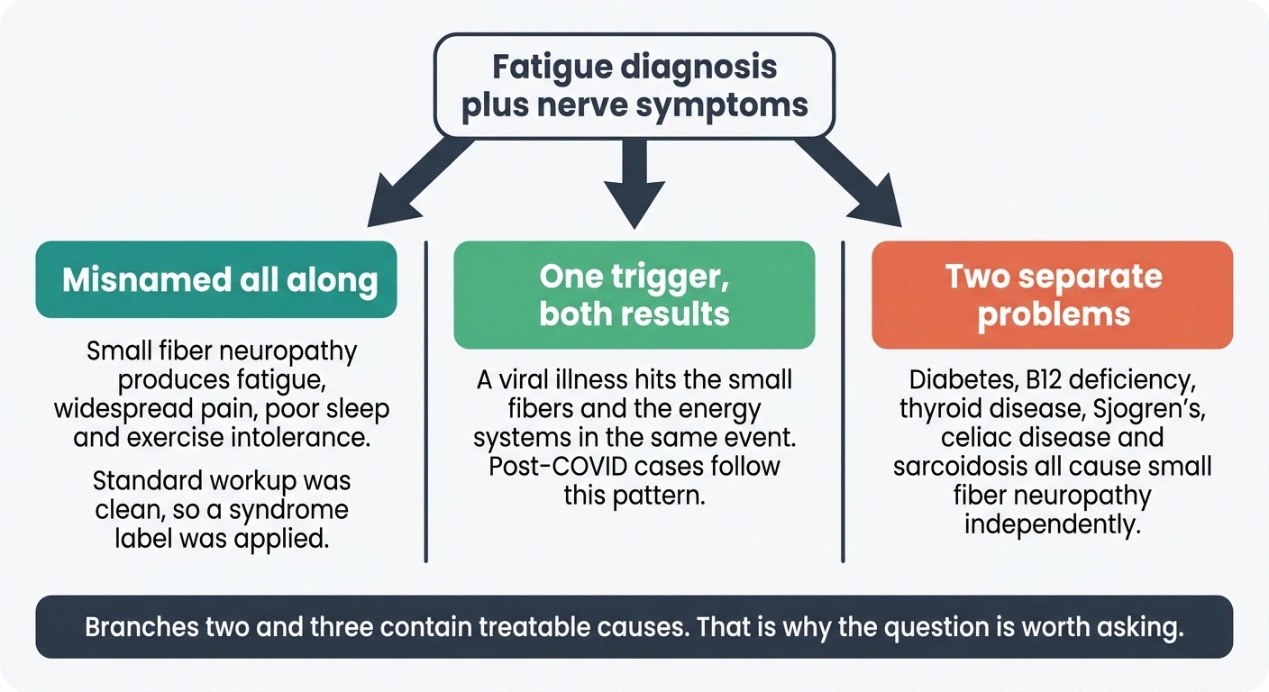
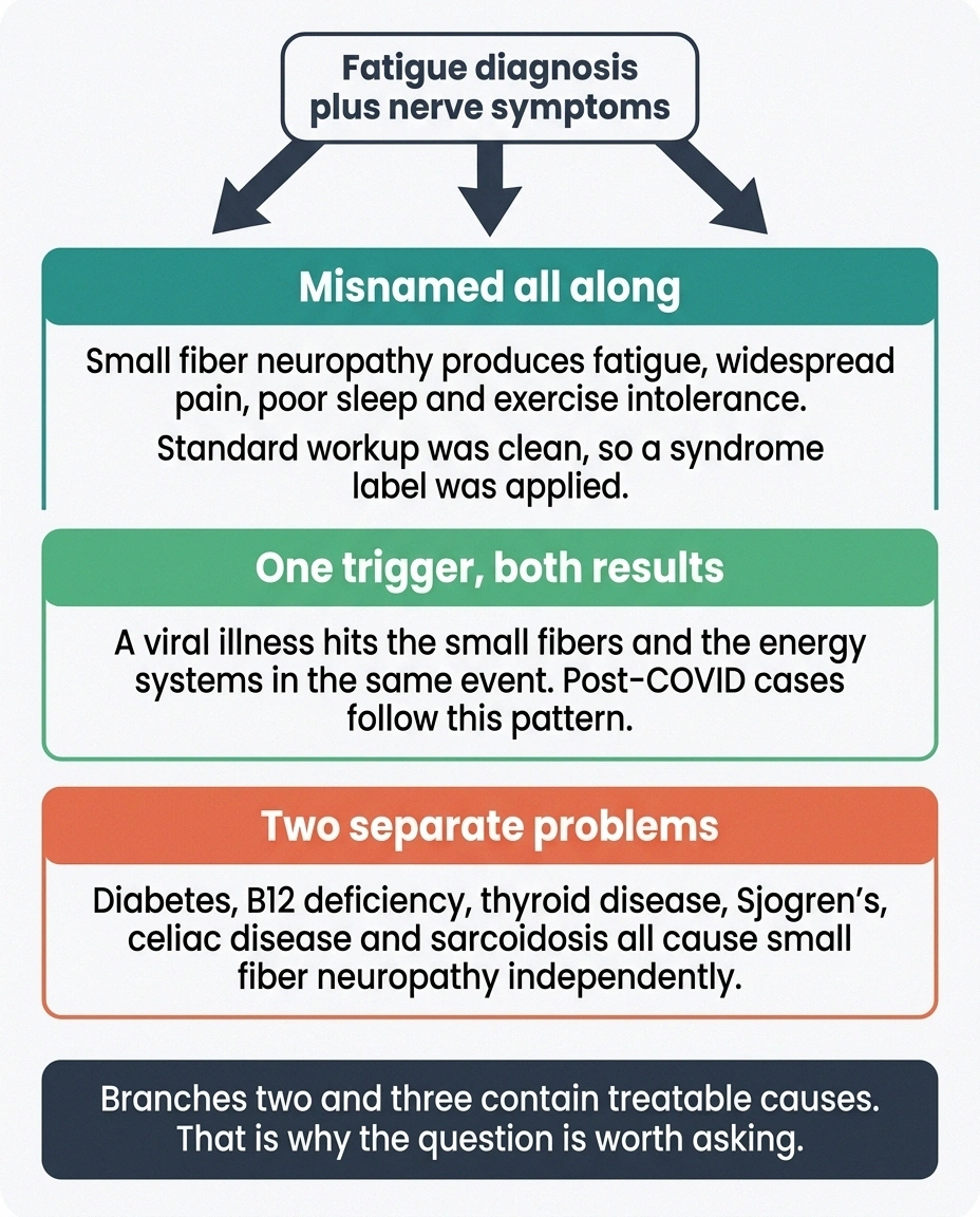
There are three ways this pairing can happen, and they are not equally common.
The nerve damage was there all along and got the wrong name. Small fiber neuropathy produces fatigue, widespread pain, poor sleep, brain fog and exercise intolerance. Those are also the diagnostic criteria for several other things. When the standard workup comes back clean, a syndrome label gets applied. This is the scenario the Oaklander work was designed to test, and it appears to account for a meaningful share of cases.
Something triggered both at once. A viral illness is the clearest example. Post-COVID condition has produced a wave of patients with new fatigue, new orthostatic intolerance and new small fiber findings appearing together after the same infection. Whatever the immune mechanism is, it seems to hit the small fibers and the energy systems in the same event rather than in sequence.
The two are genuinely separate and coincidental. Small fiber neuropathy has a long list of causes: diabetes and prediabetes, B12 deficiency, thyroid disease, Sjögren's syndrome, celiac disease, sarcoidosis, HIV, and a substantial fraction that stays idiopathic after a thorough search. Some people have ME/CFS and also happen to have one of those.
The reason this matters practically is that the second and third scenarios have treatable causes hiding in them. Finding a small fiber neuropathy is not the end of the workup. It is the start of asking why.
Getting Tested After a Normal EMG
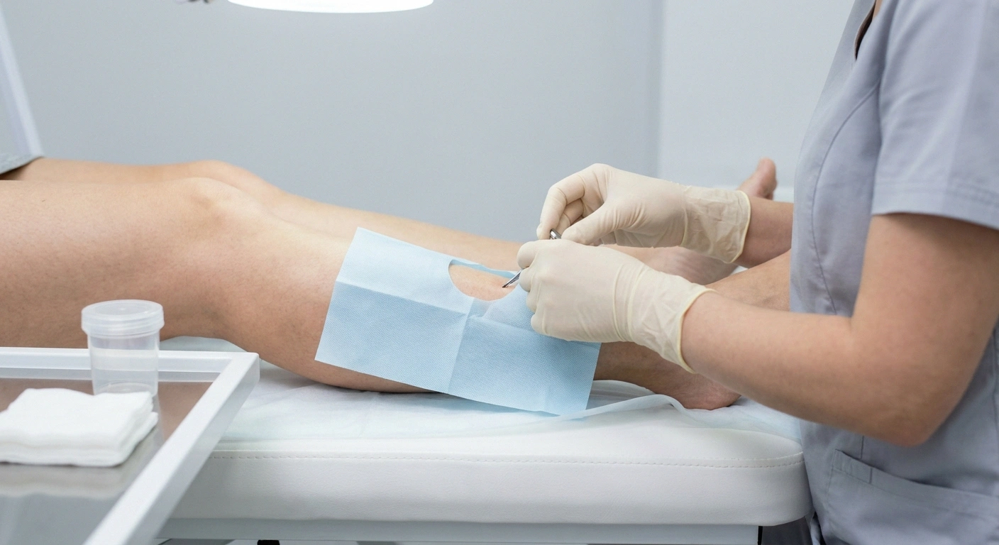
The tests that find small fiber problems are different from the ones you have already had.
Skin punch biopsy. The reference standard. A 3mm sample, usually taken from the lower leg and sometimes the thigh, under local anesthetic. It leaves a mark roughly the size of a pencil eraser. A lab counts intraepidermal nerve fiber density and compares it to age and sex matched norms. It is the test to ask for by name.
Quantitative sensory testing. Measures your detection thresholds for warmth, cold and pain. Non-invasive, but it depends on your responses, so it is supporting evidence rather than proof.
Autonomic testing. QSART measures sweat responses driven by the small fibers directly. Tilt table testing documents what your blood pressure and heart rate do when you are upright, which is the objective evidence for POTS.
Corneal confocal microscopy. A non-invasive scan of the nerve fibers in your cornea, which is the most densely innervated tissue in the body. It is the technique behind several of the newer ME/CFS findings. Availability is limited to research centers and a handful of specialist clinics.
One practical warning from the 2025 European work. Small fiber neuropathy in this population often does not follow the textbook length-dependent pattern of feet first, then hands. Non-length-dependent distributions, where symptoms start in the face, trunk or patchily across the body, are common here. If a clinician rules out small fiber neuropathy because your symptoms are “in the wrong place,” that reasoning is out of date. Our walkthrough of neuropathy diagnosis covers the full sequence of tests and what each one is looking for.
Access is the real barrier. Skin biopsy is not available everywhere, coverage varies, and many general neurologists do not order it routinely. Asking specifically, and asking for referral to a neuromuscular or autonomic specialist if the answer is no, is often what it takes.
What Changes If You Have Both
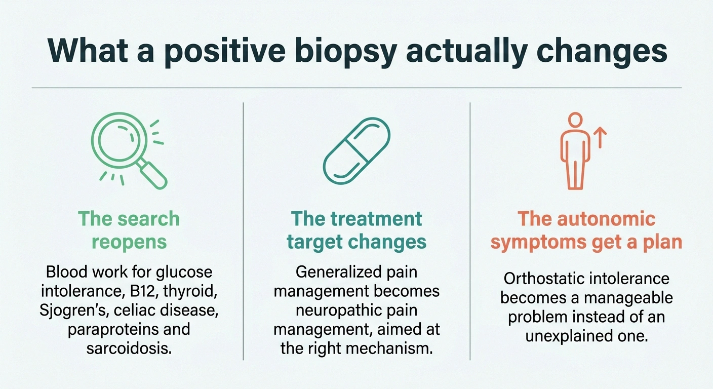
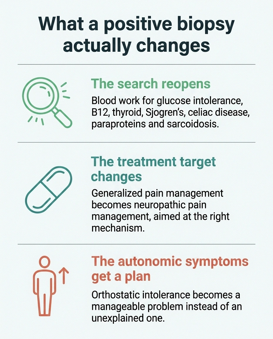
A confirmed small fiber neuropathy changes three things.
A positive biopsy is the start of the workup, not the end
Small fiber neuropathy has causes, and several of them are treatable. These are the tests worth having on the same requisition.
- A1c plus a 2-hour glucose tolerance test. Glucose intolerance short of diabetes is among the most common findings and one of the few that responds to change.
- B12 with methylmalonic acid. A B12 level alone can look normal while the functional deficiency is real.
- Thyroid function. Cheap, fast, and worth excluding.
- ANA and SSA/SSB antibodies. Sjögren's syndrome is a recognized cause and frequently presents without obvious dryness.
- Celiac serology. Nerve symptoms can precede gut symptoms by years.
- Serum protein electrophoresis and ACE. Screening for paraprotein disorders and for sarcoidosis.
It reopens the search for a cause. The blood work is worth doing properly: A1c and a two-hour glucose tolerance test, B12 with methylmalonic acid, thyroid function, ANA and SSA/SSB antibodies for Sjögren's, celiac serology, serum protein electrophoresis, ACE for sarcoidosis. Glucose intolerance short of diabetes is one of the most common findings, and it is one of the few that is genuinely modifiable.
It opens up neuropathic pain treatment. Burning that has been treated as generalized pain gets treated as nerve pain instead, which means a different set of medications with better odds in this specific problem. Duloxetine and gabapentin are the usual starting points, and neither is a cure, but they target the right mechanism.
It legitimizes the autonomic symptoms. Orthostatic intolerance stops being anxiety and becomes something with a management plan: fluid and salt intake where appropriate, compression garments, counter-pressure manoeuvres, and in some cases medication.
On immunotherapy, a note about proportion. A small post-COVID series reported that 9 of 9 patients receiving IVIG had significant improvement in neuropathic symptoms, compared with 3 of 7 who did not receive it. That is an interesting signal and nothing more. Those are tiny, uncontrolled numbers, IVIG is expensive and hard to access, and this is not standard care. It is worth knowing the research exists. It is not worth building your expectations on.
Pacing Is a Nerve Strategy Too

Anyone with ME/CFS has heard about pacing. It is worth understanding why it applies twice over when small fibers are involved.
Where the standard advice has to be rewritten
Almost every neuropathy page on the internet says walk more. With post-exertional malaise in the picture, that instruction needs replacing rather than adjusting.
Standard neuropathy advice
Build up walking gradually. Push a little further each week. Some discomfort during exercise is expected and fine.
With post-exertional malaise
Find the ceiling and stay under it. Short bouts, seated or recumbent where possible. Stop while you still feel fine. Rest before the activity, not only after.
The crash typically arrives 24 to 48 hours later, which is why the link between Tuesday and Thursday is invisible without a written record.
Post-exertional malaise means that pushing past your limit produces a delayed and disproportionate crash, often a day or two later. The standard advice everyone gives people with neuropathy, which is to walk more, does not transfer here without modification. Graded exercise prescribed the way it is prescribed for ordinary deconditioning has caused real harm in this population, and current guidance in several countries has moved away from it for exactly that reason.
What tends to work instead is finding the ceiling and staying under it. Short bouts. Recumbent or seated activity, which avoids the orthostatic load that is doing half the damage. Stopping while you still feel fine rather than when you feel finished. Horizontal rest before and after anything demanding.
Tracking helps more here than almost anywhere else, because the crash is delayed and the connection between Tuesday's activity and Thursday's collapse is invisible without a record. A simple symptom diary that logs activity, symptoms and upright time is the cheapest diagnostic tool you have.
And because the burning reliably intensifies at night in this group, the sleep half of the problem is worth attacking on its own terms rather than waiting for the pain treatment to fix it. Our notes on why neuropathy gets worse at night cover what is actually driving that pattern.
Where This Leaves You
If you have an ME/CFS or fibromyalgia diagnosis and you also have burning feet, temperature symptoms, or trouble being upright, there is a reasonable chance that a skin biopsy would find something. Somewhere between a third and a half of people in your position, based on the studies done so far.
A positive result does not undo the fatigue diagnosis and it will not fix your energy. What it does is convert a symptom that has been treated as unexplained into one with a name, a mechanism, a set of causes worth hunting, and treatments aimed at the right target.
That is a smaller win than anyone wants. It is also, for a lot of people who have spent years being told nothing is wrong, the first piece of objective evidence they have ever been handed. The years of not being believed take their own toll, and the impact of that is real enough that we have covered it separately in our piece on neuropathy and mental health.
The ask is small: a 3mm punch biopsy, or a referral to someone who does them. It is worth making.
Frequently Asked Questions
Can chronic fatigue syndrome cause small fiber neuropathy?
Causation has not been established in either direction. What research shows is that the two occur together far more often than chance would predict, with roughly 38% of studied ME/CFS patients showing small fiber polyneuropathy on skin biopsy. Whether the nerve damage is a consequence of ME/CFS, a shared result of the same trigger, or an alternative explanation for some cases remains an open question.
Why was my EMG normal if I have nerve damage?
EMG and nerve conduction studies measure only large, myelinated nerve fibers. Small fiber neuropathy affects thin A-delta and unmyelinated C fibers, which are too small and too slow to register on those tests. A normal EMG is expected in pure small fiber neuropathy and does not rule it out.
What test finds small fiber neuropathy?
A 3mm skin punch biopsy measuring intraepidermal nerve fiber density is the reference standard. Supporting tests include quantitative sensory testing, QSART sweat testing, tilt table testing for orthostatic problems, and corneal confocal microscopy where it is available.
Is fibromyalgia actually small fiber neuropathy?
Not as a whole. Studies consistently find small fiber abnormalities in roughly 40% to 60% of people carrying a fibromyalgia diagnosis, which means a large minority have a findable nerve abnormality and the remainder do not. The practical implication is that the question deserves to be asked in each individual case rather than assumed either way.
Does small fiber neuropathy explain POTS in ME/CFS?
It offers a partial mechanism. The same small unmyelinated fibers that carry pain and temperature also control blood vessel constriction. When they are damaged, leg vessels fail to tighten on standing, blood pools, and heart rate rises to compensate. In one cohort, 93% of ME/CFS patients with biopsy-confirmed small fiber neuropathy also had POTS or orthostatic intolerance.
Should I ask for IVIG?
It is not standard treatment for this situation. A small post-COVID series reported improvement in 9 of 9 patients who received IVIG versus 3 of 7 who did not, but those numbers are far too small and the study design too weak to support routine use. It is reasonable to know the research exists and to ask a specialist whether your particular case has features that would justify considering it.
Will exercise help my neuropathy if I have ME/CFS?
Standard graded exercise advice does not transfer safely to people with post-exertional malaise and has caused documented harm in this population. Movement still has value, but the approach is different: short bouts, seated or recumbent positions to reduce orthostatic load, stopping before symptoms build, and rest scheduled around activity rather than after it.
Can small fiber neuropathy be reversed?
It depends entirely on the cause. Where an underlying driver is found and treated, such as glucose intolerance, B12 deficiency or an autoimmune condition, fiber density can partially recover over time. Where no cause is identified, the realistic goal is stopping progression and managing symptoms rather than reversal.
About the Author