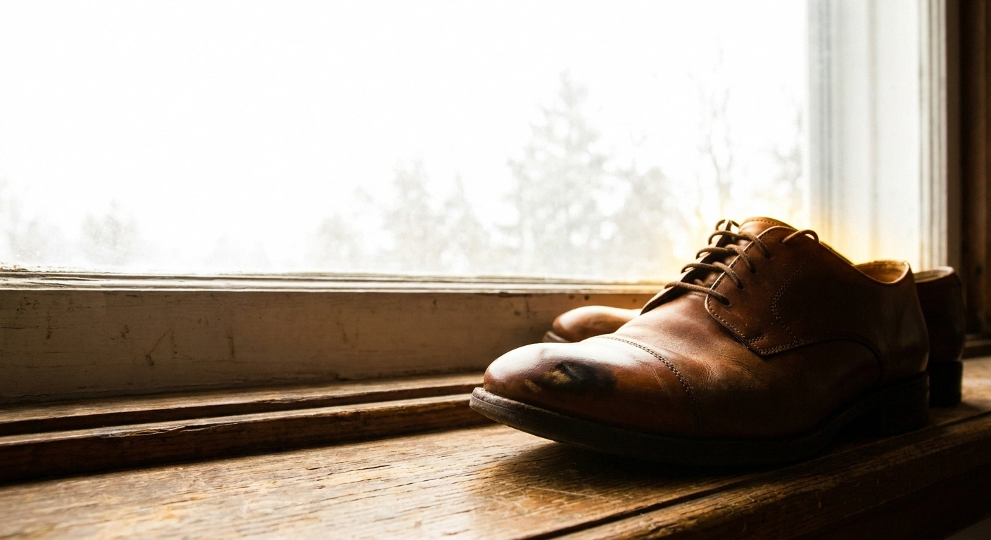
The shoes were fine in March. By September the same pair rubs across the top of the third toe, and there is a shiny patch on the leather where something underneath has been pushing up against it.
That is usually how people find out. Not by looking at their feet, which most of us do less often than we should, but by noticing that a shoe has changed shape from the inside. Then they look, and the toes are not lying flat anymore. They are bunched, gripping, curled under at the tips, with the knuckles standing proud.
Claw toes and hammertoes are extremely common in neuropathy. Reported prevalence in people with diabetes runs somewhere between 32 and 46 percent, which means a large share of anyone reading this will recognise their own feet in the paragraph above. Common enough to be normal, and common enough to be dismissed as cosmetic.
It is not cosmetic. The deformity by itself is a nuisance. The deformity combined with feet that cannot feel properly is the beginning of the most preventable serious problem in this condition, and understanding the mechanics is what makes prevention obvious rather than tedious.
What the Toes Are Actually Doing
Look at a foot with claw toes from the side and the shape is consistent: the toe lifts up at the joint where it meets the foot, then curls down at the joints beyond it.
Three numbers worth carrying around
32-46%
of people with diabetes have claw or hammer toe deformity. If your toes have changed, you are firmly in the majority experience rather than an unusual case.
20
small muscles live inside the foot itself. They spread the toes, hold them flat, and adjust your balance constantly. They sit at the very end of the longest nerves you have.
2
new pressure points appear the moment the base joint tilts up: the tip of the toe against the floor, the knuckle against the roof of the shoe. Neither existed before.
The lift at that first joint, the one at the base of the toe where it meets the long bone of the foot, is the structural key. Clinicians call it hyperextension at the metatarsophalangeal joint, and both claw toes and hammertoes share it. Everything that goes wrong afterwards follows from that single upward tilt.
Picture the toe as a lever with its fulcrum at the base. Tip the base upward and two things happen at once. The far end of the toe swings down and presses into the floor. The middle of the toe rides up and presses into the roof of the shoe.
Two new pressure points, neither of which existed before, both of them now taking load with every step you take. On a foot with normal sensation you would feel that within a day and change your shoes. On a foot with neuropathy the message never arrives.
The Half of Neuropathy Nobody Mentions
Most people understand neuropathy as a sensation problem. Numbness, burning, tingling, pins and needles. That is the sensory half.
Nerves also carry signals in the other direction, out to muscles, telling them to contract. When those motor fibers are damaged, the muscles they supply weaken and shrink. This is motor neuropathy, and it gets a fraction of the attention that the sensory side receives despite being responsible for most of the structural changes in the foot.
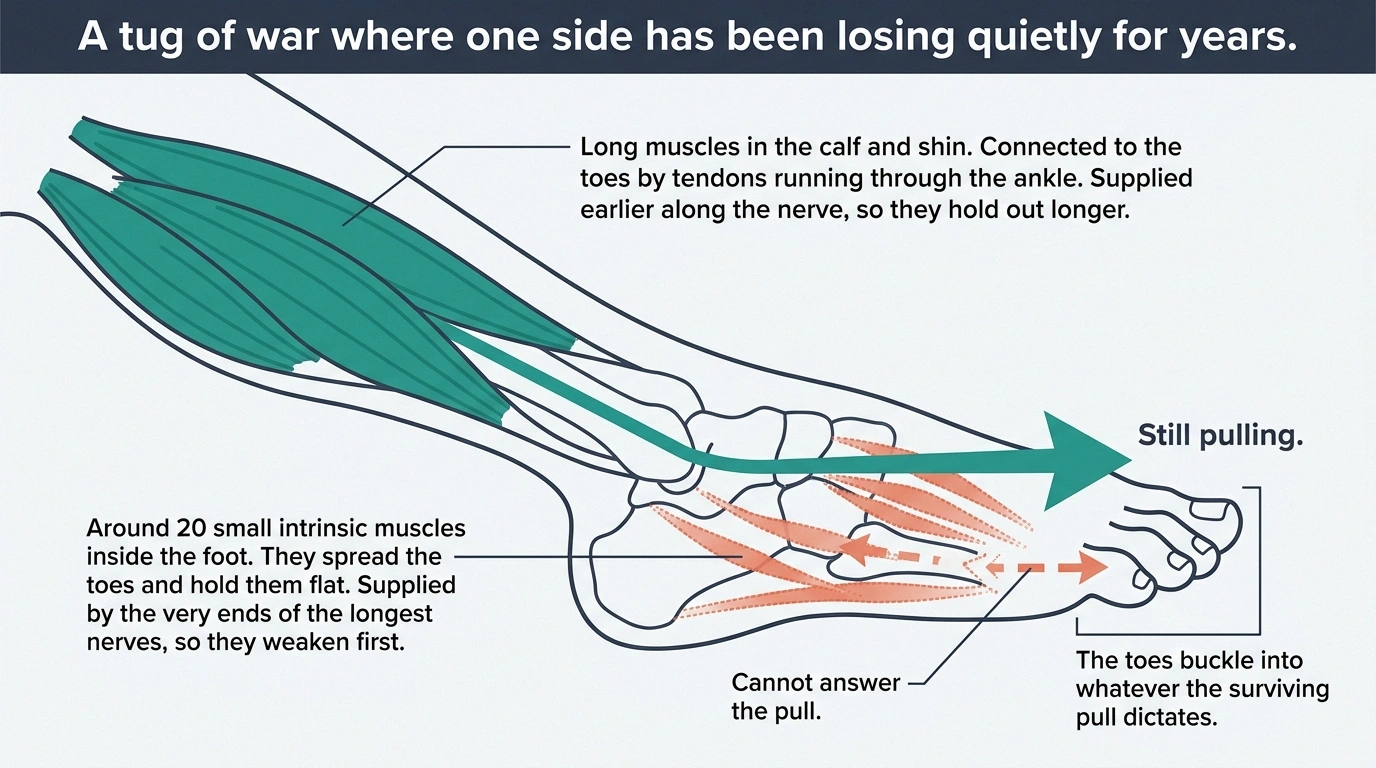
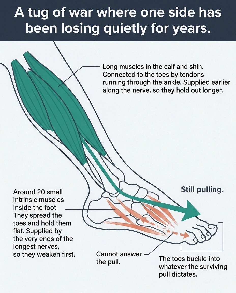
There are around twenty small muscles living inside the foot itself. They are called the intrinsic muscles, and they are small, weak individually, and collectively responsible for a lot: spreading the toes apart, holding them flat against the ground, fine-tuning your balance a hundred times a minute while you stand. They are supplied by the deepest branches of the longest nerves in the body, which puts them near the front of the queue for length-dependent damage.
Meanwhile, the big muscles that flex and extend the toes are not in the foot at all. They are up in the calf and shin, connected to the toes by long tendons running down through the ankle. Those muscles are supplied earlier along the nerve, and they hold out longer.
So you end up with a tug of war where one side has been quietly losing strength for years. The long tendons from the calf keep pulling. The small muscles that used to hold the toes flat against that pull cannot answer. The toes buckle into the shape the surviving pull dictates.
People often notice other consequences of the same weakness without connecting them: less push-off at the end of a step, difficulty spreading the toes apart on command, a foot that looks narrower or more hollowed out than it used to. Those changes belong to the same process, and they show up in the same general timeframe described in the overview of the stages of neuropathy.
Claw, Hammer, or Mallet
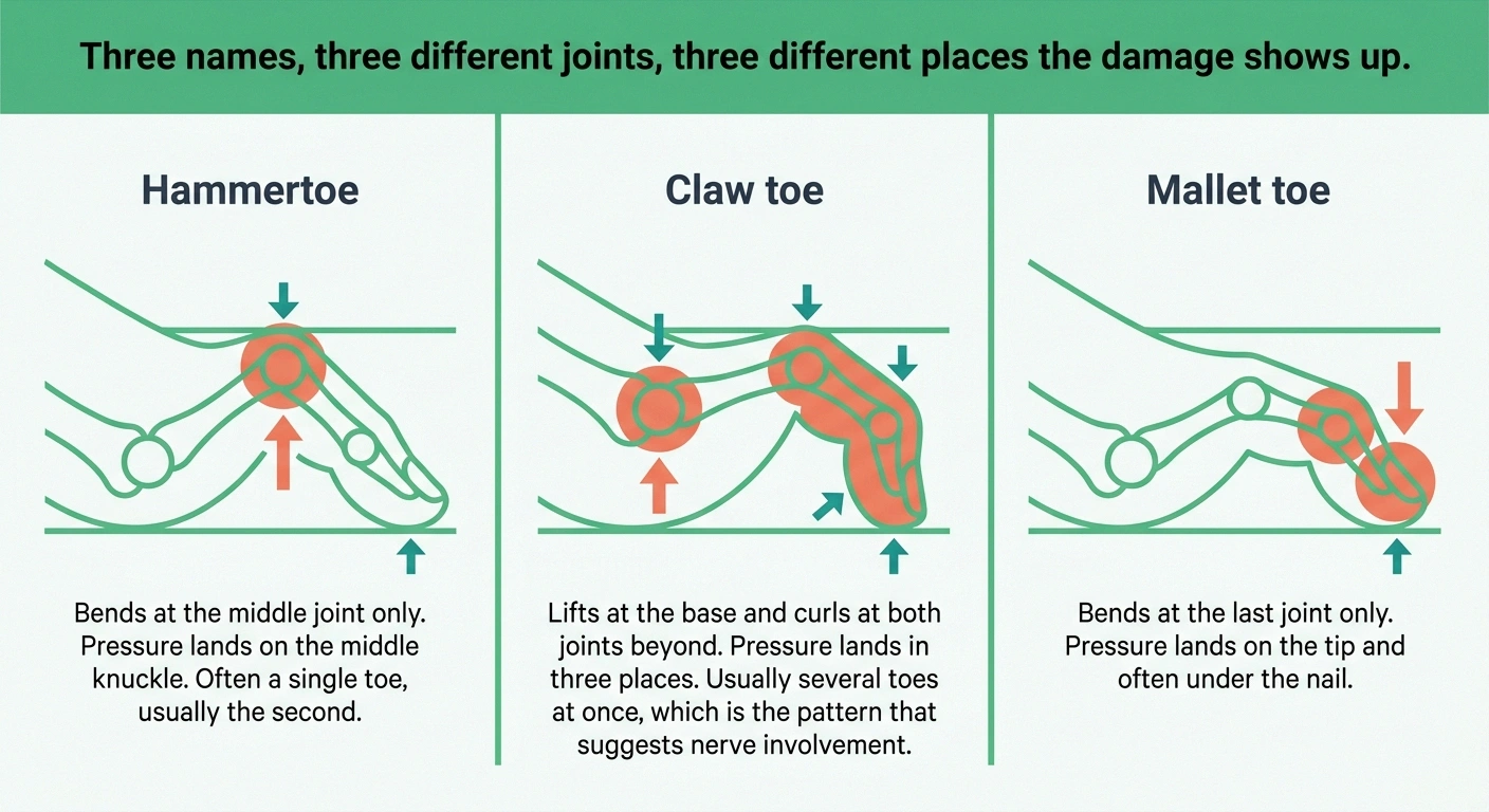
These three get used interchangeably and they are not the same thing. Each toe has three joints, and which ones are bent determines the name.
Hammertoe bends at the middle joint. The toe humps up in the middle like a knuckle, the tip points forward and down. Frequently a single toe, most often the second.
Claw toe lifts at the base joint and curls at both joints beyond it. The whole toe grips, like a hand closing. Usually several toes together, which is the pattern that points at nerve involvement rather than at a shoe or an injury.
Mallet toe bends only at the very last joint. The tip presses straight down into the floor while the rest of the toe stays flat.
The distinction is worth getting right for one practical reason: it tells you where to look for damage. Hammertoe puts pressure on the middle knuckle. Mallet toe puts it on the tip and often under the nail. Claw toe does both, plus a third spot under the ball of the foot, and that third one is the dangerous one.
Where the Cushion Went
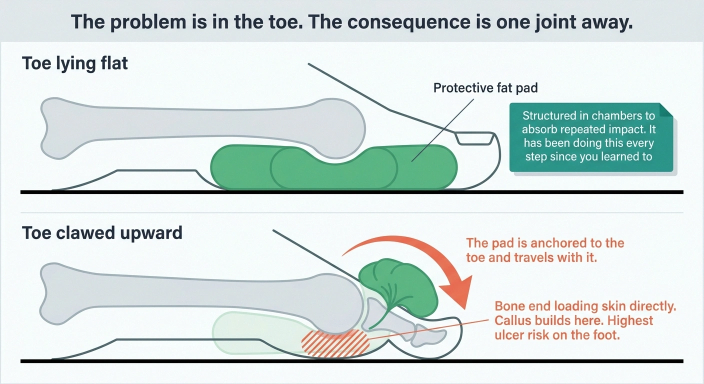
Under the ball of your foot sits a pad of specialised fat, thick and shock-absorbing, sitting directly beneath the ends of the long foot bones. It is not ordinary fat. It is structured in chambers to handle repeated impact, and it has been protecting those bone ends every step you have taken since you learned to walk.
Where the callus sits tells you what is loading it
Callus is a map of pressure. Its location is diagnostic information you can gather yourself with a mirror on the floor, and it is worth photographing so you can compare against the same spot in three months.
- Across the ball of the foot, behind the toes
- The fat pad has migrated forward and the bone ends are taking load directly. This is the site with the highest ulcer risk on the whole foot, and it is the one people least associate with their toes.
- On the very tip of a toe, sometimes under the nail
- The toe is standing on its end rather than resting along its length. This is the lesion a toe crest is designed to address.
- On top of the middle knuckle
- Shoe depth, not shoe width. Nothing about the foot will fix this one; the roof of the toe box has to move.
- Between two toes, on the facing surfaces
- Toes pressed against each other, usually a width and shape issue combined. These stay damp, which makes them the most likely to macerate and break down quietly.
- Nothing anywhere, on a foot with clear deformity
- Genuinely good news, and worth noticing. It means your current shoes are distributing load acceptably, and it is an argument for buying the same pair again rather than experimenting.
That pad is anchored to the toes.
When a toe pulls up into the clawed position, it drags the fat pad forward with it, out from under the bone end and up toward the base of the toe. The bone is left sitting on skin with a fraction of its former cushioning.
This is why the ball of the foot starts hurting or building callus in people whose deformity is up in the toes. It seems like an unrelated problem in a different part of the foot. It is the same problem, one joint away.
You can sometimes see it. Look at the sole with the toes relaxed and the pad appears bunched up near the base of the toes, with a thinner, harder-looking area behind it where the pad used to sit. Callus builds there because callus is skin’s response to repeated pressure, and it is doing exactly what it is designed to do.
The problem is that callus is stiff. A stiff layer sitting over a bony point transmits more pressure to the tissue underneath, not less. Left long enough, tissue under a thick callus breaks down from the inside, which is why the wound frequently appears without any visible injury having happened. Standard foot care exists largely to interrupt exactly this sequence, and the practical version is in the guide to neuropathy foot care.
The Research Complicates the Standard Story
Everything above is the textbook account, and it is the version you will find on every clinic page. It is mostly right. It is also not the whole picture, and the honest version is more interesting.
Four claims you will read elsewhere, sorted by how well they hold up
Almost every page on this subject states the muscle-imbalance account as settled. Here is where each piece of it actually sits, and what that means for your next decision.
| The claim | Where it stands | So |
|---|---|---|
| Motor nerve damage wastes the small muscles inside the foot | Well established | Expect other signs of it: weaker push-off, trouble spreading the toes. |
| That wasting alone explains who gets claw toes | Contradicted Atrophy did not separate those with the deformity from those without |
Advanced neuropathy does not make the deformity inevitable. |
| Thickening of the plantar fascia is also required | Current thinking Diabetic feet show measurably thicker fascia |
Individual anatomy is part of who is susceptible. Not your doing. |
| Toe exercises will straighten an established claw toe | Not supported | Still worth doing, but for balance and stability rather than toe shape. |
A study published in Diabetes Care compared people with neuropathy who had developed claw toes against people with neuropathy who had not. If muscle imbalance from intrinsic atrophy were the whole explanation, the two groups should have looked clearly different on measures of intrinsic muscle volume and imbalance.
They did not. Neither intrinsic muscle atrophy nor the degree of imbalance reliably separated who had the deformity from who did not.
Which means atrophy is part of the story rather than the entire mechanism. Later work has pointed toward the plantar fascia, the thick band of connective tissue running along the sole, which is measurably thicker in diabetic feet than in others. The current thinking is that atrophy and connective tissue change are both required, with individual anatomy determining who is susceptible.
Two things follow from this that matter to you.
The first is that having significant nerve damage does not mean claw toes are inevitable. Plenty of people with advanced neuropathy have perfectly flat toes.
The second is that toe-strengthening exercises are a reasonable idea rather than a proven fix. If atrophy alone does not determine the outcome, reversing atrophy alone will not reliably prevent it. Toe exercises are still worth doing, because intrinsic foot muscle function contributes to balance and stability regardless of what it does for toe shape, and balance is worth protecting for its own sake. Set the expectation accordingly. The general case for maintaining foot and leg strength is covered in the neuropathy exercises for seniors guide.
Flexible or Rigid, and Why It Decides Everything

Claw toes begin flexible and become rigid. Which side of that line your feet are on determines what is realistic, and you can check it at home in under a minute.
Sit down and take your shoes and socks off. Put your fingers under the ball of the foot, just behind the toes, and push upward gently, as though you were mimicking what happens when you stand on tiptoe.
If the toes straighten out as you push, the deformity is flexible. The joints still move. They are being held in position by tendon tension rather than by fused or contracted structures.
If the toes stay curled no matter what, the deformity is rigid. The joints themselves have stiffened and the toe will not come flat again.
Push gently. This is a test, not a stretch, and forcing a stiff toe is a good way to injure skin you cannot feel.
Flexible deformity is where padding, toe crests, splints, roomier shoes and stretching have something to work with, and where slowing the progression is a genuine goal. Rigid deformity changes the objective entirely: correction is off the table, and the aim becomes accommodation, meaning shoes and insoles built around the shape your foot has now rather than the shape it used to have.
That shift is not a defeat. Accommodation works. It just requires admitting the shape is permanent, which is the step people tend to postpone for a year or two while their shoes keep rubbing.
Depth Is the Specification Everyone Gets Wrong
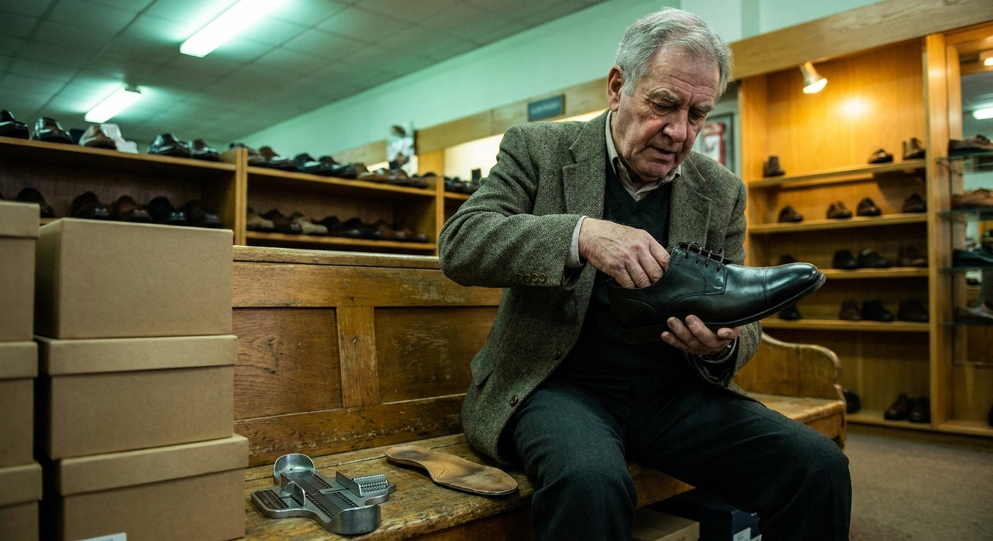
Ask anyone what shoes to buy for toe deformity and they will say wide. Width is the wrong axis.
Four things to do in the shop, in this order
All four take under two minutes combined and none of them require the assistant’s help. Do them before you put the shoe on, because a shoe already on your foot hides everything you are trying to check.
- Run a finger along the inside roof of the toe box. You are feeling for a seam. A seam that lands where a knuckle rides will win, every time, over any amount of breaking in.
- Turn the shoe over and look at the outline of the sole. Ask whether a foot is that shape. Most shoes taper to a point on the big-toe side, and toes fan outward.
- Pull the insole out and stand on it, off the shoe. If any part of your foot hangs over the edge, the shoe is too small in that dimension regardless of what the label says. This is the single most revealing check and almost nobody does it.
- Squeeze the heel counter between finger and thumb. It should resist. A heel cup that folds flat gives you nothing to stand on at the moment your toes have stopped contributing to balance.
Timing beats technique: go in the late afternoon and get both feet measured every visit. Feet are measurably larger by evening and they change shape over the years with this condition, so the size you have bought for a decade is a guess rather than a measurement.
A clawed toe rides upward. The problem is vertical clearance between the top of the knuckle and the roof of the toe box. Buying a wider shoe gives the foot more room side to side and does nothing at all about the joint that is pressing into the material above it.
What to look for instead:
- Depth, explicitly. Extra-depth shoes are a real category and they are sold under that name. The toe box is built taller. That is the specification.
- A soft, seamless upper over the toes. Stretch mesh or soft leather that yields where the knuckle pushes, rather than a stiff toe cap that pushes back.
- No seams crossing the knuckles. Run a finger inside the shoe along the top of the toe box before buying. A seam sitting exactly where a knuckle rides is a blister in preparation.
- A shape resembling a foot. Toes fan outward. Most shoes taper inward to a point. Look at the sole of the shoe from underneath and ask whether a foot is that shape.
- A removable insole, so a custom or over-the-counter orthotic can go in without stealing depth from the toe box.
- A firm heel counter that does not collapse when squeezed. Rear stability matters more once toe function is reduced, which matters for the same reasons set out in the guide to neuropathy and balance.
Two fitting rules that make more difference than the shoe model: get measured properly every time rather than buying your remembered size, since foot dimensions change with age and with this condition specifically. And shop in the late afternoon, because feet are larger then and a shoe fitted at 9am can be tight by dinner.
There is more on materials, closures and specific features in the fuller guide to choosing shoes for neuropathy.
Toe Crests, and What They Actually Do
Of the small devices sold for this, toe crests are the one with the clearest logic and some actual evidence behind them.
Four things sold in the same aisle, doing four different jobs
The packaging for all of these implies correction. Only one of the four has published support behind it, and none of them straightens a rigid toe, so check which side of that line you are on before you spend anything.
- Toe crest pad
- Job: fills the space under curled toes so load spreads along the toe instead of concentrating on the tip. Evidence: pilot study showed improvement in calluses, blood-stained lesions and ulcers at both follow-ups. Works on rigid toes: yes, as offloading, not as correction. Get the thickness checked in a real shoe.
- Toe separator
- Job: keeps adjacent toes off each other, which addresses lesions on the facing surfaces where skin stays damp. Works on rigid toes: for the between-toes problem only. It will not lift a knuckle out of the shoe roof.
- Splint or straightener
- Job: holds a toe flatter while worn. Works on rigid toes: no. On a flexible toe it may help comfort. On numb skin it is another hard object in the shoe, so treat it as something to check the skin around.
- Gel sleeve or cap
- Job: pads the top of a knuckle against the shoe. Works on rigid toes: as padding, yes, but it also takes up depth. A sleeve added to a shoe that was already shallow makes the pressure worse rather than better, which is the commonest way this one backfires.
The rule that applies to every item above: anything new inside the shoe gets checked against the skin after the first hour, then again at the end of the first day. The warning system that would normally do that job is the one that is not working.
A toe crest is a small pad that sits underneath the curled toes, filling the gap between the toes and the ground. A loop goes over one or two toes to hold it in place. Over several days of wear the pad moulds to the shape of the toes above it.
The mechanism is straightforward. A clawed toe touches down only at its tip, so all the load goes through one small point. A crest gives the underside of the toe something to rest on along its length, spreading load that was concentrated on the tip.
A pilot study of crest pads in people with lesser-toe deformities and skin lesions reported significant improvement in calluses, blood-stained lesions and ulcers, with strong statistical significance at both follow-up points. Small study, and pilot data is not a guarantee. Still, it is a cheap, low-risk, mechanically sensible intervention with published support behind it, which is more than can be said for most of the products in this category.
Practical notes if you try them. Get the fit checked rather than guessing, because a crest that is too thick becomes its own pressure source in a shoe that was already tight. Silicone versions last longer than fabric ones. And on a foot with reduced sensation, anything new inside the shoe requires checking the skin after the first hour of wear, then again at the end of the first day, since the usual warning system is not going to do that job for you.
Toe separators, splints and straightening sleeves are also sold for this. On flexible deformity they may help comfort. None of them will straighten a rigid toe, whatever the packaging suggests.
What Turns a Callus Into a Wound
This is the short section that matters most.
The same callus, four different timelines
What you see when you look changes how quickly you need to act. The distinction people most often miss is the third row, because a stained callus still looks like a callus and does not hurt.
Pale, even, dry callus with no colour inside it
Mention at your next routine foot appointment. Keep watching the spot. Do not work on it yourself.
Redness around the callus that has not faded 20 minutes after the shoes came off
Stop wearing those shoes now, not tomorrow. Report it within a week. The twenty-minute rule is what separates ordinary pressure from too much of it.
Dark, red, brown or blackish staining visible inside the callus
That is bleeding underneath, which means tissue below has already broken down. Days, not months. This one is a wound wearing a disguise, and it is the row most people walk past.
Any break in the skin, drainage, an odour, or a spot warmer than the same place on the other foot
Same day. Comparing warmth against the matching spot on the opposite foot is a better test than judging one foot alone.
Callus over a bony point on an insensate foot is the standard route to an ulcer. Not because callus is dangerous in itself, but because of what people do about it and what they fail to notice.
The visual cue that changes everything: a callus with dark, red, brown or blackish staining inside it is not a callus that needs attention soon. It is bleeding underneath, which means tissue below has already broken down, and it should be looked at by a foot specialist within days rather than at the next routine appointment. This is the single most useful thing to know about your own feet, and almost nobody is told it.
Things that reliably make matters worse:
- Cutting, shaving or filing calluses yourself. Skin you cannot feel gives no feedback about how deep you have gone. A significant proportion of foot wounds in this population start with a person tidying up a callus at home.
- Medicated corn pads and corn removers. These work by chemically burning tissue. On skin with normal sensation the burn is uncomfortable and self-limiting. On numb skin with impaired healing it is a chemical wound with no alarm attached. Avoid them completely.
- Heating pads, hot water bottles and hot soaks. Feet that cannot judge temperature accurately get burned, and the burn is discovered later.
- Barefoot, anywhere, ever. Including indoors, including briefly. Curled toes catch on carpet edges and furniture legs, and the tips are already the most exposed part of the foot.
- Wearing new shoes for a full day. An hour on day one, two hours on day two, checking the skin each time.
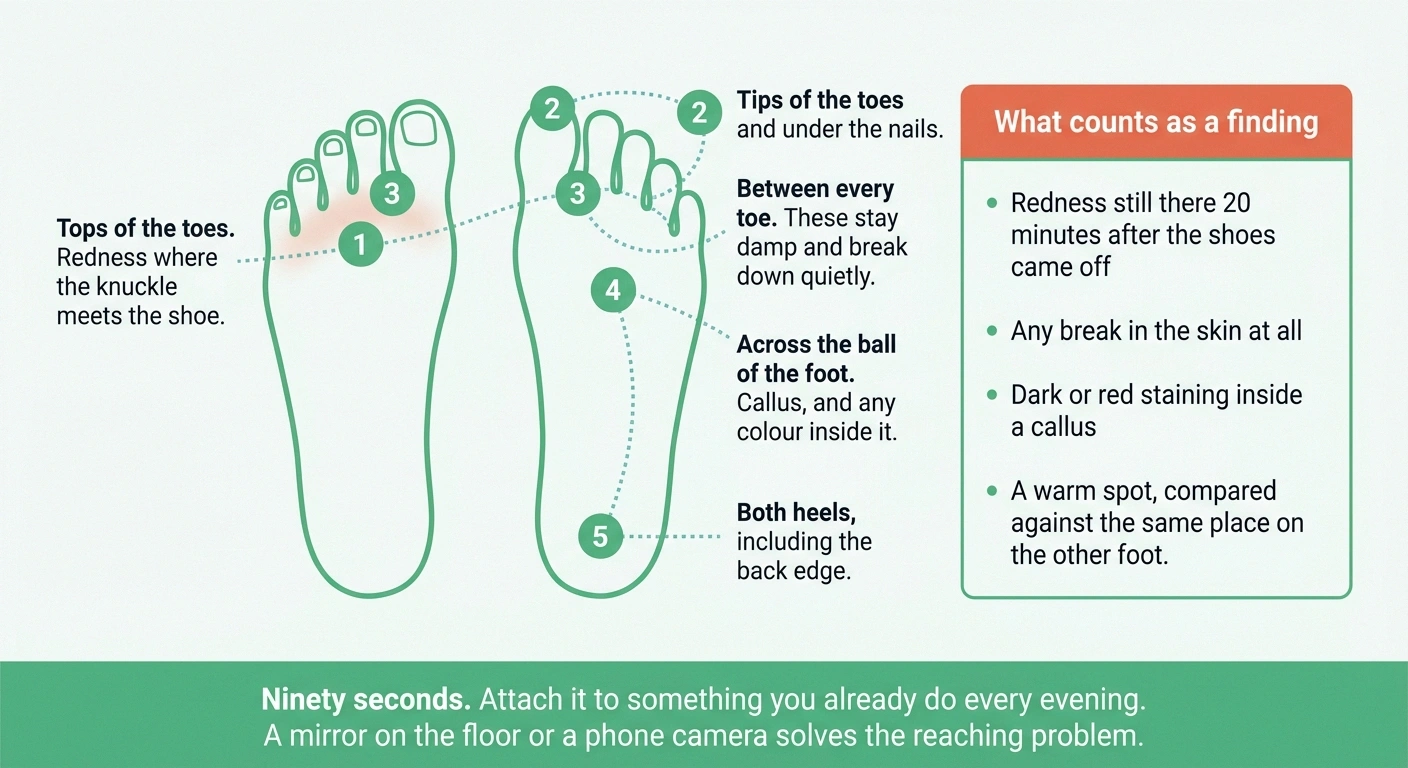
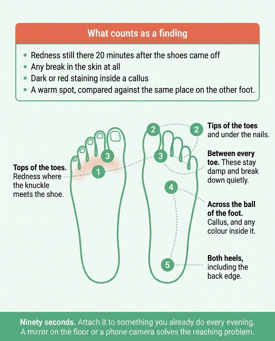
The daily check takes ninety seconds. Tops of the toes, tips of the toes, between the toes, under the ball of the foot, both heels. A mirror on the floor or a phone camera solves the reaching problem. What you are looking for is redness that has not faded twenty minutes after the shoes came off, any break in the skin, any staining inside a callus, and any warm spot that feels warmer than the same spot on the other foot.
Do it at the same time every day, attached to something you already do. Ninety seconds a day is a small price for catching a problem while it is still a red mark, and there is no substitute available for a foot that cannot report its own condition. Feet that also burn or tingle need the same inspection routine, which is worth remembering if the discomfort described in burning feet syndrome is masking what your eyes would otherwise catch.
When Surgery Comes Up
Surgical correction exists and it is a legitimate option in specific circumstances: a rigid deformity causing pain that shoes and padding cannot resolve, or a toe producing repeated ulceration despite good offloading.
Take this to the consultation
Seven questions, and why each one matters
- How many neuropathic feet do you operate on in a year? Healing and infection risk differ enough that regular experience with this population is the thing you are actually shopping for.
- Is my deformity flexible or rigid, in your assessment? It decides whether a tendon procedure or a joint fusion is on the table, and it is worth hearing whether that answer matches your own home check.
- Are you fusing a joint or releasing a tendon? These are different operations with different recoveries. The word correction covers both.
- What happens to the neighbouring toes? Fixing one toe can move load onto the next, which is why several are often addressed together.
- How long am I off the foot, and what is the plan if it heals slowly? Ask for the slow-healing plan specifically. That is the branch that matters in a neuropathic foot.
- Has my circulation been assessed? Blood supply predicts healing more than the deformity does, and it is a separate question from nerve function.
- What does my footwear look like afterwards? If the answer is still extra-depth shoes and a daily foot check, that is worth knowing before rather than after.
Sensation does not return after correction. The foot still cannot report a problem, so the routine that keeps it intact continues either way. That is the fact that turns this into a comfort-and-ulceration decision rather than a cure decision.
Procedures range from releasing or transferring a tendon in a flexible deformity, through fusing a joint to hold a rigid toe straight, to removing a section of bone to shorten the toe. Which one applies depends on the joints involved and on whether the deformity still moves.
The considerations specific to neuropathy are worth knowing before the conversation. Healing is slower where nerve supply and circulation are compromised, and infection risk is higher. Sensation does not return after correction, so the foot still cannot report a problem afterwards, and lifelong foot care continues exactly as before. Correcting one toe can also shift load onto its neighbours, which is why surgeons often address several at once.
None of that is an argument against it. It is an argument for having the conversation with a surgeon who works regularly with neuropathic feet, and for asking directly what the plan is if healing is slow.
For most people the deformity never reaches that point. It sits there, gradually, for years, and it stays manageable on shoes with enough depth, a padding solution that fits, and a ninety-second look every evening. That combination is unglamorous and it is what actually keeps feet intact. The relationship between this and the wider progression of nerve damage, including what slows it, is covered in the overview of diabetic neuropathy.
Frequently Asked Questions
What causes claw toes in neuropathy?
Motor nerve damage weakens the small intrinsic muscles inside the foot that normally hold the toes flat, while the larger muscles in the calf that pull the toes upward and downward remain relatively strong. The resulting imbalance buckles the toes. Research indicates this is not the complete explanation, since muscle atrophy alone does not predict who develops the deformity, and thickening of the plantar fascia appears to be a necessary co-factor.
What is the difference between a claw toe and a hammertoe?
A hammertoe bends at the middle joint only, humping up in the middle while the tip points forward and down. A claw toe lifts at the base joint where the toe meets the foot and curls at both joints beyond it, so the whole toe grips downward. Claw toes usually affect several toes at once, which is the pattern that suggests nerve involvement.
Can claw toes be reversed without surgery?
A flexible deformity, one where the toes straighten when you push up under the ball of the foot, may improve in comfort and position with padding, toe crests, roomier shoes and stretching, though the underlying nerve damage does not reverse. A rigid deformity, where the toes stay curled, cannot be straightened non-surgically. The realistic goal there is accommodation with footwear that fits the foot’s current shape.
Why does the ball of my foot hurt when the problem is in my toes?
The protective fat pad under the ball of the foot is anchored to the toes. When a toe pulls up into the clawed position it drags that pad forward, leaving the ends of the long foot bones sitting on much thinner tissue. Callus and pain in that spot are a direct consequence of the toe position rather than a separate problem.
Do toe exercises help claw toes?
They are worth doing, with realistic expectations. Because intrinsic muscle atrophy alone does not determine who develops the deformity, strengthening those muscles is unlikely to reverse an established claw toe. The stronger case for the exercises is balance and stability, which intrinsic foot muscles contribute to and which is worth protecting for its own sake.
What kind of shoes are best for claw toes?
Extra-depth shoes, sold under that name, because the issue is vertical clearance for the raised knuckles rather than width. Look for a soft seamless upper over the toes, no seams crossing the knuckles, a foot-shaped rather than tapered toe box, a removable insole, and a firm heel counter. Get measured each time and shop in the late afternoon when feet are at their largest.
Are toe crest pads worth trying?
They have a clear mechanism and some published support. A crest sits under the curled toes and fills the gap to the ground, spreading load that would otherwise be concentrated on the tips. A pilot study reported significant improvement in calluses and lesions at both follow-up points. Have the fit checked rather than guessing, since a crest that is too thick creates its own pressure point.
Is it safe to trim a callus on a foot with neuropathy?
Not at home. Skin that cannot feel gives no feedback about depth, and self-trimming is a common starting point for foot wounds in this population. Medicated corn pads and removers are worse, because they work by chemically burning tissue. A callus with dark or red staining inside it indicates bleeding underneath and should be seen by a foot specialist within days.
About the Author