
The first time a neurologist told me my EMG was “essentially normal,” I felt something I had not expected. Not relief. Confusion. I was sitting in that paper-covered exam chair with feet that burned so brightly at night I sometimes woke myself up gasping, and the test said my nerves were fine. It took another six months, a second opinion, and a small skin biopsy on my calf to learn that the EMG had not tested the nerves that were actually broken. It had tested the big ones. The little ones — the ones that carry burning, cold, warm, and the ache of a stove burner — were not measured that day at all.
I am Janet Ellis. If you have been told your nerves are “normal” while your feet insist otherwise, or if a doctor has used words like “small fiber” and “large fiber” and “A-delta” and “C fiber” and moved on without stopping to explain what any of that means, this is the plain conversation I wish I had gotten in that exam room. Understanding nerve fiber types changed everything about how I could ask my next doctor the right questions. I hope it does the same for you.
Your Nerves Are Not One Thing
The word “nerve” is a bit like the word “car” — useful, general, and completely hides the differences that matter. What most people picture as a single peripheral nerve is actually a bundle of many different types of nerve fibers, each with its own diameter, its own coating, its own conduction speed, and its own job.
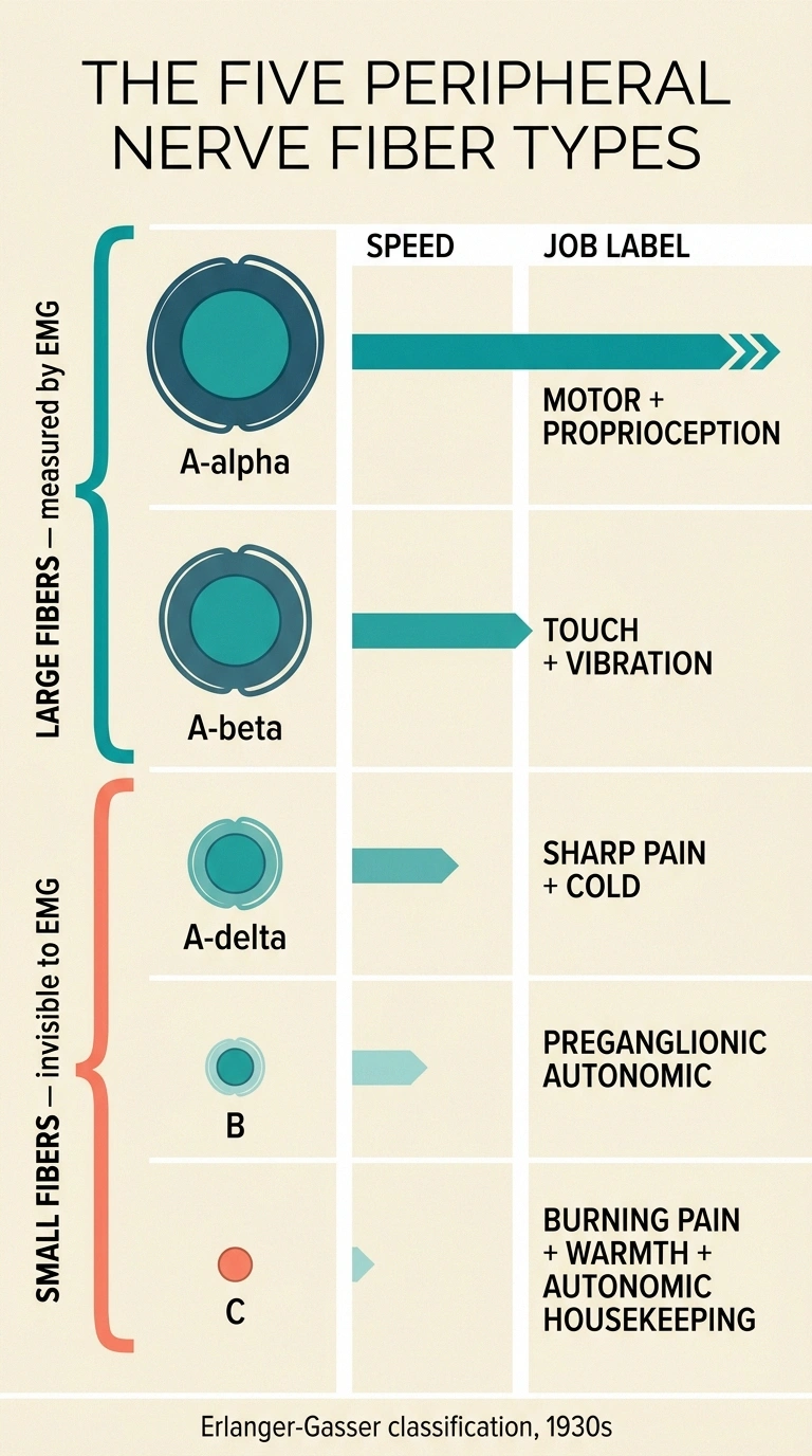
Peripheral nerves are not one thing. They are bundles of different fiber types with different sizes, coatings, and jobs. Standard EMG measures only the fast, myelinated large fibers. Burning pain, temperature issues, and autonomic symptoms come from the slow, small C fibers and A-delta fibers — which need a skin biopsy or specialized autonomic testing to evaluate.
The classification most doctors still use dates back to the 1930s, developed by two scientists named Erlanger and Gasser, who eventually won a Nobel Prize for it. They sorted fibers into groups based on diameter and conduction speed. In simpler modern language, the peripheral nerve fibers you care about most as a patient fall into two broad camps: the large fibers (myelinated, fast, carrying touch, vibration, position sense, and muscle signals) and the small fibers (thinly myelinated or unmyelinated, slow, carrying pain, temperature, itch, and autonomic signals like sweating, blood pressure, and digestion).
Different diseases attack different fiber types. Different tests measure different fiber types. Different medications work on different fiber types. This is why the language matters — not for the sake of vocabulary, but because the fiber map is the map to the right diagnosis and the right treatment.
The A-Alpha and A-Beta Fibers: The Big, Fast Nerves
Let’s start with the largest and fastest, since those are the ones the standard neurology tests are designed to catch.
A-alpha fibers are the largest of all — about 13 to 20 micrometers thick — and the fastest, conducting signals at 80 to 120 meters per second. That’s about the length of a football field every second. They are heavily coated in myelin, the fatty insulating sheath that lets electrical signals jump from gap to gap along the fiber rather than crawling continuously down it. Their main job is motor control — they carry the signals from your brain and spinal cord to your skeletal muscles and back — and they carry proprioception, which is the sense of where your body is in space without looking.
A-beta fibers are the next size down, about 6 to 12 micrometers thick, conducting at 33 to 75 meters per second. Also well-myelinated. These are the fibers that carry light touch, vibration sensation, and part of your position sense. When a doctor asks you to close your eyes and tell them whether a tuning fork buzzing on your ankle is on or off, they are testing your A-beta fibers.
These two types together — A-alpha and A-beta — are what we generally mean when we say “large fibers.” They matter for a specific reason: they are what standard EMG and nerve conduction studies measure. The technician sends a small electric pulse down a nerve and times how long it takes to arrive at a receiving electrode. Those tests are calibrated for the fast, myelinated highways. When your EMG comes back “normal,” it means those specific fibers looked fine on that specific day. It does not mean every nerve in your body is fine.
The A-Delta Fibers: Sharp, Fast, and Localized
Now we cross into small-fiber territory, and this is where a lot of the confusing pain language starts to make sense.
A-delta fibers are small — 1 to 5 micrometers in diameter — with only a thin coating of myelin. They conduct signals at 6 to 30 meters per second. Slower than the big fibers, but still fast enough that when you touch a hot pan, the sharp “ow” that lifts your hand off the burner arrives before you consciously register it.
A-delta fibers are the “first pain” fibers. Their job is to carry the sharp, well-localized, easy-to-point-to kind of pain that alerts you to acute tissue damage. Prick your finger with a pin — that first bright sting is A-delta. They also carry cold temperature signals. When you plunge a hand into ice water, the sharp cold sensation is largely A-delta.
Notice the word “well-localized.” A-delta fibers connect to specific areas of skin in a way that lets your brain map exactly where the signal is coming from. That is why you can point at the spot the pin stuck and be right within a fraction of an inch.
The C Fibers: Slow, Diffuse, and Burning
C fibers are the smallest, the slowest, and — for people with neuropathy — often the source of the most misery.
| Fiber | Size (μm) | Speed (m/s) | Myelin | Main job |
|---|---|---|---|---|
| A-alpha | 13–20 | 80–120 | Heavy | Motor to skeletal muscle, proprioception |
| A-beta | 6–12 | 33–75 | Heavy | Light touch, vibration, position sense |
| A-delta | 1–5 | 6–30 | Thin | Sharp fast pain, cold temperature |
| B | ~3 | 3–15 | Thin | Preganglionic autonomic |
| C | 0.2–1.5 | 0.5–2 | None | Burning pain, warmth, autonomic housekeeping |
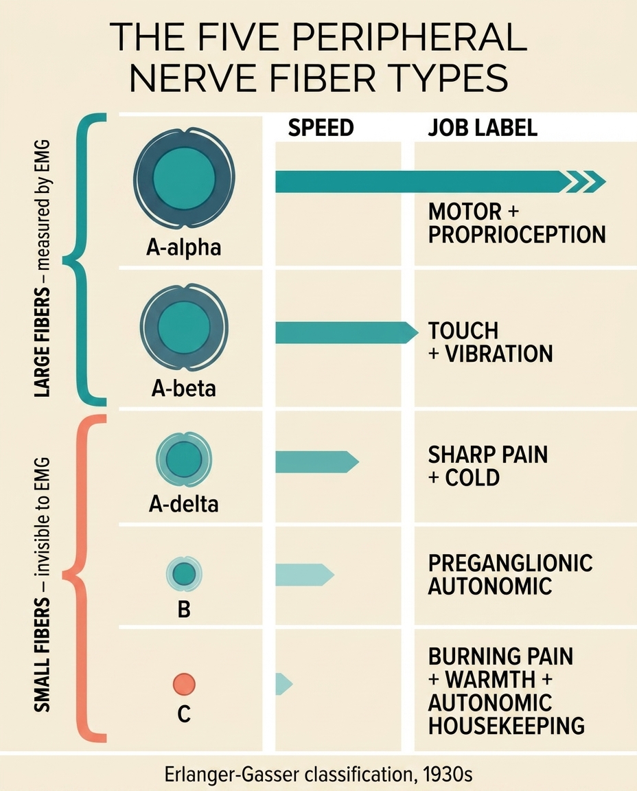
Fibers highlighted in coral (A-delta, C) are the “small fibers.” Together they carry pain, temperature, and autonomic signals — and are invisible to standard EMG.
C fibers are so small (less than 1.5 micrometers) and so slow (about half a meter per second up to about two meters per second) precisely because they have no myelin sheath at all. The electrical signal has to travel continuously along the length of the fiber rather than jumping. Slow, but energetically cheap — and it lets the body pack a very large number of C fibers into each nerve bundle.
C fibers carry three very different things:
- The “second pain” — the dull, aching, burning, throbbing kind of pain that arrives a second or two after the sharp A-delta pain and lingers. When you stub a toe, the initial sharp bright pain is A-delta. The deep miserable ache that follows for the next several minutes is C fiber.
- Warmth — the sensation of a heating pad or warm bath, as distinct from the sharp cold that A-delta fibers register.
- Post-ganglionic autonomic signals — the “housekeeping” nerve signals that control sweating, blood-vessel tone, blood pressure regulation, heart rate, digestion, bladder function, and pupil size. This is a huge job that most patients don’t realize their tiniest nerves are doing.
Because C fibers are unmyelinated and diffuse in where they connect on the skin, C-fiber pain is famously hard to point to. Patients describe it as “everywhere in this general area,” “burning across my whole foot,” “a hot poker somewhere in the sole.” That vague, spreading quality is the fiber type talking.
Two Kinds of Pain From One Injury: The Dual Pain Response
Here is a demonstration that makes the difference stick, and that I have used a hundred times in support-group conversations to help newer members understand what their nerve doctor is trying to tell them.
The Erlanger-Gasser classification that organizes peripheral nerve fibers by diameter and conduction speed dates to the 1930s. Joseph Erlanger and Herbert Gasser won the 1944 Nobel Prize in Physiology or Medicine for this work. A century later, it remains the framework doctors still use to describe which fibers are damaged in a given neuropathy.
Translation: this vocabulary is not new or fringe. It is the standard language of nerve medicine, and every neurologist you see is fluent in it.
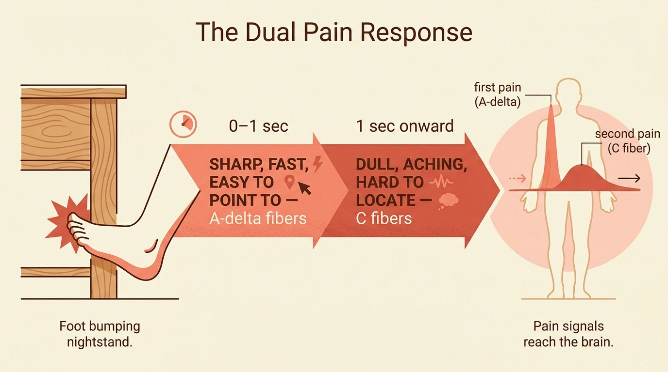
Imagine you stub your toe on the corner of a nightstand. Two waves of pain hit you, one right after the other. The first is sharp, bright, and immediate — you can point to exactly which toe and what part of it — and it fades within a second or two. The second wave arrives a moment behind the first: a duller, spreading ache that throbs for the next several minutes and is much harder to point to.
The first wave rides in on your A-delta fibers. The second rides in on your C fibers. Same injury, two different messengers, arriving at two different speeds because the fibers have two different sizes and two different coatings.
That dual pain response is universal in healthy nerves. In neuropathy, it can go strange in specific ways depending on which fibers are damaged. Damage predominantly to C fibers may amplify the second wave — the burning, the aching, the diffuse misery — while leaving the sharp first wave intact or even blunted. This is why many of us with small-fiber neuropathy describe our pain as “burning” and “deep” and “hard to locate,” and rarely as “sharp” or “stabbing.”
Small Fiber Versus Large Fiber Neuropathy: Why the Distinction Matters
Doctors sort peripheral neuropathies by which fibers are damaged, and the difference shows up in what patients notice, what tests find, and what treatments help.
| If you notice… | Fiber type likely involved |
|---|---|
| Burning pain in feet, especially at night | C fibers (small) |
| Sharp shock-like pain that’s easy to point to | A-delta fibers (small) |
| Temperature misreading, allodynia | C and A-delta (small) |
| Dry eyes/mouth, dizziness on standing, sweating changes | Autonomic C fibers |
| Numbness, “walking on cotton” | A-beta fibers (large) |
| Loss of vibration sensation, absent reflexes | A-beta fibers (large) |
| Muscle weakness, wasting | A-alpha motor fibers (large) |
| Balance problems worse in the dark | A-beta proprioception (large) |
Small fiber neuropathy means the A-delta and C fibers are damaged. The symptoms you are most likely to notice:
- Burning pain in the feet or hands, often worse at night
- Prickling, electric, or pins-and-needles sensations
- Sensitivity to temperature — feet that feel too hot, too cold, or that misread water temperature
- Allodynia (light touch, like the bedsheet, feels painful)
- Autonomic symptoms: dry eyes and mouth, changes in sweating (either too much or too little), lightheadedness when standing up, digestive changes, bladder changes, sexual dysfunction
What you may NOT notice, and what your doctor may find on exam: normal strength, normal reflexes, normal vibration sensation. Small fiber neuropathy leaves the large-fiber functions relatively intact. For more, see our dedicated page on small fiber neuropathy.
Large fiber neuropathy means the A-alpha and A-beta fibers are damaged. Different picture:
- Numbness, often described as “walking on cotton” or “wooden feet”
- Loss of vibration sensation (the tuning fork test fails)
- Loss of position sense — trouble knowing where your feet are without looking
- Reduced or absent ankle reflexes on exam
- Balance problems, especially in the dark or with eyes closed
- Muscle weakness in more advanced cases
Large fiber neuropathy is what the classic exam maneuvers were designed to catch. Standard nerve conduction studies measure large fibers directly.
Mixed fiber neuropathy — damage to both — is the most common real-world picture. Classic diabetic peripheral neuropathy usually starts as small-fiber damage first, then spreads to large fibers as the disease progresses. Chemotherapy neuropathies, alcoholic neuropathy, and many idiopathic neuropathies also tend to be mixed by the time patients seek help.
Why Your EMG Can Be Normal When Your Symptoms Are Not

This is the exact issue that sent me from the neurologist’s office to a second opinion, and it is worth understanding clearly.
An electromyography (EMG) and nerve conduction study (NCS) is a very useful test, but it is a large-fiber test. It measures how well the fastest, most heavily myelinated fibers conduct an electrical signal from Point A to Point B. It cannot, by design, measure C fibers or A-delta fibers directly. Those fibers are too slow and too small; the equipment doesn’t pick them up.
So consider what happens in pure small-fiber neuropathy: the A-alpha and A-beta fibers are healthy. The C and A-delta fibers are damaged. You have burning pain, temperature issues, and autonomic symptoms — and a normal EMG. That does not mean your doctor is wrong to run the EMG (it rules out large-fiber problems, which is important information). It means the EMG cannot rule out small-fiber neuropathy.
The tests that can assess small fibers include:
- Skin punch biopsy — a tiny 3-millimeter piece of skin, usually from the calf, examined under a microscope to count the intraepidermal nerve fiber density (IENFD). Reduced density is a diagnostic finding for small fiber neuropathy.
- QSART (Quantitative Sudomotor Axon Reflex Test) — measures how well small autonomic C fibers control sweating in response to a chemical stimulus.
- Autonomic testing — tilt-table testing, heart-rate variability, and related tests that assess how well small fibers regulate blood pressure and heart rate.
- QST (Quantitative Sensory Testing) — controlled temperature and vibration stimuli that can separate small-fiber and large-fiber deficits.
If your symptoms are classic for small-fiber neuropathy and your EMG is normal, ask specifically about a skin biopsy. That is the question that changed my diagnostic trajectory.
The B Fibers: The Autonomic Layer You Rarely Hear About
For completeness, and because they occasionally come up in medical conversations, there is a fourth category worth briefly naming.
B fibers are small, myelinated preganglionic autonomic fibers — about 3 micrometers thick, conducting at 3 to 15 meters per second. They carry autonomic signals from the central nervous system to the autonomic ganglia (the switching-station clusters where autonomic signals hand off to the C-fiber postganglionic fibers that finish the trip to organs, blood vessels, and glands).
You rarely hear about B fibers as a patient because most autonomic neuropathies affect either the postganglionic C fibers (which you can measure with QSART) or the entire autonomic pathway together. But the term shows up in medical literature and occasionally in specialist consultations, and now you know what it means when it does.
How Different Diseases Attack Different Fibers
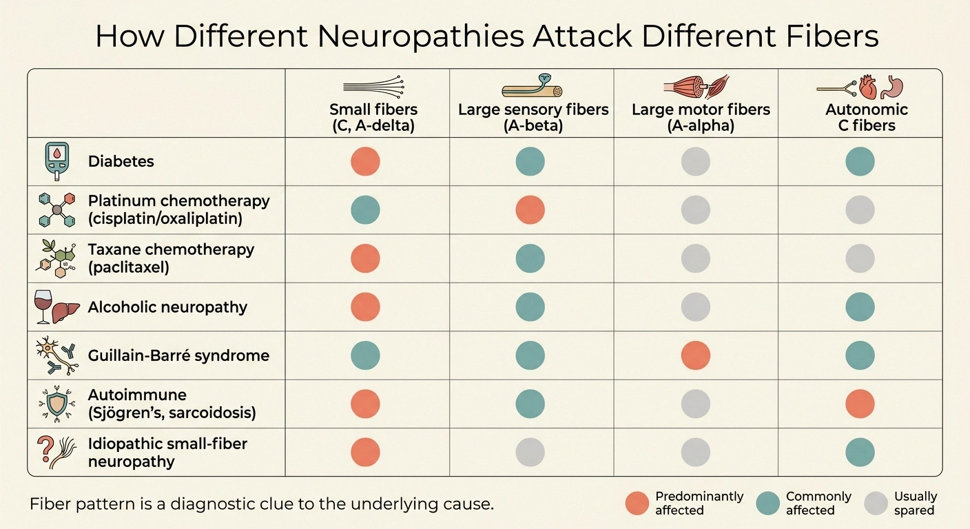
Not every neuropathy hits every fiber type equally. The pattern of which fibers are affected is often a clue to the underlying cause.
If your EMG is normal but you have burning pain, temperature sensitivity, allodynia, or autonomic symptoms, ask specifically: “Could a skin biopsy for intraepidermal nerve fiber density help evaluate small-fiber neuropathy?”
A normal EMG rules out large-fiber damage. It does not rule out small-fiber damage. This distinction has changed the diagnostic trajectory for thousands of patients.
Diabetes classically attacks small fibers first. Many people with early diabetic neuropathy have small-fiber symptoms (burning feet, temperature issues, subtle autonomic changes) for years before large-fiber findings show up on EMG. This is why “normal EMG” in a diabetic with foot symptoms is often just a snapshot of early disease.
Chemotherapy-induced neuropathy from platinum drugs (cisplatin, oxaliplatin) tends to attack large sensory fibers, producing dominant numbness and position-sense loss. Taxane chemotherapies (paclitaxel, docetaxel) tend to hit both small and large fibers, producing mixed patterns. Vincristine tends to hit both sensory and motor large fibers. For more on the chemotherapy patterns, see our chemo-induced neuropathy page.
Alcoholic neuropathy tends to be a mixed small- and large-fiber process, with burning pain and numbness both prominent — see our page on alcoholic neuropathy.
Guillain-Barré syndrome is a large-fiber (specifically motor-fiber) attack — profound weakness with relatively preserved pain and temperature sensation, at least at first. Very different presentation than the small-fiber picture.
Autoimmune small-fiber neuropathies (associated with conditions like Sjögren’s syndrome, sarcoidosis, celiac disease) selectively damage small fibers, sometimes leaving large-fiber function completely normal for years.
Idiopathic small-fiber neuropathy — the “we cannot find a cause” version — is increasingly recognized as more common than it used to be. This is where the skin biopsy has done a lot to move patients out of the “your nerves are normal, it’s in your head” category and into the “yes, your small fibers are damaged, let’s manage it” category.
Why Fiber Type Guides Treatment
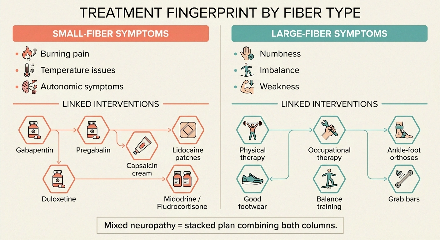
Understanding which fibers are damaged is not just intellectually satisfying. It changes what actually helps.
For small-fiber (C and A-delta) pain, the medications with the best evidence work on the pain-signaling machinery of those fibers. Gabapentin and pregabalin (Lyrica) reduce the excitability of small pain fibers. Duloxetine (Cymbalta) works on descending pain-modulating pathways that heavily involve C-fiber inputs. Topical capsaicin (as a cream or patch) works directly on the TRPV1 receptor found on C fibers, essentially depleting the fiber’s ability to signal for a period of weeks. See more in our roundup of the best neuropathy creams.
For large-fiber deficits (A-alpha and A-beta) that produce numbness, imbalance, and weakness, medication does much less. What helps is physical and occupational therapy — retraining the body to compensate for the sensory information that is no longer arriving reliably. Good footwear (see our best shoes for neuropathy page), grab bars and lighting adjustments to prevent falls, ankle-foot orthoses for foot drop, walking sticks and canes when needed, and dedicated balance training. Our page on balance and fall prevention covers this in depth.
For autonomic (small fiber) symptoms, treatment gets specific to what is affected. Orthostatic hypotension may need salt and fluid adjustments, compression garments, or medications like midodrine or fludrocortisone. GI symptoms may need prokinetic medications and dietary changes. Sweating irregularities may need specific interventions. Our page on autonomic neuropathy walks through this territory.
For mixed neuropathies — the majority of real-world cases — the treatment plan combines the small-fiber pain regimen, the large-fiber compensation strategies, and the autonomic-specific interventions as needed. This is one reason why neuropathy care is rarely a single prescription. It is a stack.
How to Talk to Your Doctor About Fiber Types

This vocabulary can help you have a much sharper conversation at your next appointment. Some questions that opened doors for me:
- My EMG was normal. Could this be small-fiber neuropathy that wouldn’t show on it?
- Would a skin biopsy for intraepidermal nerve fiber density be reasonable here?
- Which fiber types do you think are most affected in my case?
- Are there autonomic symptoms — dry eyes, dizziness on standing, sweating changes, GI issues — that should trigger small-fiber testing?
- Am I dealing with small fiber, large fiber, or both? How does that change the treatment plan?
- “My EMG was normal, but I still have burning pain. Could this be small-fiber neuropathy that wouldn’t show on the EMG?”
- “Would a skin biopsy for intraepidermal nerve fiber density be reasonable here?”
- “Which fiber types do you think are most affected in my case, and what would that suggest for treatment?”
- “Do I have any autonomic symptoms — dry eyes, dizziness on standing, sweating changes, GI issues — that should be tested for small-fiber involvement?”
- “Are we looking at a small-fiber problem, a large-fiber problem, or both? How does that change the plan?”
These are not adversarial questions. They are specific questions that show your doctor you are engaged and thinking, and they usually get more thoughtful answers than the general “so what do you think is going on.”
Where Fiber Types Fit in the Bigger Neuropathy Picture
Understanding fiber types is one layer of understanding neuropathy overall. It sits alongside the other big framing questions: how far along the process has gone (see what are the stages of neuropathy), what caused it in the first place (see idiopathic neuropathy and medications that can cause neuropathy), and whether any of it is reversible (see can neuropathy be reversed). Distinguishing nerve pain from vascular pain is another useful reframe — our page on nerve pain versus vascular pain walks through that.
But knowing whether it is your small fibers, your large fibers, or both that are struggling is the single most useful piece of vocabulary I have picked up in this journey. It reframes the exam findings. It reframes the test results. It reframes what medications are worth trying. And it reframes the frustrating conversation where a doctor says “your EMG is normal” — because now you know that sentence means something specific, not something total.
Frequently Asked Questions
What is the difference between A-delta fibers and C fibers?
A-delta fibers are small myelinated fibers that carry sharp, well-localized “first pain” and cold temperature at 6 to 30 meters per second. C fibers are the smallest unmyelinated fibers, carrying dull, burning, hard-to-localize “second pain,” warmth, and autonomic housekeeping signals (sweating, blood pressure regulation) at less than 2 meters per second. Both are considered small fibers.
Can you have small fiber neuropathy with a normal EMG?
Yes. Standard EMG and nerve conduction studies measure large fibers only. C fibers and A-delta fibers are too small and too slow to register on those tests. A normal EMG in a patient with burning feet, temperature sensitivity, or autonomic symptoms does not rule out small fiber neuropathy. A skin biopsy for intraepidermal nerve fiber density is the standard next step.
Which nerve fibers does diabetes damage first?
Diabetes classically damages small fibers first. Many patients with early diabetic neuropathy have burning pain, temperature issues, and subtle autonomic changes for years while their standard nerve conduction studies remain normal. Large fiber damage often develops later as the disease progresses.
Why does my neuropathy pain feel burning rather than sharp?
Burning pain is characteristic of C fiber damage. C fibers are unmyelinated small fibers that carry dull, spreading, poorly-localized pain and warmth sensations. When C fibers are irritated or damaged, the brain interprets their misfiring signals as burning. Sharp, well-localized pain would be more suggestive of A-delta fiber involvement.
Do gabapentin and pregabalin work on all nerve fibers?
Gabapentin and pregabalin primarily reduce the excitability of pain-signaling fibers — mostly small fibers (C and A-delta) but also some pain-related large-fiber input. They do not repair fibers or restore lost function such as numbness or muscle weakness. Their target is pain-signal reduction, which is why they help burning and stabbing symptoms more than numbness or imbalance.
What test measures C fibers?
Several tests can assess C fiber function. Skin punch biopsy with intraepidermal nerve fiber density counting is the most common. QSART (Quantitative Sudomotor Axon Reflex Test) measures the ability of small autonomic C fibers to trigger sweating. Autonomic testing (tilt table, heart rate variability) also assesses C fiber function indirectly. Standard EMG does not.
Can small fiber neuropathy progress to large fiber neuropathy?
Yes, in many cases the same underlying process that damages small fibers eventually damages large fibers as well. This is the typical trajectory in diabetic neuropathy — small-fiber symptoms appear first, then large-fiber findings (numbness, reduced reflexes, imbalance) develop over months to years. Not every small fiber neuropathy progresses this way, however. Some remain pure small-fiber conditions for the long term.
Are motor nerves considered large fibers?
Yes. Motor nerves that carry signals to skeletal muscles are A-alpha fibers, the largest and fastest of all peripheral nerves. When a neuropathy affects motor function — producing weakness or muscle wasting — large motor fibers are involved. This is why standard EMG can detect motor nerve involvement well.
About the Author