
The story is almost always the same when someone finally lands in a neurology office with lumbosacral plexopathy. It started with pain — deep, boring pain in one hip or thigh, worse at night, not fitting a “pulled muscle” or “pinched nerve in the back” story. A few weeks in, the leg started giving out. Standing from a chair got harder. The thigh visibly wasted. Along the way, three or four clinicians offered three or four explanations — sciatica, disc problem, diabetic neuropathy, “your age” — none of which quite fit.
I’m Janet Ellis. I write for NeuropathyResource because I live with peripheral neuropathy myself, and I’ve spent years translating dense neurology papers into language other patients can actually use. I’m not a medical professional. What I can do is walk you through a distinction that gets missed constantly — the difference between length-dependent peripheral neuropathy (the “stocking and glove” burning that starts in the feet) and lumbosacral plexopathy (a very different animal that hits one leg proximally and hard). Getting the label right matters because the causes, tests, and treatments diverge sharply from a typical neuropathy protocol.
What Lumbosacral Plexopathy Actually Is
Your lower body is wired by a nerve network called the lumbosacral plexus. It sits inside your pelvis and it’s where the nerve roots from your lower spine (L1 through S4) merge, cross, and reorganize before they leave the pelvis as the major peripheral nerves of the leg — the femoral nerve that powers hip flexion and knee extension, the sciatic nerve that runs down the back of the leg, the obturator nerve on the inner thigh, the gluteal nerves for hip abduction, and the pudendal nerve for pelvic-floor function.
Lumbosacral plexopathy is injury to the nerve network inside the pelvis — not the spine (radiculopathy) and not the peripheral nerve in the leg (peripheral neuropathy). It hits asymmetrically, hits the thigh muscles first, and produces pain out of proportion to the weakness — exactly the opposite of the symmetric burning-feet pattern most people picture with “neuropathy.”
“Plexopathy” means injury at the plexus level itself — after the nerve roots exit the spine but before the individual named nerves have fully separated into the leg. That location is what makes it its own thing. Injury one step further up the chain (at the nerve root, inside the spinal canal) is called radiculopathy. Injury one step further down (at a specific peripheral nerve, in the limb) is called mononeuropathy or entrapment. Plexopathy sits in the middle, and because the plexus is such a densely interwoven structure, a single lesion at that level can affect several downstream nerves simultaneously — which is what produces the distinctive clinical picture.
Why this matters practically: the workup, the imaging, and the treatment for a plexopathy don’t look like the workup for a length-dependent peripheral neuropathy or for a lumbar disc problem. Miss the plexus as the site of injury and the tests will come back clean and the diagnosis will keep bouncing around, sometimes for months.
How It Differs From Peripheral Neuropathy
The classic peripheral neuropathy that most people picture — the one diabetes is the most common cause of — is length-dependent. It starts in the longest nerves first, which is why the feet go before the calves and the calves go before the thighs. It’s symmetric, meaning both feet are affected roughly equally. It’s distal-predominant, meaning toes and soles bother you long before knees or hips. The symptoms tend to be burning, tingling, numbness, and pins-and-needles, in a “stocking” distribution that gradually creeps upward over years.
Lumbosacral plexopathy looks nothing like that. It is asymmetric — one leg, not both. It is proximal-predominant — the thigh muscles hit before the foot, exactly the opposite of length-dependent PN. Pain is prominent early and often severe, in a deep, boring, aching quality rather than the burning tingle of PN. Weakness is prominent early too, and often outpaces the sensory changes. Multiple nerve territories get hit in the same limb at once, because a single lesion at the plexus affects multiple nerves that emerge from it. And the reflex loss is patchy — the knee jerk often disappears (femoral involvement) while the ankle jerk stays; nothing like the symmetric loss of ankle reflexes typical of length-dependent PN.
If you or a family member has been diagnosed with “diabetic neuropathy” but the pattern is proximal thigh weakness on one side rather than burning feet on both, someone needs to reconsider whether the label fits. Length-dependent diabetic neuropathy and diabetic lumbosacral radiculoplexus neuropathy are both driven by diabetes, but they’re very different diseases with very different courses and treatments.
How It Differs From Sciatica and Radiculopathy — the Frequently Missed Distinction
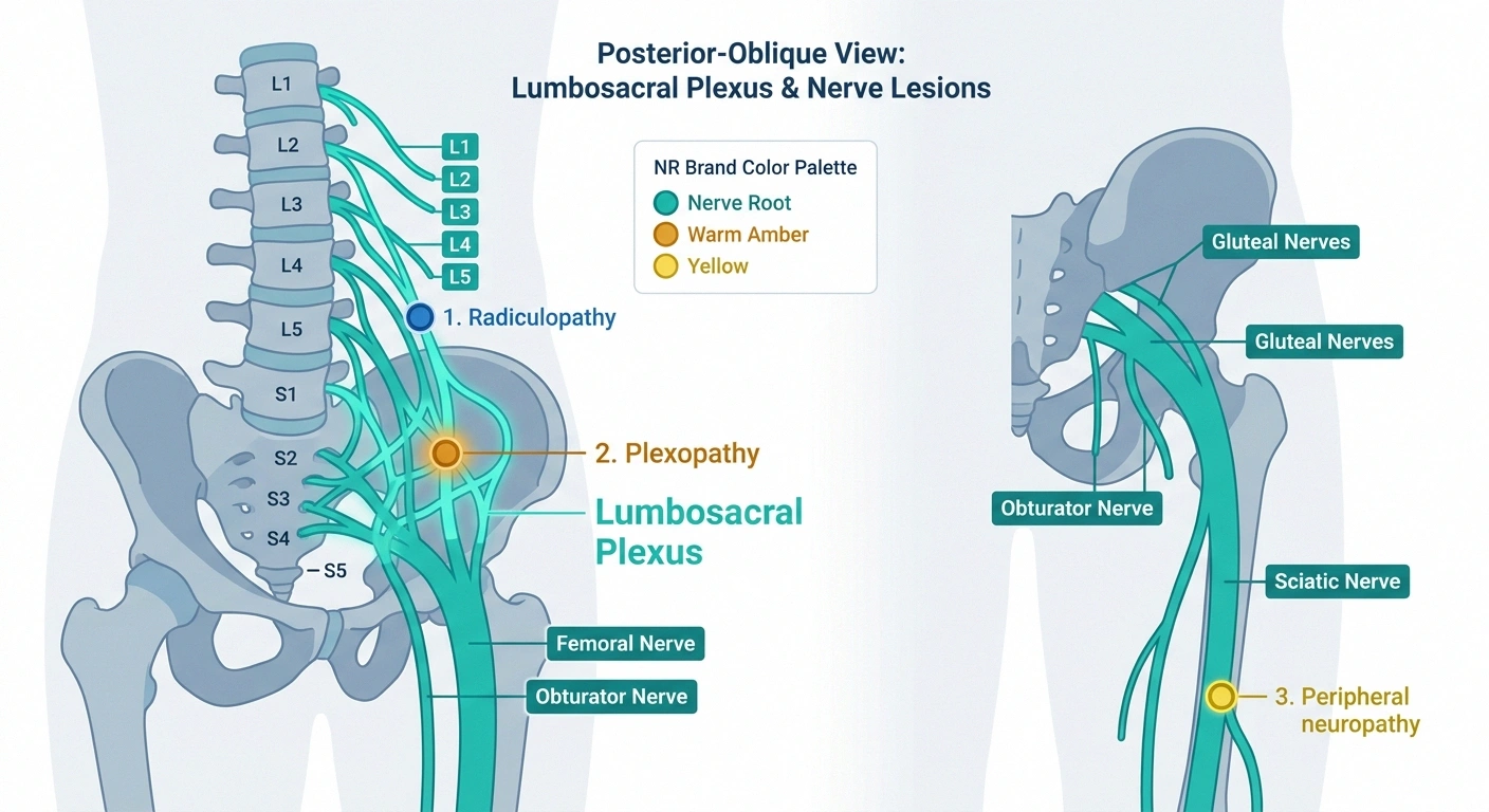
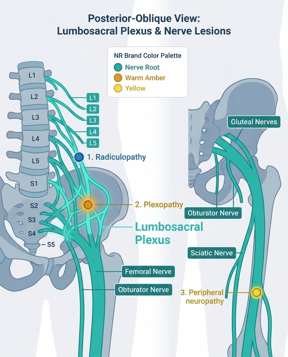
Radiculopathy is injury at the nerve root, inside or right at the spinal canal — usually from a disc herniation, bone spur, or foraminal stenosis pressing on one nerve root. Sciatica is the popular name for radiculopathy affecting the L5 or S1 nerve root, producing pain that radiates from the buttock down the back of the leg along that root’s dermatome.
Telling the Three Apart — Plexopathy vs Radiculopathy vs Peripheral Neuropathy
The confusion with plexopathy is understandable — both cause leg pain and can cause weakness. But the anatomy is different, the pattern of weakness is different, and the imaging findings are different. Radiculopathy affects one nerve root at a time, producing a specific dermatomal pain pattern and weakness limited to what that root supplies. Plexopathy affects the tangled network downstream, producing pain and weakness spanning multiple root levels in a single limb. A lumbar MRI often looks impressively abnormal in radiculopathy (herniated disc, foraminal narrowing) and is often unremarkable in plexopathy — because the lesion is in the pelvis, not the spine.
There’s a diagnostic pearl that separates the two decisively on nerve conduction study, and it’s worth understanding because you can advocate for the right test if you know it exists. Sensory nerve action potentials — the SNAPs measured on nerve conduction studies — are preserved in radiculopathy but reduced or absent in plexopathy. The reason is anatomic: the dorsal root ganglion, which contains the sensory nerve cell bodies, sits outside the spinal cord. In radiculopathy the lesion is upstream of the ganglion, so the peripheral sensory nerve is still connected to a living cell body and generates a normal SNAP even when the patient has obvious sensory symptoms. In plexopathy the lesion is downstream of the ganglion, so the peripheral sensory nerve degenerates, and the SNAP shrinks or disappears. That single finding is what an electromyographer uses to localize root versus plexus when the physical exam alone can’t tell them apart.
Diabetic Amyotrophy — the Most Common Recognizable Form

The most-recognized presentation of lumbosacral plexopathy has multiple names for essentially the same syndrome: diabetic amyotrophy, Bruns-Garland syndrome, and diabetic lumbosacral radiculoplexus neuropathy (DLRPN). Ludwig Bruns first described the picture in 1890; Hugh Garland added his name to it in 1955; P. James B. Dyck’s group at Mayo Clinic published the definitive modern series and microvasculitis biopsy data in early-2000s Brain and Neurology papers.
of patients with diabetic lumbosacral radiculoplexus neuropathy (DLRPN) develop mirror-image involvement in the contralateral leg over the months following the initial presentation — one of the features that distinguishes DLRPN from an isolated mononeuropathy.
Dyck PJB et al., Mayo Clinic — Brain and Neurology series
The classic patient is a middle-aged or older adult with type 2 diabetes — and this is a counterintuitive detail — often not poorly controlled. Many patients have HbA1c values in a reasonable range. A striking proportion have had significant unintentional weight loss in the months before symptoms began. Then the pain starts: severe unilateral hip, buttock, or thigh pain, deep and boring, often worst at night, sometimes with lancinating jolts through the leg. The pain phase dominates the first several weeks — patients rarely sleep, and opioids alone don’t touch it.
Weakness follows the pain by days to weeks. The thigh visibly wastes. Standing from a chair becomes a two-hand event. The knee jerk disappears. Roughly half of patients develop mirror-image involvement in the opposite leg over the following months. The course is monophasic — it worsens over weeks to months, plateaus, then slowly recovers over a year or two.
The pathology, per the Dyck biopsy work, is microvasculitis — inflammation of the tiny blood vessels supplying the plexus, producing ischemic injury to the nerve fibers. It is the same mechanism that drives postsurgical inflammatory neuropathy, just triggered by metabolic stress rather than surgical stress. Corticosteroids and IVIG have been used to modulate the immune process; the data are mixed on whether they change the ultimate deficit, but many neuromuscular specialists use them for pain and for what appears to be some acceleration of recovery. Recovery over 12-24 months is usually meaningful but often incomplete — a lot of patients regain independent walking but retain some residual weakness, and foot drop can persist.
Cancer, Radiation, and Tumor-Related Plexopathy
Two very different cancer-related causes deserve separate mention because the workup and the prognosis diverge sharply between them.
The first is direct infiltration or compression of the plexus by a pelvic tumor — most commonly prostate, colorectal, cervical, endometrial, ovarian, sarcoma, or lymphoma. The presenting feature is almost always pain, and it’s often severe and progressive over weeks before any objective weakness is measurable. Any patient with a history of pelvic cancer who develops new proximal leg pain and weakness needs pelvic imaging, urgently. MRI of the lumbosacral plexus typically shows a discrete mass or diffuse plexus enhancement. The prognosis depends almost entirely on the underlying oncologic control — the plexus deficit follows the tumor.
The second is radiation-induced lumbosacral plexopathy — a delayed complication that shows up years, sometimes decades, after pelvic radiotherapy for the same cancers listed above. The distinguishing clinical feature is that radiation plexopathy is typically painless — progressive weakness without the severe pain that dominates tumor infiltration. That single feature (pain versus no pain) is often what clinicians rely on to distinguish the two when a patient with a treated cancer develops new plexopathy years later. EMG has a specific finding for radiation injury called myokymic discharges — spontaneous grouped motor unit firing — that is relatively specific and, when present, helps clinch the diagnosis. Radiation plexopathy has no reversal treatment. The goal is stabilization, supportive care, and rehabilitation.
Hematomas, Anticoagulants, and Emergency Causes
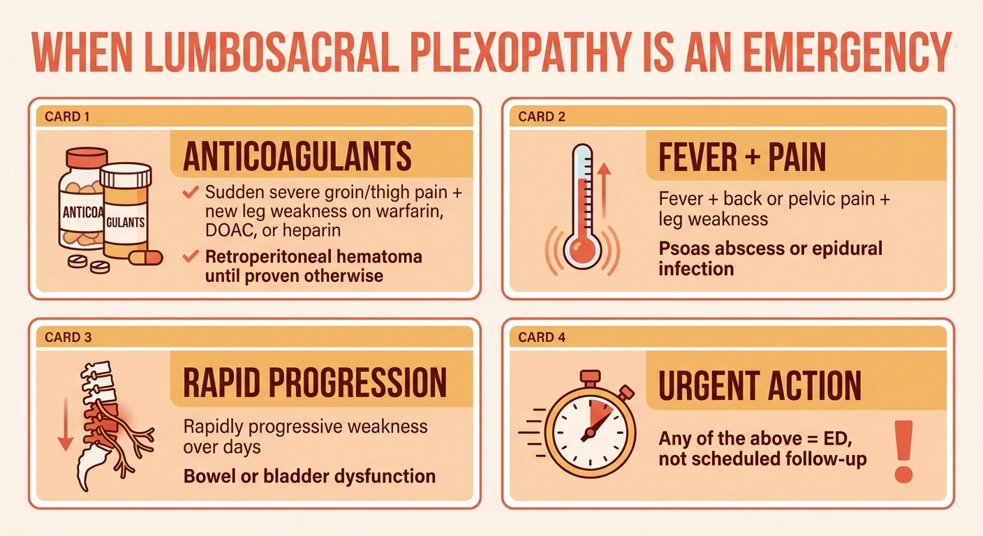
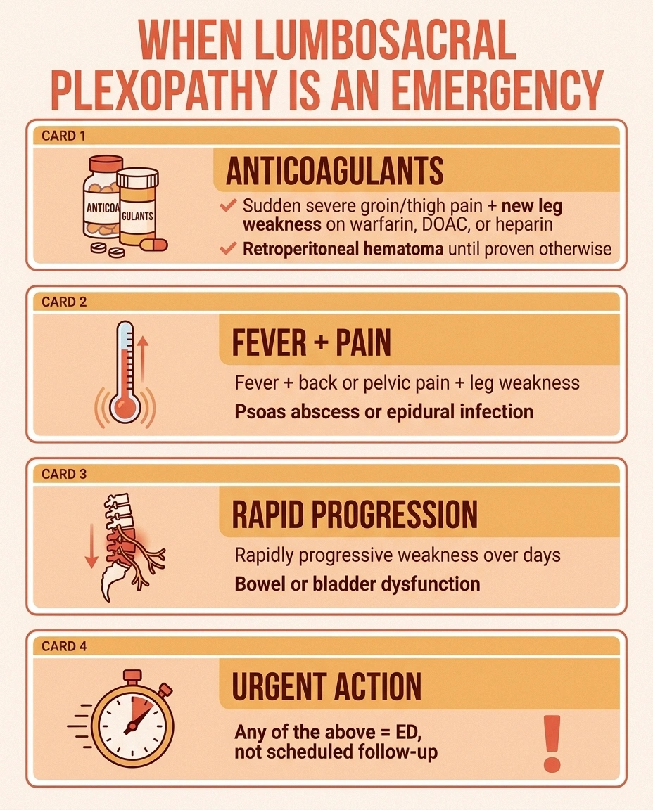
Not every cause of lumbosacral plexopathy has a slow subacute course. Some are true emergencies, and the highest-stakes one is retroperitoneal or psoas hematoma in a patient on anticoagulation.
Most causes of lumbosacral plexopathy unfold over days to weeks. A few unfold in hours and destroy nerve if not treated urgently.
Go to the emergency department, not the primary-care voicemail, for any of these:
- Sudden severe groin or thigh pain with new leg weakness while on warfarin, a DOAC, heparin, or a strong antiplatelet — retroperitoneal hematoma until proven otherwise
- Fever with back or pelvic pain and new leg weakness — possible psoas abscess or epidural infection
- Rapidly progressive weakness over days, not weeks
- New bowel or bladder dysfunction with the leg symptoms
The scenario is a patient on warfarin, a direct oral anticoagulant, heparin, or a strong antiplatelet, sometimes with a supratherapeutic INR, sometimes after minor trauma or vigorous exertion, sometimes spontaneously. Blood accumulates in the psoas muscle sheath or the retroperitoneal space and compresses the femoral nerve or the LS plexus. The onset is measured in hours, not days. Severe groin and thigh pain, weakness of hip flexion and knee extension in a femoral-nerve distribution, and reduced sensation over the anterior thigh are the hallmarks. There’s often a drop in hemoglobin on labs that flags the bleeding.
This is time-critical. Reversal of the anticoagulation and, in significant cases, drainage of the hematoma can preserve nerve function. Delay produces permanent femoral neuropathy. If you or a family member is on any anticoagulant and develops sudden severe groin or thigh pain with new leg weakness, this needs the emergency department, not a phone-tag chain with primary care.
Other rapidly progressive presentations that warrant urgent evaluation include psoas abscess (severe pain, fever, elevated inflammatory markers — often in a patient with prior tuberculosis exposure, an intravenous drug history, or immunocompromise) and, rarely, an abdominal aortic aneurysm compressing the plexus. The common thread is that any rapidly progressive weakness, weakness accompanied by bowel or bladder dysfunction, or acute severe pelvic pain in the setting of leg weakness is an emergency-department problem — not “call the office Monday morning.”
Postpartum Plexopathy and Other Less-Common Causes
A newer mother who walks out of the labor and delivery unit with a foot drop is very likely dealing with postpartum lumbosacral plexopathy. The mechanism is compression of the lumbosacral trunk — the fibers that eventually contribute to the peroneal nerve — against the sacral ala during prolonged second-stage labor, especially when the fetal head is large relative to the maternal pelvis or when forceps have been used. The classic presentation is unilateral foot drop and weakness of lateral leg muscles immediately after delivery, sometimes with numbness across the lateral lower leg. Recovery is usually near-complete over weeks to a few months, but the walk back can be slow and frustrating, and physical therapy is essential.
Idiopathic lumbosacral plexitis is the non-diabetic version of DLRPN — same subacute, painful, asymmetric proximal weakness in a middle-aged adult without diabetes. Treatment mirrors DLRPN and the course is similar.
Rarer contributors include retroperitoneal fibrosis, pelvic abscesses, aortoiliac vascular disease, endometriosis involving sacral nerves, and iatrogenic injury from pelvic surgery. Some medications with known nerve toxicity can contribute or mimic — always worth reviewing the medication list, because the drugs with plausible neuropathy risk are more numerous than most patients realize.
How Doctors Actually Diagnose It — MRI, EMG, and the Sensory-Nerve Trick
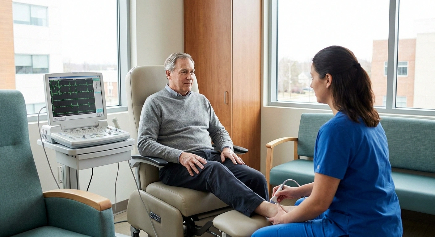
The workup for suspected lumbosacral plexopathy has a specific shape, and knowing that shape helps you spot when part of it has been skipped. The five essential pieces are the neurologic exam, MRI of the lumbosacral plexus with contrast, MRI of the lumbar spine, EMG with nerve conduction studies, and targeted lab work.
The Diagnostic Pearl: Why SNAPs Tell Root From Plexus
This single finding on nerve conduction study is how an electromyographer localizes the lesion when the clinical exam alone can’t distinguish root from plexus.
The neurologic exam is where localization begins. A skilled examiner maps out exactly which muscles are weak, which sensory territories are affected, and what the reflexes are doing. The distribution — asymmetric, proximal, multi-nerve-territory in one leg — is what points at the plexus rather than at a root or a peripheral nerve.
MRI of the lumbosacral plexus with intravenous contrast is the imaging gold standard. This is not a lumbar spine MRI. It’s a dedicated pelvic study that images the plexus itself, looking for a mass, plexus enhancement (suggesting inflammation), or a hematoma. A standard lumbar spine MRI does not visualize the plexus. If the workup has included a spine MRI but not a dedicated plexus MRI, and the diagnosis is still uncertain, that’s the missing piece to ask about. MRI of the lumbar spine is done alongside to rule out radiculopathy from disc disease.
Nerve conduction study and EMG do two jobs. First, they localize the lesion — root versus plexus versus peripheral nerve — using the SNAP-preserved-in-radiculopathy pearl described above. Second, they identify specific patterns: myokymia flags radiation injury, a widespread multifocal axonal pattern flags DLRPN or plexitis, and focal amplitude loss in a femoral distribution can flag a hematoma-related injury. Understanding the full logic of the standard neuropathy workup pathway helps you walk into these tests knowing what they’re looking for.
Lab work is targeted to the suspected cause. Blood sugar and HbA1c for DLRPN. CBC, coagulation studies, and imaging for suspected hematoma. ESR, CRP, ANA, ANCA panels if a systemic vasculitis is on the differential. Tumor markers and cancer screening for suspected malignancy. Cerebrospinal fluid analysis is sometimes added when leptomeningeal spread of cancer is a concern.
Treatment: Cause-Specific and Time-Sensitive
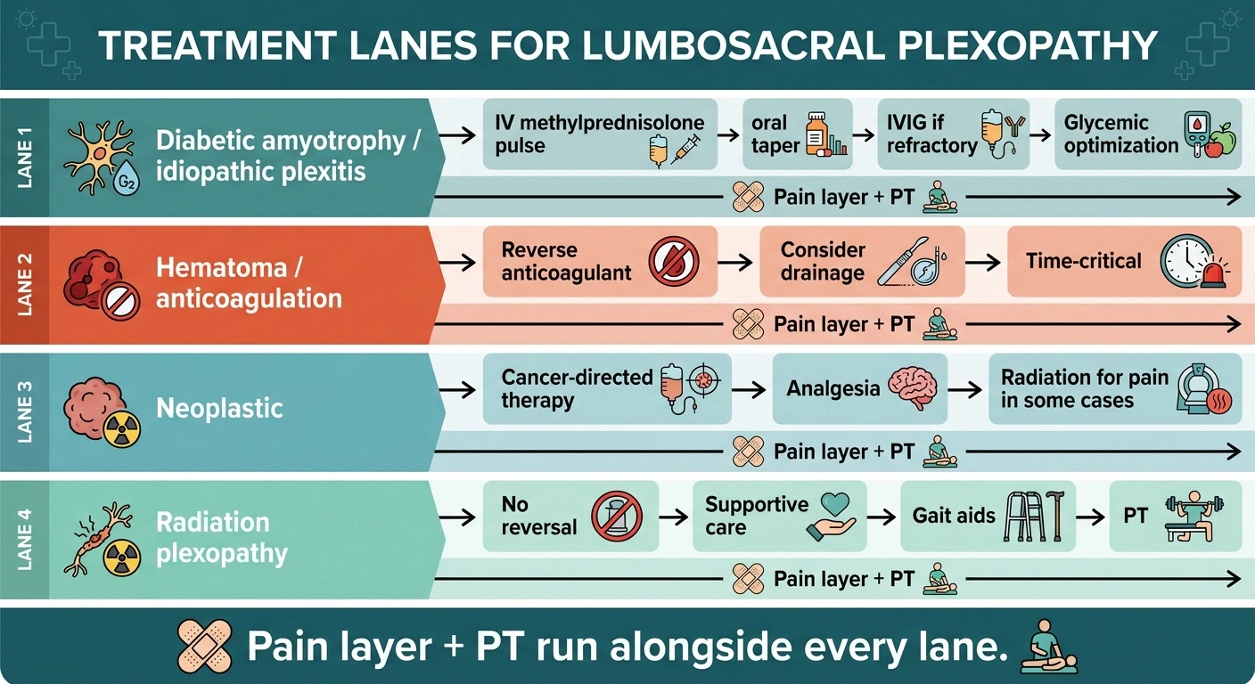
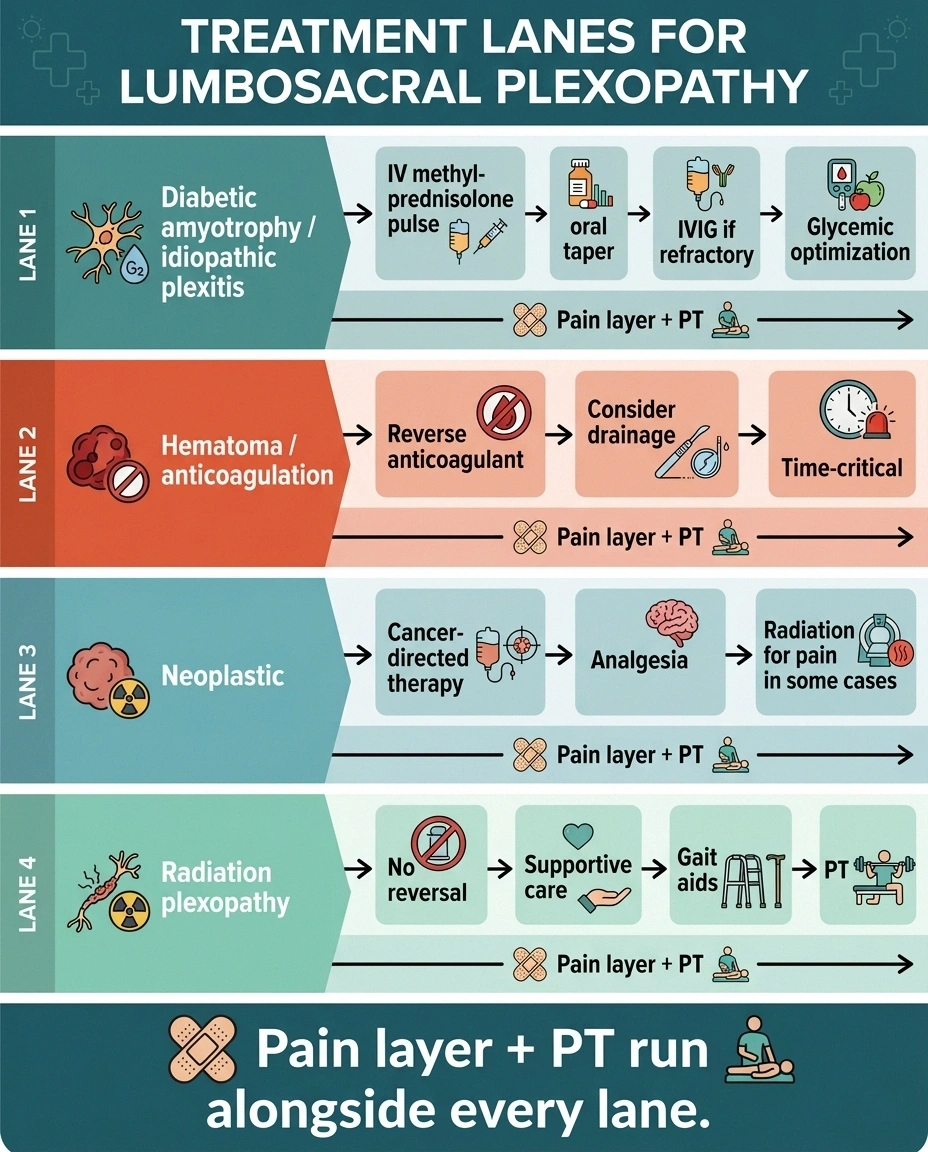
Because the causes of lumbosacral plexopathy diverge so widely, treatment is dictated by which cause is actually driving the picture. There is no single “plexopathy protocol” — the workup exists precisely to determine which lane the patient is in.
Treatment Depends On Cause
Pain layer (gabapentin, pregabalin, duloxetine, tricyclics) and physical therapy run alongside every treatment lane.
For DLRPN and idiopathic plexitis, the layer that changes the course, when it changes anything, is immunotherapy. IV methylprednisolone pulses followed by an oral prednisone taper are the most common first-line choice — the Mayo work suggests they help with pain and may modestly accelerate recovery, though the effect on the ultimate deficit is debated. IVIG is used in some centers, particularly for severe or refractory cases. Glycemic optimization matters even when baseline control is reasonable.
For hematoma from anticoagulation, treatment is time-critical: reverse the anticoagulant, address the bleeding source, and drain the hematoma in significant cases to relieve compression on the plexus. Delay is the enemy.
For neoplastic plexopathy, treatment is oncologic — the plexus deficit follows the tumor, and cancer-directed therapy (surgery, chemotherapy, radiation, hormonal therapy) is what changes the trajectory. Analgesia is essential alongside. For radiation-induced plexopathy, there is no reversal — focus shifts to symptom management, gait aids, and preserving whatever function remains.
The pain layer runs alongside every one of these. Neuropathic pain in acute plexopathy can be severe, and it often responds only partially to the standard agents — but a systematic trial is worth it. Gabapentin is a common starting point with careful titration, pregabalin covers similar territory, and duloxetine works through a different mechanism and treats co-occurring depression at the same time. Low-dose tricyclics, topical lidocaine patches, and a short-term opioid course for severe acute pain are all reasonable in the right context. Physical therapy is not optional — denervated muscles contract and lose range of motion quickly if not moved, and weakness plus reduced sensation makes the risk of falls a real concern that has to be addressed proactively.
What Recovery Actually Looks Like Over 1-2 Years

Setting realistic expectations about recovery is one of the most important conversations you can have with your care team, because the timeline for lumbosacral plexopathy is measured in months to years, not weeks — and the endpoint is often “meaningfully recovered” rather than “back to baseline.”
The DLRPN Recovery Arc
Diabetic amyotrophy is the paradigm case. The pain phase dominates the first weeks to a few months and gradually fades. Weakness continues to progress after the pain starts to improve, which can be demoralizing — patients often feel worse in month two than in month one even though the underlying inflammation is settling. The plateau lands somewhere in months two to six. Slow reinnervation then takes over, driven by axon regrowth at roughly one millimeter per day. Motor recovery continues for 12-24 months. Most patients regain functional walking. A meaningful minority retain some residual weakness — a persistent foot drop, thigh atrophy that doesn’t fill back in, or fatigue with prolonged standing.
Neoplastic plexopathy recovery depends on tumor control. Hematoma recovery is good when the compression is relieved promptly and poor when it isn’t. Radiation plexopathy usually doesn’t recover — the goal is preservation of the function that remains. Postpartum plexopathy is the outlier and usually recovers nearly fully over weeks to a few months.
The question of whether the deficit is fully reversible is on almost every patient’s mind. The honest answer is that reversibility depends on how much axonal damage occurred and how quickly the driving process was addressed. Nerves that were injured but not killed can recover. Nerves that died do not.
What to Tell Your Doctor If You Suspect It
If the pattern looks like lumbosacral plexopathy — asymmetric proximal leg pain and weakness, not the burning stocking distribution of length-dependent PN — the language you bring to the appointment matters. Non-neurologists may not think about the plexus routinely, and using specific terminology helps steer the conversation.
Start with the timeline: when did the pain begin, when did the weakness begin, and how has the pattern evolved. Describe the distribution in bodily terms — “the pain is deep in my right hip and thigh, worse at night, and now I can’t stand up from a chair without pushing off with my arms.” That single sentence tells a neurologist far more than “leg pain” does.
Ask for a neurology referral and use the terms “lumbosacral plexopathy” and, if you have diabetes, “diabetic amyotrophy” by name. Ask whether MRI of the lumbosacral plexus with contrast has been ordered — not just a lumbar spine MRI, because those are different studies. Ask whether EMG with nerve conduction studies has been scheduled, and mention that sensory nerve action potentials help distinguish plexus from root involvement.
Escalate immediately for red flags. Rapidly progressive weakness over days, new bowel or bladder dysfunction, sudden severe pain with weakness in the setting of anticoagulation, or fever with the leg symptoms all warrant same-day evaluation. The stakes on missing an acute hematoma, an epidural abscess, or a rapidly infiltrating tumor are high enough that erring on the side of urgency is the right call.
Frequently Asked Questions
Is lumbosacral plexopathy a form of peripheral neuropathy?
Broadly yes — the plexus is part of the peripheral nervous system, so injury to it falls under the peripheral neuropathy umbrella. But the pattern is very different from the length-dependent, symmetric, distal-first PN most people picture with the term. Plexopathy is asymmetric, proximal-predominant, and painful, and the causes and workup diverge sharply. Grouping them together clinically leads to the wrong tests being ordered.
What’s the difference between lumbosacral plexopathy and sciatica?
Sciatica is pain along the sciatic nerve distribution, usually caused by a herniated disc pressing on a single nerve root at the spine (radiculopathy). Lumbosacral plexopathy is injury to the plexus itself inside the pelvis, affecting multiple nerves from a single limb at once. Sciatica typically follows a dermatomal pattern along one root; plexopathy produces multi-territory weakness that doesn’t map to any single root. Imaging differs too — sciatica often shows disc herniation on lumbar MRI, while plexopathy needs a dedicated pelvic MRI.
How is diabetic amyotrophy different from regular diabetic neuropathy?
Regular diabetic peripheral neuropathy is the symmetric, distal, burning-feet pattern most people associate with diabetes — length-dependent, worst in the toes. Diabetic amyotrophy (Bruns-Garland syndrome or lumbosacral radiculoplexus neuropathy) is a very different disease that happens to also be driven by diabetes. It’s asymmetric, hits the proximal thigh muscles hard, has severe early pain, and tends to strike patients whose diabetes is not particularly poorly controlled, often after unintentional weight loss. The mechanism appears to be microvasculitis rather than metabolic nerve damage.
Can lumbosacral plexopathy be reversed?
Partial to substantial recovery is common, but complete reversal is not the norm. The outcome depends on the cause, the severity of the initial damage, and how quickly the driving process was addressed. Diabetic amyotrophy typically recovers meaningfully over 12-24 months but often leaves some residual weakness. Hematoma-related plexopathy recovers well when the compression is relieved promptly. Radiation plexopathy usually does not recover. Postpartum plexopathy usually recovers nearly fully.
How long does diabetic amyotrophy last?
The pain phase typically lasts weeks to a few months, followed by a longer phase of weakness that plateaus somewhere between two and six months, then a slow recovery phase that continues for 12-24 months. Most patients regain functional walking within a year, but some residual weakness or a persistent foot drop can remain. The full trajectory is measured in a year or two, not weeks — patience matters, and setting realistic expectations early prevents the demoralization that comes from expecting a rapid turnaround.
Do I need special imaging for lumbosacral plexopathy?
Yes. A dedicated MRI of the lumbosacral plexus with intravenous contrast is the imaging gold standard, and it is a different study from a lumbar spine MRI. The spine MRI images the vertebrae, discs, and nerve roots inside the spinal canal. The plexus MRI images the network of nerves inside the pelvis. If your workup so far has only included a lumbar spine MRI, ask specifically whether a plexus MRI has been considered — because the two studies show very different anatomy and answer very different questions.
Is lumbosacral plexopathy an emergency?
Most causes are subacute — they unfold over days to weeks and warrant urgent but not emergent evaluation. However, several presentations are true emergencies: sudden severe pain with new leg weakness in a patient on anticoagulation (possible retroperitoneal hematoma), fever with severe back or leg pain and weakness (possible epidural abscess or psoas abscess), or rapidly progressive weakness with bowel or bladder dysfunction. Any of those warrants same-day emergency-department evaluation. When in doubt, the emergency department is the right choice — the stakes on delayed recognition of an acute compressive or infectious cause are high.
Can lumbosacral plexopathy happen without diabetes or cancer?
Yes. Idiopathic lumbosacral plexitis is the non-diabetic version of diabetic amyotrophy — the same subacute painful proximal weakness in a patient without diabetes or malignancy. Postpartum plexopathy affects new mothers after prolonged labor. Retroperitoneal fibrosis, psoas abscess, aortic aneurysm, and iatrogenic injury from pelvic or vascular surgery are all documented non-diabetic causes. The workup is designed to sort through these possibilities, which is why the imaging, EMG, and lab pieces all matter — the treatment for each cause is different.
About the Author