
Most nerve damage that happens around cancer is collateral. Chemotherapy drugs are toxic to nerve fibers. Tumors press on nerves from outside. The immune system, roused by a tumor somewhere else entirely, occasionally turns on nerve tissue by mistake. In all three cases the nerve is a bystander.
Neurolymphomatosis is not that. In neurolymphomatosis the lymphoma cells are inside the nerve. They have migrated into the nerve tissue itself and are growing there, the way they would grow in a lymph node. The nerve is not being squeezed or poisoned or caught in crossfire. It is the tumor site.
That single distinction drives everything else about this condition: why the symptoms are so painful, why they arrive in an odd asymmetric pattern, why one scan finds it and another misses it, and why the drug chosen to treat it is one that most lymphoma patients never receive.
Before going further, one thing needs saying plainly, because a page like this can frighten people it should not. This is rare. If your neuropathy is the familiar kind, symmetric tingling and numbness that started in both feet and crept upward slowly over years, this is almost certainly not what you have. The pattern described below looks quite different, and the difference is recognisable.
A Word That Means Something Very Specific
Neurolymphomatosis is defined as the direct infiltration of nerve tissue by malignant lymphocytes. The involved structures can be cranial nerves, the nerve roots as they leave the spine, the plexuses where nerves bundle and reorganise at the shoulder and hip, or peripheral nerve trunks out in the limbs.
Four different problems that get called the same thing
Drug toxicity
Chemotherapy agents damaging nerve fibers directly.
Tell: symmetric, both sides, starts in toes and fingertips, tracks cumulative dose.
Immune misfire
Antibodies raised against a tumour cross-reacting with nerve tissue elsewhere.
Tell: can precede the cancer diagnosis by months; often affects sensory nerves and balance heavily.
Compression
A mass pressing on a nerve from outside it.
Tell: confined to one nerve's territory; imaging shows the mass rather than the nerve.
Infiltration
Malignant cells growing inside the nerve tissue itself. This is neurolymphomatosis.
Tell: asymmetric, painful out of proportion, changes over weeks rather than years.
Sorting which of the four is in play is most of the diagnostic work, and the three tells in each box are what a clinician is weighing when they ask how it started and how fast it changed.
The overwhelming majority of cases arise from B-cell non-Hodgkin lymphoma, with diffuse large B-cell lymphoma the most frequent single source. It also occurs in leukemia. It does not occur out of nowhere in someone with no lymphoid malignancy anywhere, although in a substantial share of cases the nerve involvement is what reveals a lymphoma nobody knew about.
It helps to hold this alongside the alternatives, because the four ways cancer touches nerve get conflated constantly and they are genuinely different problems. Chemotherapy-induced neuropathy is drug toxicity, and it is symmetric, dose-related, and usually starts in the toes and fingertips. Paraneoplastic neuropathy is an immune misfire, where antibodies raised against a tumor cross-react with nerve tissue at a distance. Compression is mechanical, a mass pressing on a nerve from outside. Infiltration, which is this, is the tumor growing within the nerve.
Those four produce different patterns, need different tests, and respond to different treatments. Sorting out which one is in play is most of the diagnostic work.
The Patterns It Takes
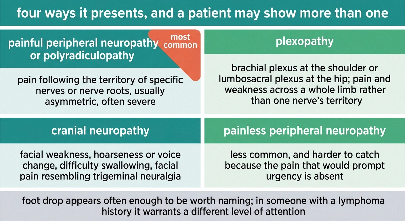
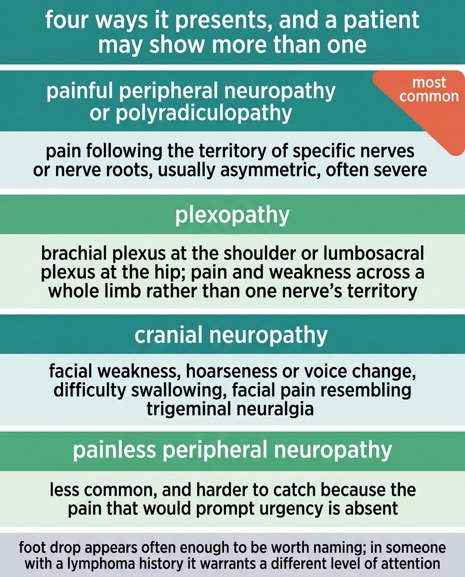
Neurolymphomatosis presents in four recognised ways, and a given patient may show more than one.
Painful peripheral neuropathy or polyradiculopathy. Pain in the distribution of specific nerves or nerve roots, usually asymmetric, often severe. This is the most common presentation.
Plexopathy. Involvement of the brachial plexus at the shoulder or the lumbosacral plexus at the hip, producing pain and weakness across a whole limb rather than in one nerve's territory.
Cranial neuropathy. Facial weakness, hoarseness or voice change, difficulty swallowing, or facial pain that can look like trigeminal neuralgia. Vagus nerve involvement has been documented.
Painless peripheral neuropathy. Less common, and harder to catch precisely because the pain that would prompt urgency is absent.
Foot drop turns up often enough to be worth naming separately. A sudden foot drop has many possible causes, most of them far more common than this, but a foot drop appearing in someone with a lymphoma history deserves a different level of attention than one appearing after a long car journey.
What Makes a Doctor Suspect It

Three features move this from the bottom of a differential list toward the top.
The first is pain that is out of proportion. Ordinary length-dependent neuropathy is uncomfortable, often deeply so, but it tends to be a burning or aching that fits the amount of nerve involved. Infiltrative disease produces pain that clinicians describe as disproportionate, the kind that does not settle with the usual measures and is hard to sit with.
The second is asymmetry. Nerve damage from metabolic causes affects the longest fibres first and both sides roughly equally, which is why it starts in both feet. Infiltration follows whichever nerve the cells landed in. One shoulder. One side of the face. A single limb. If you want the fuller distinction, our explanation of polyneuropathy versus mononeuropathy covers why pattern is such a strong diagnostic signal.
The third is pace. This progresses over weeks to a few months. Diabetic neuropathy takes years. A neuropathy that has meaningfully changed between one clinic visit and the next is behaving differently from the common causes, and that alone justifies a harder look.
Add a known or suspected lymphoma and the suspicion rises sharply. But it is worth repeating that in primary neurolymphomatosis there is no known lymphoma. The nerve problem comes first, and the search that follows is what finds the disease.
Primary and Secondary Are Not the Same Situation
Two people can carry the same diagnosis and be in quite different positions.
Primary
Nerve involvement is the first and sometimes the only manifestation. No lymphoma was known beforehand.
Independent predictor of better treatment response, odds ratio 5.0 on multivariable analysis.
Secondary
A known lymphoma has relapsed into nerve tissue, often after treatment cleared it everywhere else.
Poorer baseline performance status also predicted weaker response, odds ratio 0.2.
Almost every survival figure published for this condition pools both groups together. Before accepting a number someone has quoted, establish which group it describes and which group you are in, because the pooled average understates one of them.
Primary neurolymphomatosis means the nerve involvement is the first and sometimes the only manifestation. There was no prior lymphoma diagnosis. The disease announced itself through nerve.
Secondary neurolymphomatosis means a known lymphoma has relapsed into nerve tissue, often after treatment that cleared it elsewhere.
The distinction is not merely descriptive. In the published cohort analysis, primary disease was an independent predictor of better treatment response on multivariable analysis, with an odds ratio of 5.0. Performance status mattered too, with poorer baseline function predicting weaker response.
If you are trying to make sense of a prognosis someone has quoted at you, this is one of the first things to establish. Numbers pooled across both groups understate the outlook for one of them.
Why PET Finds What MRI Misses
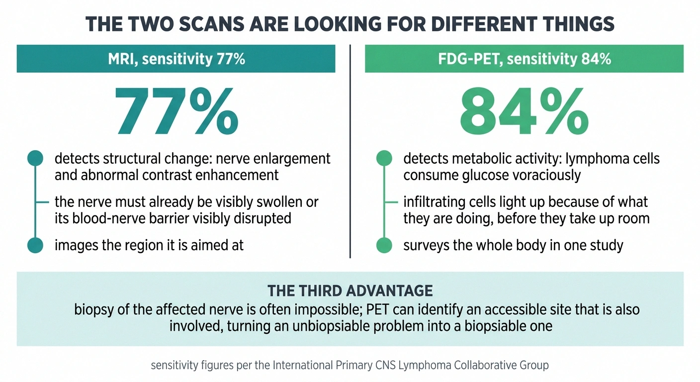
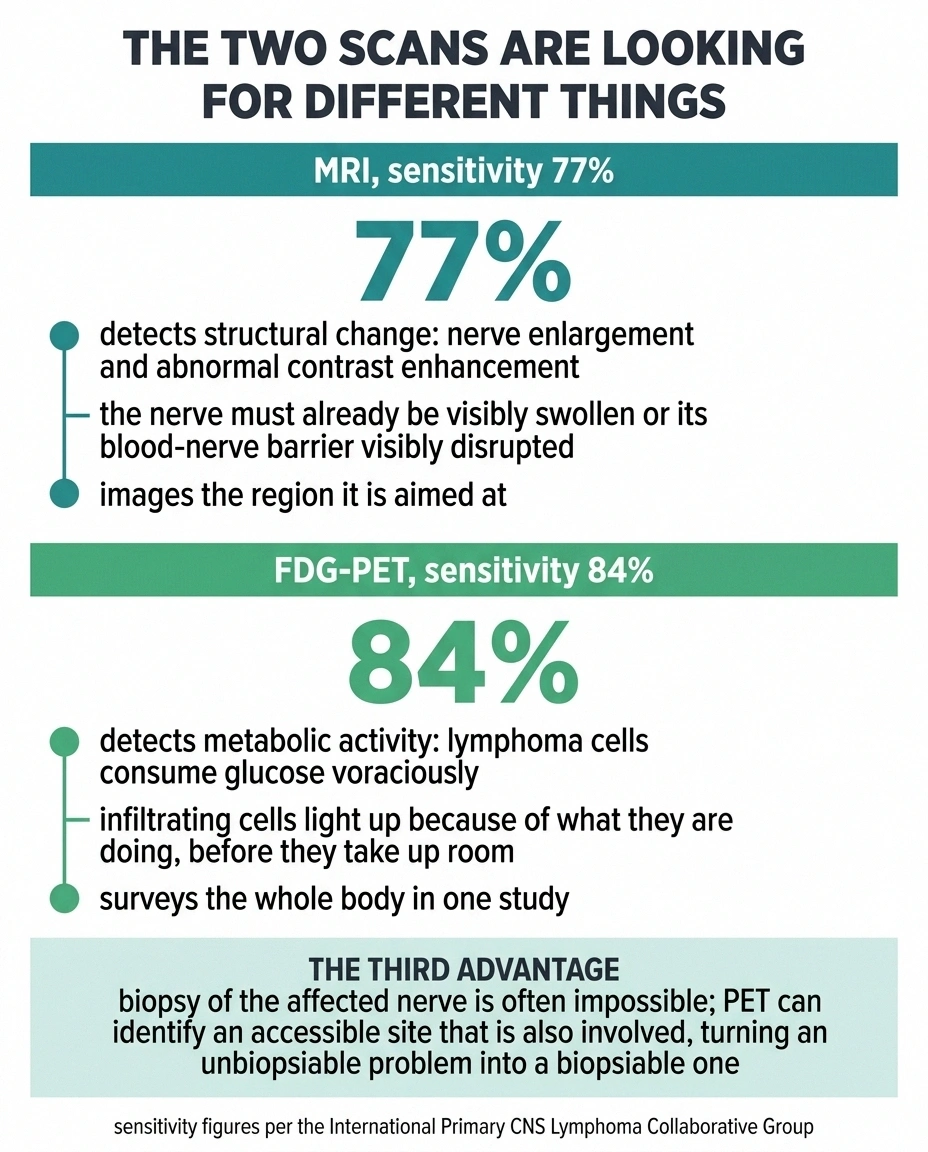
This section contains the most practically useful fact on the page.
MRI is the reflex imaging choice for nerve problems, and it does detect neurolymphomatosis. It shows enlargement of the affected nerve and abnormal enhancement after contrast, meaning the nerve lights up in a way healthy nerve does not. When it works, it works well.
FDG-PET works better. According to the International Primary CNS Lymphoma Collaborative Group, the sensitivity figures are 84 percent for PET against 77 percent for MRI. The gap is not enormous, but it is the difference between finding this and not finding it in a meaningful number of people.
The reason is mechanistic rather than technical. MRI is looking for a structural change, so the nerve has to be visibly swollen or its blood-nerve barrier visibly disrupted before anything shows. FDG-PET is looking for metabolic activity, and lymphoma cells consume glucose voraciously. They light up because of what they are doing, not because of how much room they take up. Infiltrating cells can be metabolically obvious well before they are structurally obvious.
PET also surveys the whole body at once. Published cases document involvement picked up across cranial nerves, spinal nerve roots, the brachial plexus, the cervicothoracic ganglion, intercostal nerves, branches of the vagus nerve, the lumbosacral plexus and the sciatic nerves. A targeted MRI of one shoulder finds disease in that shoulder. It cannot tell you the other plexus is involved too.
There is a third advantage that gets less attention. Biopsy of an affected nerve is definitive but frequently impossible, because the involved nerve is often somewhere you cannot safely take tissue from. PET can identify an accessible site that is also involved, turning an unbiopsiable problem into a biopsiable one.
If a lymphoma-related nerve problem is genuinely suspected and only an MRI has been done, asking whether PET is indicated is a reasonable question.
A Normal Spinal Tap Does Not Settle It
Cerebrospinal fluid analysis is a standard part of the workup, and it is frequently negative in neurolymphomatosis.
A negative result is not symmetrical with a positive one
Both the spinal tap and MRI carry this asymmetry, and for the same underlying reason. A positive finding confirms. A negative finding only tells you the test did not detect anything, and each test misses this disease at a known rate: CSF cytology frequently, MRI in roughly one case in four.
A sentence that keeps the conversation open, when results come back clear but the symptoms have not changed: “Given how negative tests perform in this specific condition, does this result actually lower the suspicion, or does it just leave us where we were?” That question is asking about a test's negative predictive value, and it is a fair thing to ask about any result that closes a line of investigation.
The explanation follows directly from what the disease is. CSF cytology detects malignant cells floating free in spinal fluid. In neurolymphomatosis the cells are embedded in nerve tissue, not swimming in fluid. They can stay there without shedding into the CSF in detectable numbers.
This matters because a negative tap is easy to over-read as reassurance. It is genuinely useful when positive and much weaker evidence when negative. The published work on non-biopsiable cases exists precisely because the standard confirmatory routes so often come back empty and the diagnosis has to be assembled from clinical picture, imaging, and whatever tissue can be reached.
The formal requirement is integration: clinical findings, imaging, histopathology from nerve or from non-neural tissue, and CSF analysis, weighed together. No single one of them carries the diagnosis alone.
Why Treatment Reaches for Methotrexate
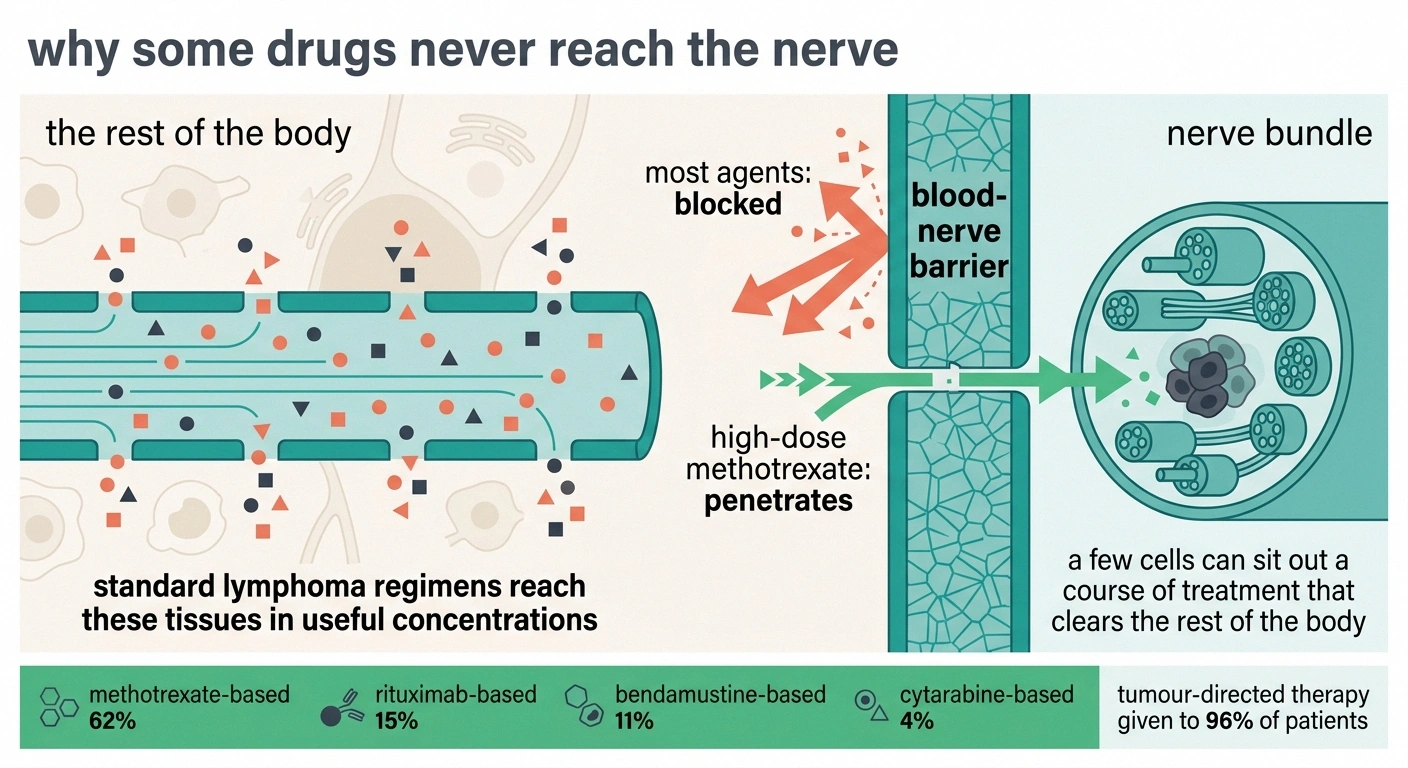
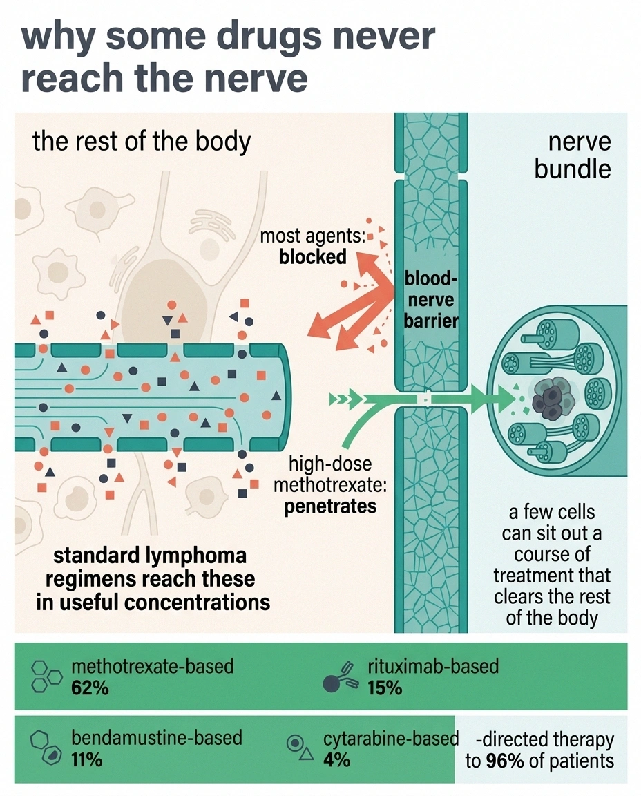
The treatment choice puzzles people, and the logic behind it is worth understanding because it explains an otherwise strange situation: someone whose lymphoma responded well to standard therapy, whose scans were clean, developing nerve disease anyway.
Peripheral nerve and the central nervous system are partially protected from the bloodstream. Many effective lymphoma drugs simply do not cross into them in useful concentrations. The result is a sanctuary, a place where a few cells can sit out a course of treatment that clears the rest of the body.
High-dose methotrexate penetrates those compartments. That is why it dominates the treatment data despite not being the first choice for lymphoma elsewhere. In the reported cohorts, first-line regimens broke down as methotrexate-based in 62 percent of patients, rituximab-based in 15 percent, bendamustine-based in 11 percent, and cytarabine-based in 4 percent. Tumor-directed therapy of some kind was given to 96 percent.
Response rates by regimen, in the reported series, were 92 percent for bendamustine, 86 percent for rituximab, and 78 percent for methotrexate. Those figures come from unequal group sizes and different patient situations, so they should not be read as a ranking. The overall response rate across all treatments was 67 percent, of which 52 percent were complete responses and 16 percent partial.
Two thirds of patients responding, and half of them completely, is a genuinely meaningful number for a disease this aggressive.
What the Outcome Numbers Actually Say
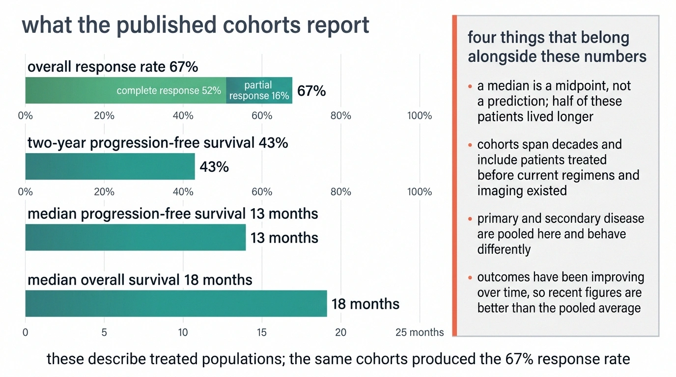
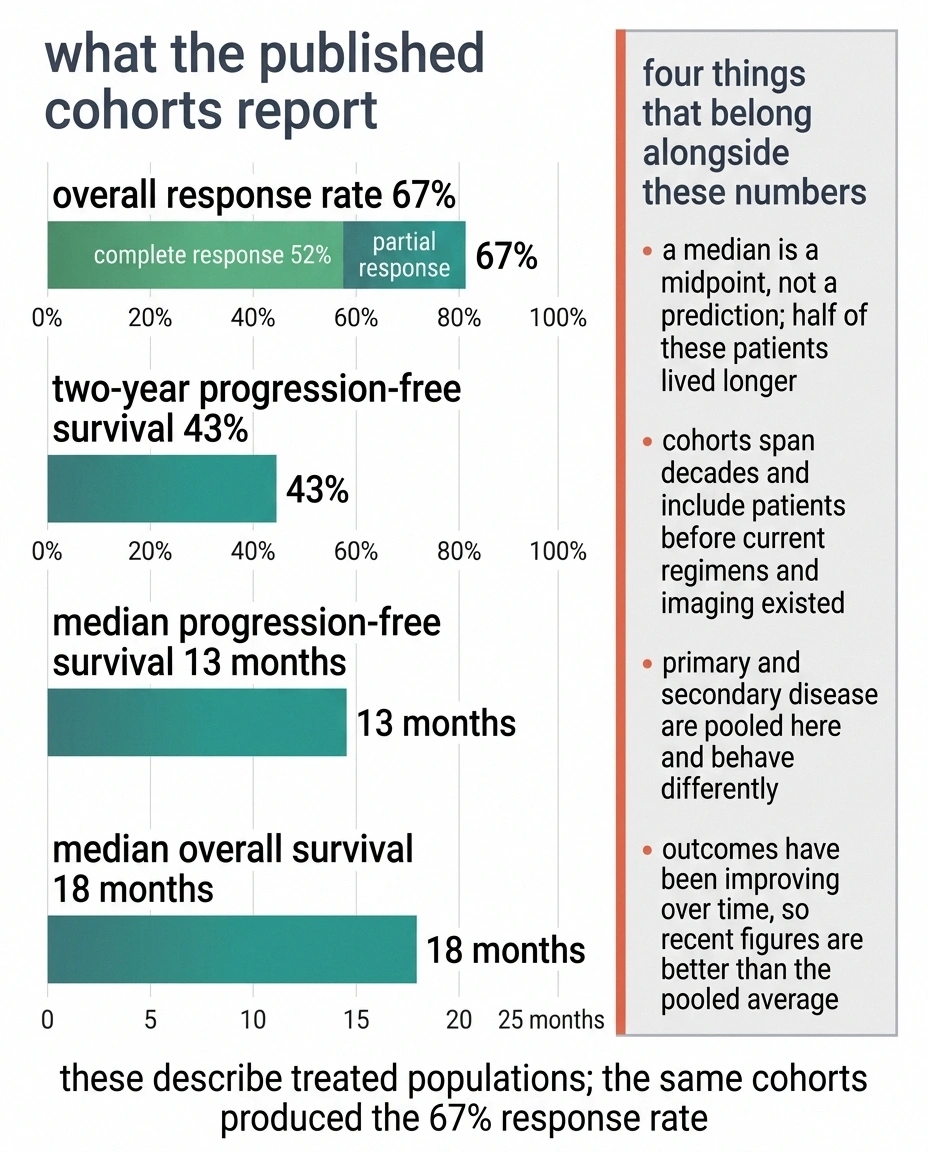
The survival data deserve honest handling, because they are sobering and they are also more nuanced than a single figure suggests.
Median overall survival in the published cohorts is around 18 months. Median progression-free survival is about 13 months, with two-year progression-free survival at 43 percent.
Four things belong alongside those numbers.
A median is a midpoint, not a prediction. Half of the people in those cohorts lived longer, some substantially so, and nothing in a median tells you which half any individual falls into.
The cohorts span decades. A 22-year study includes patients treated long before current regimens and imaging existed. The published work notes explicitly that outcomes have been improving over time, which means recent figures are better than the pooled average.
Primary and secondary disease are combined in most headline numbers, and they behave differently.
And these are treated populations, which is the point of the treatment data above. The 67 percent response rate and the 18-month median describe the same group.
The most consequential variable is how quickly it is found. Nerve damage that has already destroyed axons does not fully recover even when the lymphoma responds, because axon regrowth runs at roughly a millimetre a day and often stalls. Delay costs function permanently, independent of what it costs in disease control. That is the argument for pushing hard on an unexplained, painful, asymmetric, fast-moving neuropathy in anyone with a lymphoma history rather than waiting to see how it develops.
If Someone Has Just Said This Word to You

A few practical things are worth knowing.
What “baseline documentation” needs to contain to be useful later
Asking for a baseline is easy. Asking for one specific enough to detect change six weeks later is the part that gets skipped, and a note reading “neurologically intact” is worth nothing when you need to compare.
- Strength graded muscle by muscle on the standard 0 to 5 scale, named individually, not summarised as a limb
- The upper border of sensory change, recorded against a landmark such as the ankle bone or mid-calf
- Reflexes at both ankles and knees, each side written separately
- A timed walk over a set distance, with the distance and the time both written down
- Your own pain score at rest and at its worst, dated, because it is the measure that moves first
Keep your own copy. Records move between departments slowly, and the comparison is often needed before the notes arrive.
Find out whether yours is primary or secondary. It changes the outlook and it changes how any quoted statistic should be read.
Ask what imaging has been done. If the answer is MRI only and suspicion is real, ask about FDG-PET, including whether it might identify a site that can actually be biopsied.
Ask whether the treatment plan accounts for nerve as a sanctuary site. It is a specific question with a specific answer, and it distinguishes a plan built for this from a standard lymphoma protocol.
Get pain managed properly and early, in parallel with the cancer treatment rather than after it. Infiltrative nerve pain is severe and it responds poorly to over-the-counter measures. This warrants a real pain plan from the outset, and it is reasonable to insist on one.
Ask for baseline neurological documentation. Strength, sensation, and reflexes recorded now give you something to measure against later, which turns “I think it might be a bit worse” into information.
And if you have arrived here worried rather than diagnosed, hold on to the shape of the thing. This condition is asymmetric, painful out of proportion, and moves over weeks. Neuropathy that is symmetric, gradual, and has been creeping up from both feet for years is a different animal, and our overview of how ordinary neuropathy progresses describes what that far more common course actually looks like. If your symptoms genuinely do not fit that pattern, seeing a neurologist is the right next step, and asymmetry plus severe pain plus rapid change is a combination worth naming explicitly when you book.
Frequently Asked Questions
What is neurolymphomatosis?
It is the direct invasion of nerve tissue by lymphoma cells. Cranial nerves, nerve roots, plexuses, or peripheral nerve trunks can be affected. It differs from chemotherapy nerve damage, from immune-mediated paraneoplastic neuropathy, and from a tumor pressing on a nerve, because in this case the malignant cells are growing inside the nerve itself. Most cases arise from B-cell non-Hodgkin lymphoma.
How is it different from chemotherapy-induced neuropathy?
Chemotherapy neuropathy is drug toxicity. It is symmetric, related to cumulative dose, and typically begins in the toes and fingertips on both sides. Neurolymphomatosis is the cancer itself inside nerve tissue, so it is usually asymmetric, often severely painful, and progresses over weeks to months rather than following the chemotherapy schedule. Pain that is out of proportion and confined to one region is the distinguishing feature.
What scan finds neurolymphomatosis?
FDG-PET is the most sensitive single test, reported at 84 percent sensitivity compared with 77 percent for MRI. PET detects the high glucose consumption of lymphoma cells, so involvement can be visible before the nerve is structurally enlarged, and it surveys the whole body rather than one region. MRI remains useful and shows nerve enlargement with abnormal contrast enhancement.
Does a normal MRI rule it out?
No. MRI sensitivity is around 77 percent, meaning roughly one in four cases is missed. A normal MRI in someone with a strongly suspicious clinical picture is a reason to consider FDG-PET rather than a reason to stop looking.
Does a normal spinal tap rule it out?
No. CSF cytology is frequently negative because the malignant cells are embedded within nerve tissue rather than circulating freely in spinal fluid. A positive result is helpful, but a negative one carries much less weight and does not exclude the diagnosis.
Why is high-dose methotrexate used?
Nerve and central nervous system tissue are partially shielded from the bloodstream, so many lymphoma drugs never reach useful concentrations there. High-dose methotrexate penetrates those sanctuary sites. This explains why nerve disease can appear in someone whose lymphoma elsewhere responded well to standard treatment, and why methotrexate-based regimens make up around 62 percent of first-line therapy in the published series.
Is neurolymphomatosis curable?
Complete responses do occur. Across reported cohorts the overall response rate was 67 percent, with 52 percent complete responses. Median overall survival was around 18 months, though those figures pool decades of patients and both primary and secondary disease, and outcomes have been improving over time. Primary neurolymphomatosis carries a better prognosis than relapse into nerve.
Can nerve damage recover after treatment?
Partly, and it depends heavily on how much axon loss occurred before treatment started. Nerve fibers regrow at roughly a millimetre a day when they regrow at all, so damage that has been present for months may leave permanent deficits even when the lymphoma responds fully. This is the main argument for investigating a rapidly progressive asymmetric neuropathy quickly rather than observing it.
About the Author