One sentence from the 2026 AANEM task force review is worth more to most patients than the rest of the paper combined:
No test offers enough sensitivity or specificity to serve as a stand-alone gold standard.
If you have been told your skin biopsy was normal and therefore you do not have small fiber neuropathy, that sentence is your rebuttal, published by the professional body whose members perform the test.
The full document sits behind a journal paywall, written for neurologists. What follows is a translation: what the task force reviewed, what each test is actually worth, which blood tests belong in a workup, and how to use any of it in a fifteen-minute appointment.
What the Task Force Was Asked to Do
The American Association of Neuromuscular and Electrodiagnostic Medicine convened a Small Fiber Neuropathy Task Force to answer two questions. First, how good are the tests used to diagnose small fiber neuropathy. Second, which laboratory tests are worth running to find a cause once the diagnosis is made.
The review was published in Muscle & Nerve in 2026 under the lead authorship of Katzberg. Thirteen diagnostic studies and two laboratory screening studies met the inclusion criteria, which is a small number and tells you something about the state of the field. This is not a domain with a deep evidence base.
An important structural note before going further. This was an evidence review, not a prescriptive protocol. The task force graded what exists. It did not issue a list saying order these fourteen tests on every patient. That distinction matters, and I will flag clearly below where the material shifts from the task force review to a separate published lab list, because they are different documents with different weight.
Skin Biopsy: The Best Test We Have, With Real Numbers
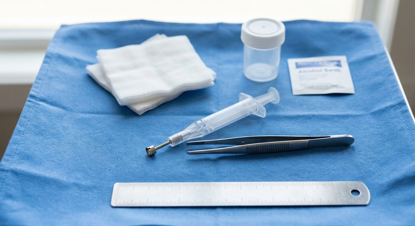
Skin punch biopsy came out as the most validated diagnostic option, and it was the only test to reach Class II evidence.
74 percent sensitivity, converted into people
Take 100 people who genuinely have small fiber neuropathy and biopsy all of them.
74 to 78
get an abnormal result. The test worked, and it points where their symptoms already pointed.
22 to 26
get a normal result while having the condition. Nothing about their nerves changed. The test simply did not catch it.
This is why the phrase to use is “my biopsy was normal”, not “I don’t have it”. The first is a result. The second is a conclusion the numbers do not support on their own.
The procedure takes a small circle of skin, typically from the lower leg, roughly three millimeters across. A lab stains the nerve fibers and counts how many are crossing up into the epidermis, giving a figure called intraepidermal nerve fiber density. Fiber length and the spacing between fibers are also measured. Density gets compared against normative values for your age and sex.
Now the numbers that rarely get said out loud in the appointment.
- Sensitivity: 74 to 78 percent. Of people who genuinely have small fiber neuropathy, roughly three quarters will have an abnormal biopsy. Which means somewhere between one in four and one in five will have a normal result despite having the condition.
- Specificity: 65 to 80 percent. Of people who do not have it, most but not all will test normal. A meaningful fraction of abnormal results occur in people without the condition.
Sit with the sensitivity figure. A negative skin biopsy shifts the odds. It does not close the question, and treating it as a verdict is a misuse of the test that the task force’s own numbers do not support.
The specificity figure cuts the other way and gets discussed even less. An abnormal biopsy is not automatically proof either. It is strong evidence that has to fit with the clinical picture.
Why a Normal EMG Settles Nothing
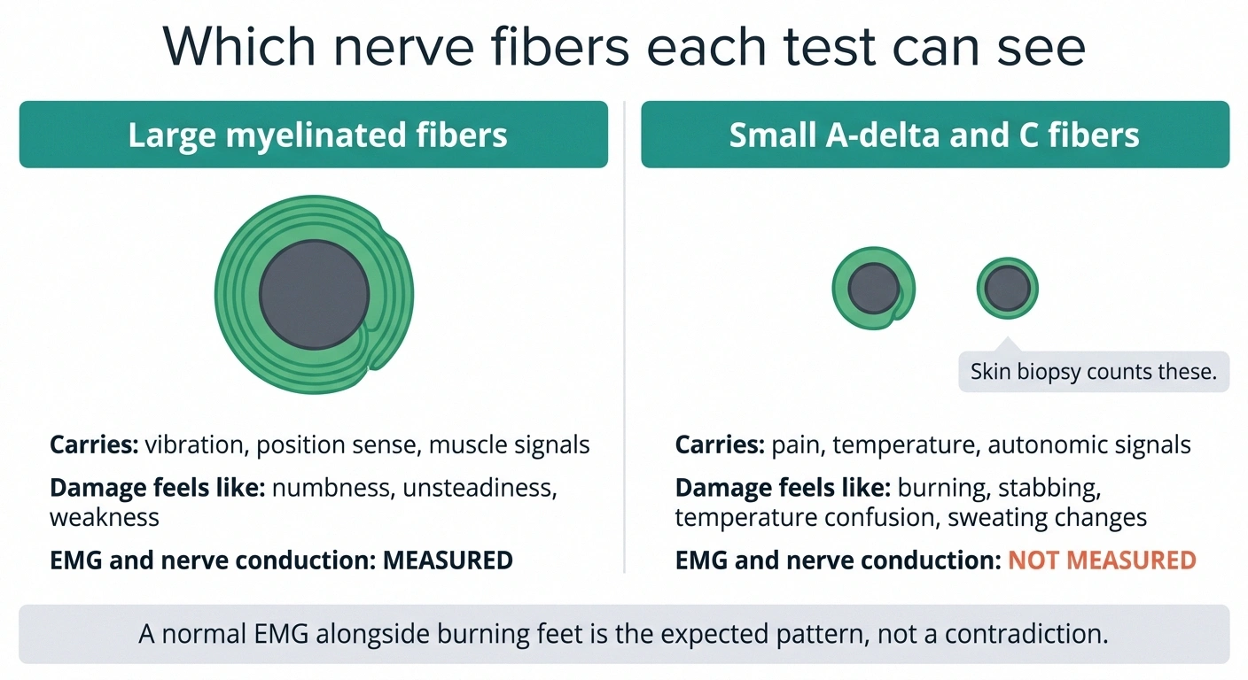
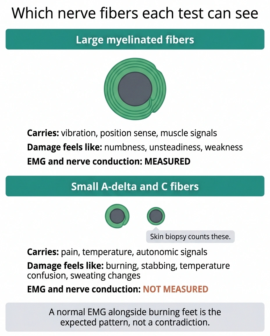
This is the most common source of misdirection in the whole area, and it is worth being precise about.
Two nerve populations, two sets of tests
A test can only report on the fibers it is capable of measuring.
| Large myelinated fibers | Small A-delta and C fibers | |
|---|---|---|
| They carry | Vibration, position sense, muscle signals | Pain, temperature, autonomic signals |
| Symptoms when damaged | Numbness, unsteadiness, weakness | Burning, stabbing, temperature confusion, sweating changes |
| Seen by EMG and NCS | Yes | No, not at all |
| Seen by skin biopsy | Not the target | Yes, this is what it counts |
A normal EMG alongside burning feet is not a contradiction. It is the pattern.
Nerve fibers come in different sizes. Large myelinated fibers carry vibration, position sense, and motor signals. Small fibers, the thinly myelinated A-delta and the unmyelinated C fibers, carry pain, temperature, and autonomic signals.
Nerve conduction studies and EMG measure large fiber function. That is what the technology does. Small fibers are too small and too slow to register on the equipment at all.
So in pure small fiber neuropathy, a normal EMG is the expected result. It is the finding consistent with the diagnosis, not the finding that rules it out.
People are told “your nerve test was normal” and reasonably hear “your nerves are fine.” Understanding what the study measured is often the difference between accepting a dead end and asking the next question. If you have a report in hand, it is worth learning how to read what an EMG report actually says rather than relying on the summary sentence.
The Tests That Did Not Clear the Bar
Several tests you may encounter were reviewed and landed lower than their reputations suggest.
Corneal confocal microscopy reached only Class III. This is the technique that images the dense nerve plexus in the cornea as a window onto small fiber health, and it is genuinely appealing: non-invasive, repeatable, no cutting. The task force concluded the validation is insufficient and it should not be used as a standalone diagnostic in isolated small fiber neuropathy. It remains a promising research tool. If a clinic offers it to you as the definitive answer, that goes past the current evidence.
Indirect functional tests, including laser evoked potentials, contact heat evoked potentials, and the cutaneous silent period, showed variable sensitivity with generally high specificity, all at Class III. Practically, that combination means an abnormal result is fairly informative and a normal result is not. They can add to a picture. None of them decides it.
What the task force did not do is as informative as what it did. It did not name a definitive test. It did not endorse corneal confocal microscopy for isolated cases. And it did not endorse any specific screening panel at a high level of evidence, because the evidence to support one at that level does not exist yet.
What Evidence Classes Actually Mean
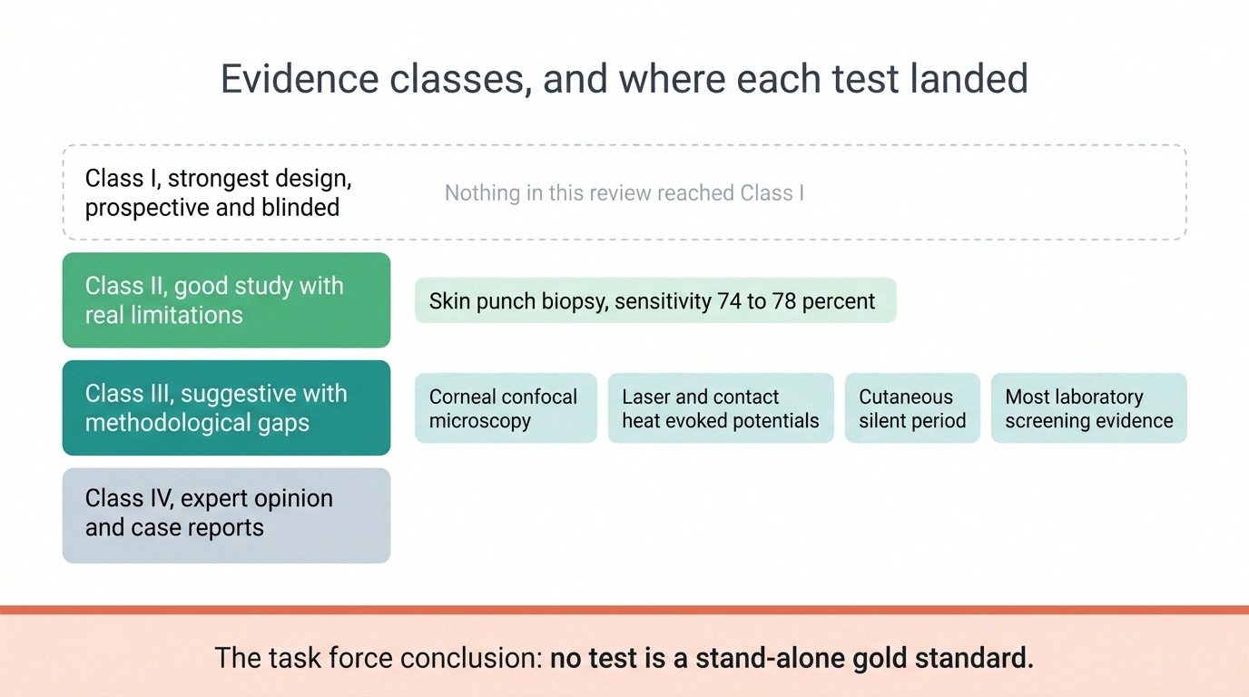
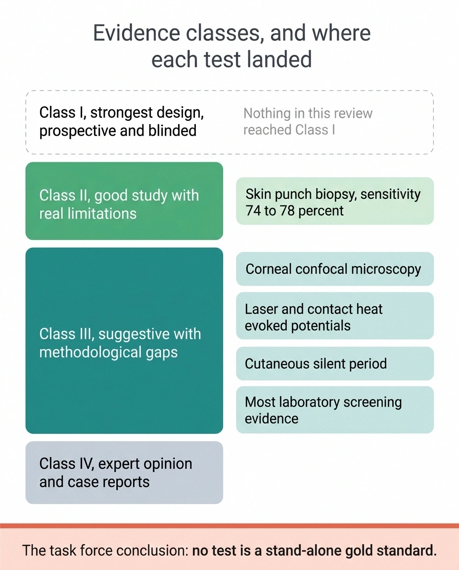
These labels appear throughout the review and are rarely explained to the people they affect.
- Class I: the strongest design. Prospective, blinded, broad enrollment, low risk of bias.
- Class II: a good study with real design limitations. Skin biopsy sits here.
- Class III: suggestive, with methodological gaps that could plausibly account for the findings. Most of this field sits here.
- Class IV: expert opinion, case reports, uncontrolled observation.
Nothing in this review reached Class I. When a well-informed neurologist says the diagnostic picture is uncertain, that is not evasion. It is an accurate description of the literature.
The First-Round Blood Panel

Here is the shift I flagged earlier. The following list does not come from the task force review itself, which found that laboratory screening identified a metabolic or immune cause in up to 64 percent of patients but graded most of that evidence Class III. The structured list below comes from a separate open-access paper on lab screening in clinical practice, and it reflects current practice rather than a high-evidence mandate.
That caveat stated, this is the most complete first-round list in the published literature, and it is the one worth bringing to an appointment.
Metabolic and endocrine
- Fasting plasma glucose and HbA1c
- Triglycerides, HDL cholesterol, fasting insulin
- Serum uric acid
- Pyridoxal 5-phosphate, the active form of vitamin B6
- Total vitamin B12
- Creatinine and eGFR
- Liver enzymes: GGT, AST, ALT, ALP
- Ferritin and transferrin saturation
- TSH
- Complete blood count
Immune and inflammatory
- Antinuclear antibodies and anti-extractable nuclear antigen antibodies
- Anti-tissue transglutaminase IgA, for celiac disease
- Serum protein electrophoresis, immunofixation, and free light chains
- Serum ACE and calcium
- Hepatitis C and HIV serology
Three items on that list deserve a note because they get skipped.
Vitamin B6 cuts both ways. Deficiency causes neuropathy and so does excess, and B6 toxicity from supplements is more common than most people expect given how many nerve formulas contain generous doses. Ordering the active form rather than assuming deficiency is the point.
The paraprotein trio matters more than its yield suggests. Monoclonal gammopathy turns up in only 1.4 to 3.9 percent of these patients, but finding one changes the entire direction of the workup. That is a test you run for consequence, not for probability.
Ferritin and transferrin saturation are screening for hemochromatosis, which is treatable and routinely missed. It is one of the quieter wins on the list.
The Second Round, and When It Is Worth Running
The second tier is not for everyone. These get added when the first round comes back borderline, or when something in your history points a direction.
The list, condensed for an appointment
Go down the left column and mark what has already been run. The gaps are the conversation.
First round, run for most people
Metabolic: fasting glucose, HbA1c, triglycerides, HDL, fasting insulin, uric acid, B6 as pyridoxal 5-phosphate, B12, creatinine and eGFR, GGT, AST, ALT, ALP, ferritin, transferrin saturation, TSH, full blood count
Immune: ANA, anti-ENA, anti-tissue transglutaminase IgA, serum protein electrophoresis, immunofixation, free light chains, serum ACE, calcium, hepatitis C, HIV
Second round, run when something points there
Borderline glucose: 75g tolerance test, 1hr and 2hr values
Borderline B12: holotranscobalamin, methylmalonic acid, homocysteine, folate
Alcohol: carbohydrate-deficient transferrin, phosphatidylethanol
Autoimmune: anti-dsDNA, complement, cryoglobulins, CRP, rheumatoid factor, anti-CCP, ANCA, newer antibodies including anti-TS-HDS and anti-FGFR3
Sarcoid: 1,25-dihydroxy vitamin D, chitotriosidase, soluble IL-2 receptor
Genetic: TTR, alpha-galactosidase A, SCN9A, SCN10A, SCN11A
Source note: the AANEM task force reviewed screening evidence and found a metabolic or immune cause identified in up to 64 percent of patients, mostly at Class III evidence. This tiered list is drawn from a separate open-access paper on clinical practice, not from the task force review itself.
If glucose results were borderline: a 75 gram oral glucose tolerance test with one-hour and two-hour values. This is worth pressing for. Fasting glucose and HbA1c both miss impaired glucose tolerance, and prediabetes turns up in something like 10 to 25 percent of small fiber neuropathy cohorts. A normal HbA1c is not a clean bill of metabolic health.
If B12 was borderline: holotranscobalamin, methylmalonic acid, homocysteine, and folate. Total B12 can sit in the normal range while functional deficiency is present at the tissue level.
If alcohol may be a factor: carbohydrate-deficient transferrin and phosphatidylethanol.
If autoimmune disease is suspected: anti-dsDNA, complement studies, cryoglobulins, CRP, rheumatoid factor, anti-CCP, systemic sclerosis antibodies, and ANCA. Lupus has been reported in as much as 27.5 percent of some cohorts, and Sjögren’s syndrome is among the more frequent findings in this group.
The newer antibodies: anti-TS-HDS, anti-FGFR3, anti-plexin D1, anti-Caspr2, and anti-LGI1. These are the frontier. Availability varies by lab, interpretation is still being worked out, and their presence does not automatically establish causation.
If sarcoidosis is on the table: 1,25-dihydroxy vitamin D, chitotriosidase, and soluble IL-2 receptor.
Genetic testing, when the presentation fits: TTR for hereditary amyloidosis, alpha-galactosidase A and lyso-Gb3 for Fabry disease, and the sodium channel genes SCN9A, SCN10A, SCN11A along with TRPA1. Sodium channel variants have been reported in as many as 16.7 percent of some series. Whether a given variant is causing your symptoms is a separate and harder question than whether you carry it.
That last point generalizes across this entire panel. An abnormal result is a lead. Causation has to be argued, and several of the associations above are still debated.
What Idiopathic Means After All This

Run the full workup and a substantial number of people still end up labeled idiopathic. It is a demoralizing word, and it is worth understanding what it does and does not mean.
It means: nothing on the current list of testable causes came back positive.
It does not mean: there is no cause, your symptoms are not real, or nothing can be done.
Two things keep the label from being final. The tests themselves are imperfect, which is where this article started. And the list of identifiable causes keeps growing. The sodium channel genes were not routinely tested a decade ago. The newer antibodies are newer than that. People carrying an idiopathic label from a workup done five or more years ago may simply have been tested before the relevant test existed.
Which is a concrete reason to ask a specific question at your next visit: what has become testable since my workup was done.
How to Use This at Your Next Appointment

Four moves, in order of value.
Four sentences, in order of usefulness
- “Can we go through which of these have already been run? I have results from three different offices and I don’t think anyone has the whole set.”
- “My glucose testing stopped at HbA1c. Is a two-hour tolerance test worth adding?”
- “I understand the biopsy sensitivity is around 74 to 78 percent. Given my symptoms haven’t changed, does a normal result end the question?”
- “My workup was done in [year]. Has anything become testable since then?”
The tone that works is curious rather than combative. You are asking about the limits of a test, which is a question every neurologist has thought about more than you have.
Ask which of the first-round tests have already been run. Not whether you had bloodwork. Which tests. Panels get ordered in fragments across different offices over years, and nobody has assembled the complete picture. Bring the list above and go down it. If you have old results, knowing how to read neuropathy blood test results helps you spot the gaps yourself before you get there.
If glucose testing stopped at HbA1c, ask about the tolerance test. This is the single highest-yield gap in most workups, because impaired glucose tolerance is common, treatable, and invisible to the tests most often ordered.
If a skin biopsy came back normal and the conversation ended there, quote the sensitivity. Seventy-four to seventy-eight percent, per the AANEM task force review published in Muscle and Nerve in 2026. You are not arguing with your neurologist. You are asking whether a test that misses roughly a quarter of cases should be the end of the inquiry when your symptoms have not changed.
Ask what has become testable since your last workup. Particularly if it was more than three years ago.
None of this requires you to become an expert or to arrive adversarial. It requires one page of notes and a willingness to ask a second question. If you want a structure for that conversation, there is a reasonable approach to preparing for a neurology appointment that applies here directly, and the broader neuropathy diagnostic pathway gives context for where these tests sit relative to everything else.
The task force’s central finding is genuinely useful to a patient, even though it reads as a disappointment. No single test decides this. That means a normal result does not close your case, and it means the process is supposed to be a conversation across several pieces of evidence rather than one verdict delivered from a lab report.
Frequently Asked Questions
What blood tests are recommended for small fiber neuropathy?
The first-round panel described in the published literature includes fasting glucose and HbA1c, a lipid panel with fasting insulin, uric acid, pyridoxal 5-phosphate for vitamin B6, total B12, creatinine and eGFR, liver enzymes, ferritin and transferrin saturation, TSH, complete blood count, antinuclear and anti-ENA antibodies, anti-tissue transglutaminase IgA for celiac disease, serum protein electrophoresis with immunofixation and free light chains, serum ACE, calcium, and hepatitis C and HIV serology. The AANEM task force found that laboratory screening identifies a metabolic or immune cause in up to 64 percent of patients, though most of that evidence was graded Class III.
Can small fiber neuropathy show up on an EMG?
No, and a normal result is expected. Nerve conduction studies and EMG measure large myelinated nerve fibers. Small fiber neuropathy affects thinly myelinated A-delta fibers and unmyelinated C fibers, which are too small and too slow to register on this equipment. A normal EMG in someone with small fiber neuropathy is the finding consistent with the diagnosis, not the finding that excludes it.
How accurate is skin biopsy for small fiber neuropathy?
According to the AANEM task force review, skin punch biopsy measuring intraepidermal nerve fiber density has a sensitivity of 74 to 78 percent and a specificity of 65 to 80 percent, at Class II evidence. It is the most validated test available. It also means roughly one in four people who genuinely have the condition will have a normal biopsy, so a negative result reduces the likelihood without ruling the diagnosis out.
Is there a gold standard test for small fiber neuropathy?
No. The task force concluded that no test offers enough sensitivity or specificity to serve as a stand-alone gold standard, and that a multimodal approach combining clinical findings with several tests is required. No test reviewed reached Class I evidence.
Is corneal confocal microscopy used to diagnose small fiber neuropathy?
It was reviewed and graded Class III. The task force concluded that validation is currently insufficient and that it should not be used as a standalone diagnostic test in isolated small fiber neuropathy. It is a non-invasive technique that images corneal nerve fibers and remains an active research tool, but a clinic presenting it as a definitive answer is going beyond the current evidence.
My HbA1c was normal. Could blood sugar still be the cause?
Yes. Fasting glucose and HbA1c can both miss impaired glucose tolerance, which is why a 75 gram oral glucose tolerance test with one-hour and two-hour values sits in the second-tier list for anyone with borderline results. Prediabetes has been reported in roughly 10 to 25 percent of small fiber neuropathy cohorts, making it one of the more common findings and one of the more actionable.
What does it mean if every test comes back normal?
The label applied is idiopathic, meaning no cause was identified among the causes currently testable. It does not mean there is no cause. Two things keep it from being permanent: the tests themselves are imperfect, and the list of identifiable causes continues to expand. Sodium channel gene variants and several newer antibodies were not routinely tested a decade ago. If your workup is more than a few years old, ask what has become available since.
Should I ask for genetic testing?
It sits in the second tier, ordered when the clinical presentation points that way rather than routinely. Relevant targets include TTR for hereditary amyloidosis, alpha-galactosidase A and lyso-Gb3 for Fabry disease, and the sodium channel genes SCN9A, SCN10A and SCN11A. Sodium channel variants have been reported in up to 16.7 percent of some series, though whether a given variant is causing symptoms is a separate question from whether it is present. This is a discussion to have with a neurologist who knows your full presentation.
About the Author