A woman in our support group spent nearly two years being told she probably had MS before anyone checked her B12.
She had numbness in both feet that had crept up to her ankles, a strange band of tightness around her ribs, and terrible fatigue. Her first doctor heard “numbness and tingling in a woman under fifty” and reached for multiple sclerosis. It took a neurologist, an MRI, and eventually a simple blood test to sort out what was actually happening — which turned out to be a severe B12 deficiency doing damage to both her peripheral nerves and her spinal cord at the same time.
Her story stuck with me because it captures why this question matters. MS and peripheral neuropathy both produce numbness and tingling. They are otherwise almost nothing alike — different location, different mechanism, different pattern, different tests, different treatments. And the features that separate them are things you can actually understand and notice, which is what I want to lay out here.
The usual explanation you will find online is “MS is central, neuropathy is peripheral,” and then the article stops. That is true and it is not enough. What I want to do is show you why that anatomical fact produces a set of differences you can recognize.
The Anatomy That Drives Every Other Difference
Your nervous system has two compartments.
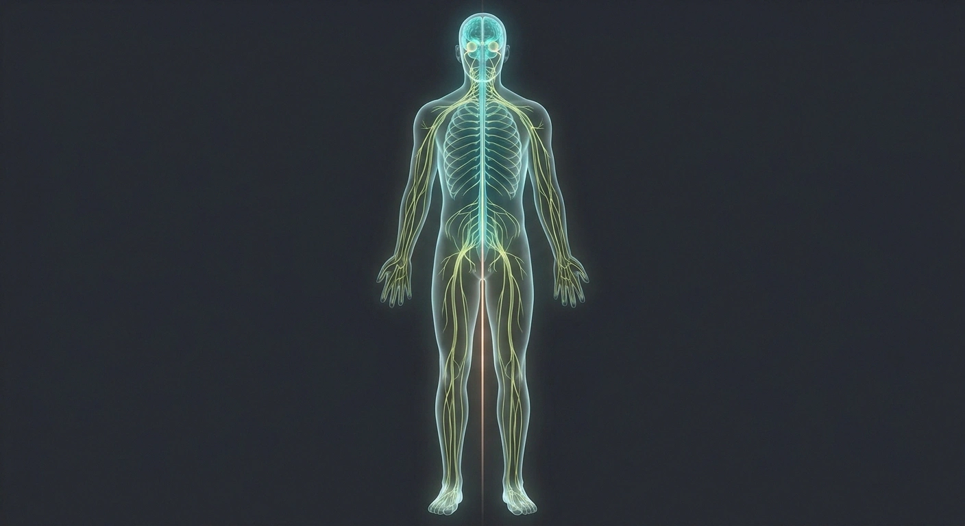
The central nervous system is your brain, spinal cord, and optic nerves. The peripheral nervous system is everything else — the nerves running out from the spinal cord to your hands, feet, organs, and skin.
Multiple sclerosis is a chronic inflammatory demyelinating disease of the central nervous system. The immune system attacks myelin, the insulating sheath around nerve fibers, producing scattered patches of damage — lesions — in the brain, spinal cord, and optic nerves. Peripheral neuropathy is damage to nerves in the other compartment entirely.
Here is a detail that explains a great deal: the two compartments make their myelin with different cells. In the central nervous system, myelin comes from cells called oligodendrocytes. In the peripheral nervous system, it comes from Schwann cells. They are different cell types with different surface proteins, which is a large part of why an autoimmune attack aimed at one compartment usually leaves the other alone. MS and peripheral neuropathy are not two versions of the same disease. They are attacks on different tissue.
Everything below follows from that.
Difference One: The Pattern on Your Body
Peripheral neuropathy is typically length-dependent. The longest nerves in the body fail first, which means the feet, symmetrically, both at once. Later the hands. If you shaded it on a body outline it would look like socks and gloves. It creeps upward gradually and stays roughly symmetric the whole way.
Symmetric, distal, ascending, sock-shaped points toward peripheral neuropathy. Patchy, regional, following a spinal level, or oddly distributed points toward something central.
The two compartments even build their myelin with different cells — oligodendrocytes centrally, Schwann cells peripherally. That is a large part of why an autoimmune attack on one usually leaves the other alone. These are not two versions of the same disease.
MS does not follow that map at all. MS numbness is multifocal and patchy — a lesion in one spot of the spinal cord produces numbness in whatever region that spot serves. That might be one thigh. Half the face. One side of the trunk. A band across the ribs. The left arm and the right leg simultaneously.
One MS pattern with no neuropathy equivalent at all is the “MS hug” — a band-like tightness or squeezing around the chest or abdomen, caused by a spinal cord lesion affecting the nerves that wrap around the torso. Length-dependent peripheral neuropathy essentially never produces that, because your torso is not at the end of a long nerve.
So: symmetric, distal, ascending, sock-shaped points toward neuropathy. Patchy, regional, respecting a spinal level, or oddly distributed points toward something central.
Difference Two: The Time Course
This may be the most useful discriminator for someone tracking their own symptoms, because you already have the data.
Peripheral neuropathy creeps
Appears over months and years. Does not remit. Your feet do not wake up numb on Tuesday and normal a month later. One direction, slowly.
MS relapses and remits
A symptom appears over hours to days, lasts weeks, then partly or fully resolves. Months or years later, something different happens somewhere else.
Three weeks of thigh numbness two years ago that fully resolved, now blurred vision in one eye, is a different story from feet that have gone slowly numb since 2022 and never once improved.
Most peripheral neuropathies creep. They appear over months and years and they do not go away. Your feet do not wake up numb on Tuesday and normal by the following month. The trajectory is slow and one-directional. Our overview of the stages of neuropathy describes that typical slow course in detail.
Classic MS relapses and remits. A symptom appears over hours to days, persists for weeks, then partly or fully resolves — sometimes completely, as though it never happened. Then months or years later, something different happens somewhere else.
That episodic pattern is genuinely distinctive. If you had three weeks of numbness in your left thigh two years ago that entirely resolved, and now you have blurred vision in one eye, you are describing a pattern of separate episodes in separate places. That is a fundamentally different story from feet that have been slowly going numb since 2022 and never once improved.
Difference Three: What Your Reflexes Do
This one requires a doctor, and it is one of the fastest bedside discriminators there is.
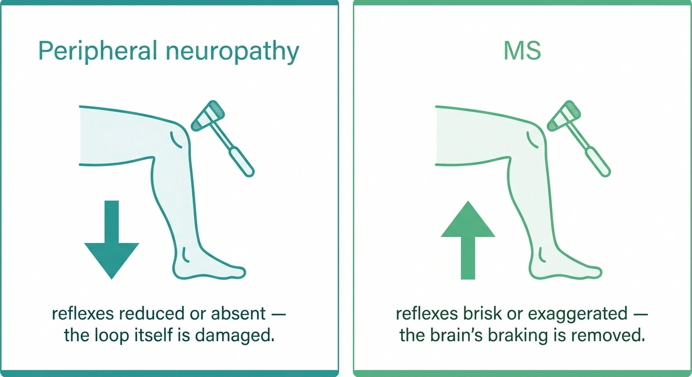
In peripheral neuropathy, the reflex loop itself is damaged. Reflexes go down. Reduced or absent ankle jerks are among the most consistent exam findings in length-dependent neuropathy.
In MS, spinal cord and brain lesions remove some of the inhibition the central nervous system normally applies. Reflexes go up. Brisk, exaggerated responses. Often spasticity — a stiff, catching quality when a limb is moved. Sometimes an abnormal toe response.
Same rubber hammer, opposite results. That is why the neurological exam is not a formality, and why a good one gathers more information in ten minutes than weeks of symptom searching.
Difference Four: Vision
Peripheral neuropathy does not affect your vision. The optic nerve is part of the central nervous system, so it is simply not in the territory that peripheral neuropathy damages.
The 2024 revision of the McDonald criteria added the optic nerve as a fifth anatomical location counting toward dissemination in space — alongside periventricular, juxtacortical/cortical, infratentorial and spinal cord. It can be confirmed by orbital MRI, optical coherence tomography, or visual evoked potentials.
The same revision allows the kappa free light chain index as an equivalent to spinal-fluid oligoclonal bands. Practically: people whose first MS attack is visual can now be diagnosed sooner. This is a framework clinicians apply — not something to apply to yourself.
MS very much can. Optic neuritis — inflammation of the optic nerve — is one of the classic MS presentations. It typically produces vision loss or blurring in one eye over hours to days, often with pain on eye movement, and a characteristic washing-out of color, especially red. For many people with MS it is the first attack they ever have.
This got more diagnostically important recently. The 2024 revision of the McDonald criteria — the framework neurologists use to diagnose MS — added the optic nerve as a fifth anatomical location that can count toward “dissemination in space,” alongside the four that were already there: periventricular, juxtacortical or cortical, infratentorial, and spinal cord. Optic nerve involvement can now be confirmed by orbital MRI, optical coherence tomography, or visual evoked potentials. Practically, that means people whose MS begins with vision symptoms can be diagnosed sooner than they would have been under the older framework. The same 2024 revision also allows the kappa free light chain index as an equivalent to the older spinal-fluid oligoclonal band test.
None of that is something you apply to yourself. It is context for why a neurologist may order an eye-focused test that seems unrelated to your numb feet.
Difference Five: Heat

Here is a small, oddly specific one that people find clarifying.
Many people with MS experience Uhthoff's phenomenon — a temporary worsening of symptoms when body temperature rises. A hot shower, a fever, a summer afternoon, hard exercise. Vision blurs, weakness increases, fatigue deepens. Then it resolves when they cool down. The mechanism is that demyelinated central nerve fibers conduct less reliably at higher temperatures.
Peripheral neuropathy has its own relationship with temperature, but a different one. Many people find their symptoms worse at night and worse in the cold, and warmth is often soothing rather than aggravating. There is no true Uhthoff equivalent.
If a hot shower reliably and temporarily makes your neurological symptoms worse and then they recover, that is worth mentioning to your doctor specifically. It is a small detail with real diagnostic weight.
What Each Test Is Actually Looking At
This is where people get confused most often, and the fix is simple: the two tests examine two different compartments.
MRI → brain and spinal cord
Where MS lives. Shows T2/FLAIR lesions in characteristic locations. Does not routinely image peripheral nerves — a normal brain MRI tells you nothing about whether you have neuropathy.
NCS and EMG → peripheral nerves
The workhorses for neuropathy. In uncomplicated MS they are typically normal, because the disease is not in that compartment. MS does not show up on a standard nerve conduction study.
Being sent for one and not the other is not an oversight. It reflects which question your doctor is asking.
MRI images the brain and spinal cord. That is where MS lives. MRI of the neuroaxis in MS typically shows multiple T2 and FLAIR lesions in characteristic locations, interpreted through the McDonald criteria. MRI does not image your peripheral nerves in any routine way, which is why a normal brain MRI tells you essentially nothing about whether you have peripheral neuropathy.
Nerve conduction studies and EMG test the peripheral nerves — how fast and how strongly they conduct. These are the workhorses for neuropathy. And in uncomplicated MS, they are typically normal, because the disease is not in that compartment. MS does not show up on a standard nerve conduction study.
That is the whole reason both tests exist, and why being sent for one and not the other is not an oversight — it reflects which question your doctor is asking. Our guide to neuropathy diagnosis and the tests your doctor may order covers the peripheral side in detail, including skin biopsy for small fiber neuropathy, which standard nerve conduction studies can miss entirely.
Can You Have Both?
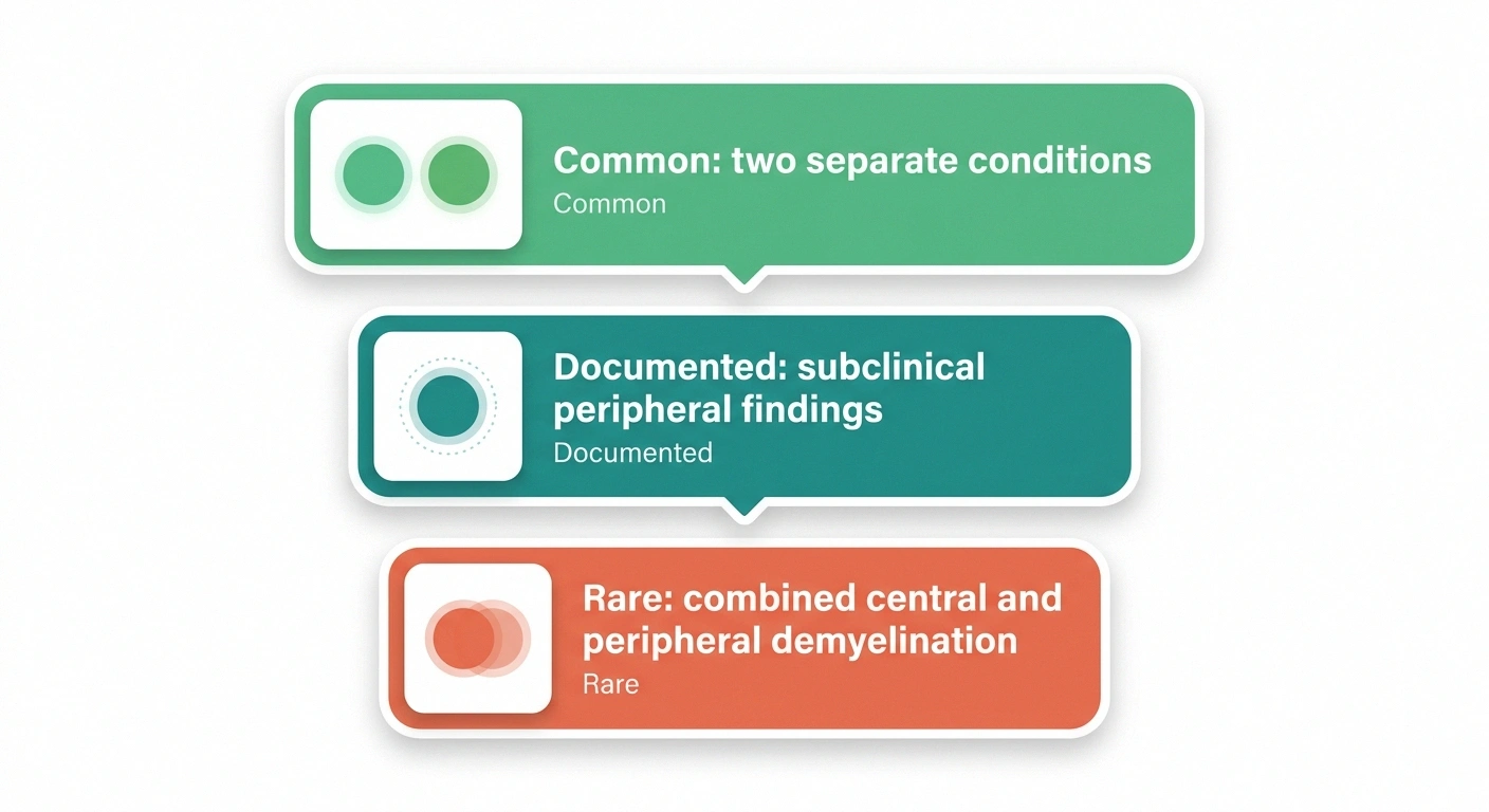
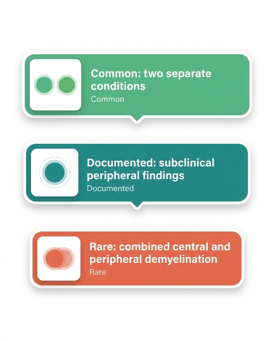
Yes, in three quite different ways, and separating them matters.
The common way: two separate conditions. A person with MS gets diabetes, or a B12 deficiency, or takes a medication that damages nerves — and now has a peripheral neuropathy alongside their MS. Two independent problems in one body. This is by far the most frequent version, and it is under-recognized, because new numbness in someone with MS tends to get attributed to the MS automatically.
The subtle way: subclinical peripheral involvement. Research using nerve conduction studies has found abnormal findings suggesting peripheral neuropathy in some people with MS and with neuromyelitis optica. Often mild and not causing obvious symptoms, but real and documented.
The rare way: combined central and peripheral demyelination. This is an uncommon clinical entity in which both compartments are demyelinating at once, described most often where features of MS and CIDP coexist. It exists in the literature as case reports and small series. It is genuinely rare, and it is not the explanation for most people's overlapping symptoms.
The practical takeaway: if you have MS and develop new symptoms that look like classic neuropathy — symmetric, distal, burning, both feet — that deserves its own workup rather than being folded into the MS diagnosis by default. Some of the causes are correctable.
The Shared Imitator: Vitamin B12
I want to give B12 its own section because of the story I opened with, and because it sits in a genuinely unusual position.
Vitamin B12 deficiency damages nerves in both compartments at once.
It causes peripheral neuropathy — numb, tingling feet. It also causes subacute combined degeneration of the spinal cord, producing brisk reflexes, spasticity, balance loss and MRI changes that can genuinely resemble MS. Untreated, one person can show up with numb feet, an abnormal cord MRI and fatigue, reading like MS on first pass.
If your B12 sits in the low-normal range, ask about a methylmalonic acid level — it detects functional deficiency a normal-looking B12 can hide. Most people with MS symptoms do not have a B12 problem, and correcting B12 does not treat MS. But it is cheap, fast, and occasionally changes everything.
B12 deficiency damages nerves in both compartments. It causes peripheral neuropathy — numb, tingling feet, the ordinary presentation. It also causes something called subacute combined degeneration of the spinal cord, which damages specific tracts in the cord and can produce spasticity, brisk reflexes, balance loss, and MRI changes that can genuinely resemble MS.
So a person with untreated B12 deficiency can present with numb feet, an abnormal spinal cord MRI, brisk reflexes, and fatigue — a combination that reads like MS on first pass. And it is correctable with a blood test and treatment.
This is the single most important reason to make sure B12 has actually been checked, and checked properly. If your level is in the low-normal range, ask about a methylmalonic acid level, which detects functional deficiency that a normal-looking B12 can hide. Our guide to vitamin deficiencies that cause neuropathy covers the testing in more depth.
To be clear: most people with MS symptoms do not have a B12 deficiency, and correcting B12 does not treat MS. But it is cheap, fast, and occasionally changes everything.
Who Tends to Get Which

Demographics are a soft signal — never diagnostic, but they shape which possibility a doctor considers first.
MS typically begins between roughly 20 and 40 years of age and is substantially more common in women. It is more prevalent farther from the equator. It rarely announces itself for the first time in someone's seventies.
Length-dependent peripheral neuropathy skews strongly older and tracks closely with diabetes, prediabetes, age, alcohol use, chemotherapy exposure, and nutritional deficiency. It becomes considerably more common past sixty.
So numbness starting in both feet in a 68-year-old with type 2 diabetes and numbness starting in one thigh in a 31-year-old woman are two very different starting points, even though both people would type “numbness and tingling” into a search bar.
Symptoms Only One of Them Produces
A quick sorting list. None of these is definitive on its own.
Points toward MS
- Vision loss in one eye with pain on movement
- Double vision
- Electric shock down the spine on neck flexion
- Band-like tightness around the torso
- Predictable heat worsening that recovers
- New bladder urgency or retention
- A symptom that resolved after weeks
Points toward neuropathy
- Symmetric burning feet
- Lost ankle reflexes
- Reduced vibration sense in the toes
- Skin that hurts to light touch
- Calluses or unnoticed foot wounds
- Worse at night
- Gradual, never remitting, over years
Shared by both
- Numbness
- Tingling
- Fatigue
- Balance trouble
The overlap sits in the four most-searched symptoms. That is why these get confused.
Points toward MS: vision loss or blurring in one eye with pain on eye movement; double vision; an electric-shock sensation running down the spine when you bend your neck forward (Lhermitte's sign); band-like tightness around the torso; symptoms that worsen predictably with heat and recover with cooling; new bladder urgency or retention; a symptom that appeared over days and then substantially resolved.
Points toward peripheral neuropathy: symmetric burning feet; loss of ankle reflexes; reduced vibration sense in the toes; skin that hurts to light touch in a stocking distribution; calluses, foot deformity, or an unnoticed wound from unfelt pressure; symptoms worse at night; gradual, never-remitting progression over years.
Shared by both: numbness, tingling, fatigue, and balance trouble. Which is precisely why these two get confused in the first place — the overlap sits in the four most-searched symptoms.
Balance Trouble Comes From Different Places
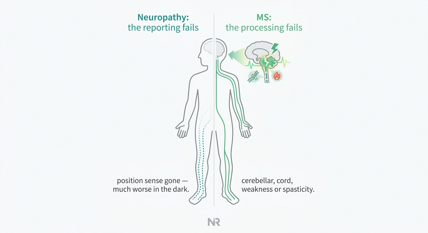
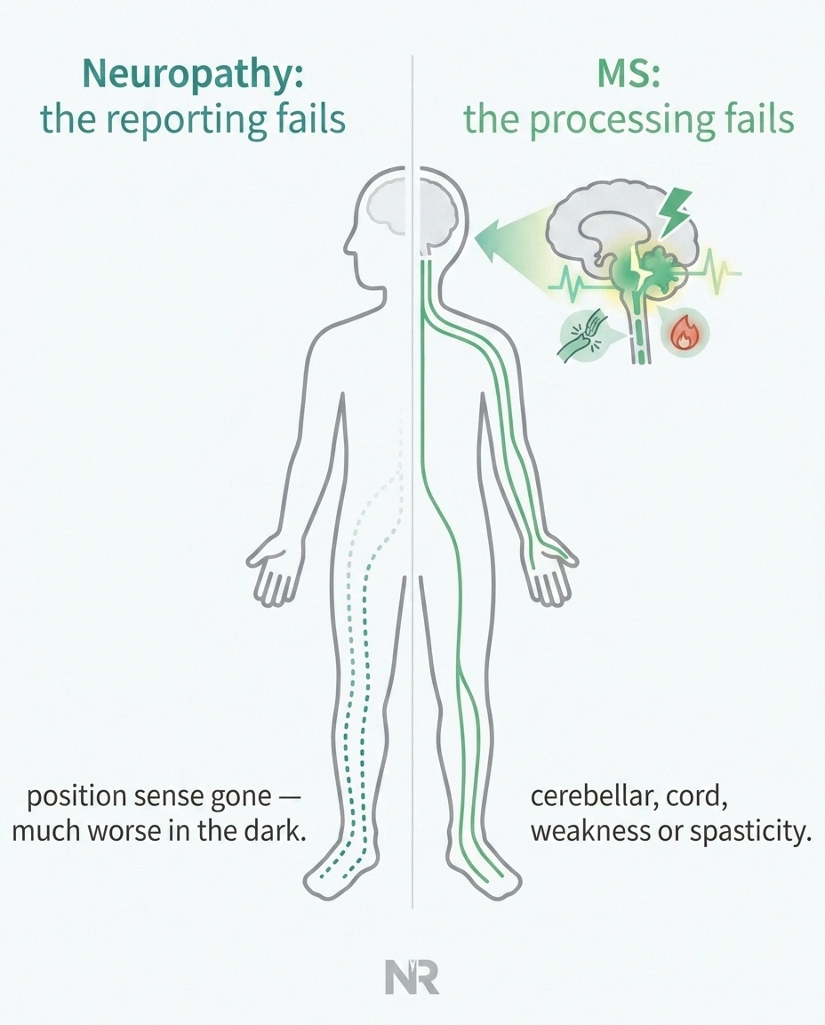
Both conditions cause unsteadiness, and the reason differs in a way that affects what helps.
In peripheral neuropathy, balance fails because position sense is gone. The nerves reporting where your feet are in space have stopped reporting. Your brain is fine; it is receiving no data. That is why people with neuropathy get much worse in the dark or with eyes closed — vision was doing the job the feet stopped doing.
In MS, balance can fail from cerebellar involvement, spinal cord lesions, weakness, or spasticity — problems in the processing and control side rather than the reporting side.
Both raise fall risk substantially, and both benefit from balance training and home modification, though the specific programs differ. Our guide to neuropathy and fall prevention covers the sensory-loss version, and it is worth reading regardless, because the home safety half applies to anyone unsteady on their feet.
What to Bring to the Appointment
The single most valuable thing you can hand a neurologist is a clean timeline. Not a symptom list — a timeline.
- What, where, when, and did it resolve. “March 2024: numbness left thigh, three weeks, fully resolved.” That one line does more diagnostic work than a paragraph about how it felt.
- Whether it is symmetric. Both feet equally, or one side?
- Any vision episodes, ever — even brief ones you dismissed at the time.
- Any relationship to heat — showers, fever, exercise, hot weather.
- Bladder and bowel changes, which people frequently omit out of embarrassment and which carry real weight.
- Your full medication and supplement list, with doses.
- Family history of MS, autoimmune disease, or neuropathy.
Write it before you go. Nobody recalls their own chronology accurately under the pressure of a fifteen-minute appointment.
When Not to Wait
Some symptoms belong at the top of the schedule regardless of which condition turns out to be behind them:
- Sudden vision loss or blurring in one eye, especially with pain on eye movement
- New band-like tightness around the chest or abdomen
- New bladder retention or loss of bowel control
- Rapidly progressive weakness, particularly if ascending
- New numbness in the groin or inner thighs
- Any foot wound, ulcer, redness or swelling if you have reduced sensation — same-day call
Several of these are emergencies for reasons that have nothing to do with either MS or neuropathy. That is exactly why they get evaluated rather than searched.
- Sudden vision loss or blurring in one eye, especially with pain on eye movement
- New band-like tightness around the chest or abdomen
- New bladder retention — inability to urinate — or loss of bowel control
- Rapidly progressive weakness, particularly if it is ascending
- New numbness in the groin or inner thighs
- Any foot wound, ulcer, redness, or swelling if you have reduced sensation — same-day call
Several of these are emergencies for reasons that have nothing to do with either MS or neuropathy, which is exactly why they get evaluated rather than researched.
What I'd Tell a Friend Who Asked

If your symptoms are symmetric, in both feet, slowly worsening over years, burning at night, with no vision episodes and no periods of recovery — that is the shape of peripheral neuropathy, and there is a long list of causes worth chasing, several of them correctable. Burning feet alone has a dozen ordinary explanations.
If your symptoms came in episodes, appeared in odd patches rather than symmetrically, involved your vision, got temporarily worse in the heat, or resolved and later returned somewhere else — that is a different shape, and it belongs with a neurologist rather than an internet search.
And either way, make sure someone has actually checked your B12.
The waiting is genuinely hard — that stretch between noticing something and knowing what it is. The uncertainty tends to be heavier than most diagnoses turn out to be, and it takes a real toll; our piece on neuropathy and mental health covers that weight honestly. Bring your timeline, bring someone with you, and let the exam do the work it is genuinely good at.
Frequently Asked Questions
Can peripheral neuropathy be a sign of multiple sclerosis?
Not typically. MS is a disease of the central nervous system, meaning the brain, spinal cord and optic nerves, while peripheral neuropathy affects nerves outside those structures. The two involve different tissue and even different myelin-producing cells. However, people with MS can separately develop peripheral neuropathy from an unrelated cause such as diabetes, vitamin B12 deficiency or a medication, and research has documented mild subclinical peripheral nerve findings in some people with MS. New symmetric burning or numbness in someone with MS deserves its own evaluation rather than automatic attribution to the MS.
What is the main difference between MS numbness and neuropathy numbness?
The pattern and the time course. Peripheral neuropathy numbness is usually symmetric and length-dependent, beginning in both feet and slowly ascending over months or years without remitting. MS numbness is typically patchy and regional, following the area served by a specific brain or spinal cord lesion, and it often appears over hours to days, lasts weeks, then partly or fully resolves. MS can also produce a band-like tightness around the torso, sometimes called the MS hug, which length-dependent peripheral neuropathy does not cause.
Does MS show up on a nerve conduction study?
Generally no. Nerve conduction studies and EMG test the peripheral nerves, and in uncomplicated MS those nerves are not affected, so results are typically normal. MS is diagnosed primarily with MRI of the brain and spinal cord, interpreted through the McDonald criteria, sometimes supported by spinal fluid analysis and evoked potential testing. Conversely, a normal brain MRI says essentially nothing about whether peripheral neuropathy is present. The two tests examine two different compartments of the nervous system.
Can vitamin B12 deficiency look like MS?
Yes, and this is one of the most important things to rule out. B12 deficiency causes peripheral neuropathy and also causes subacute combined degeneration of the spinal cord, which can produce brisk reflexes, spasticity, balance loss and MRI changes resembling MS. Someone with untreated deficiency can present with numb feet, an abnormal spinal cord MRI and fatigue, a picture that superficially reads like MS. If your B12 level is low-normal, ask about a methylmalonic acid level, which detects functional deficiency that a normal-appearing B12 result can conceal.
Do reflexes help tell MS and neuropathy apart?
They are one of the fastest bedside discriminators. In peripheral neuropathy the reflex loop itself is damaged, so reflexes are reduced or absent, particularly at the ankle. In MS, central nervous system lesions remove some of the brain's normal inhibition, so reflexes become brisk or exaggerated, often with spasticity and sometimes an abnormal toe response. The same reflex hammer produces opposite results, which is a significant part of why an in-person neurological examination gathers more information in ten minutes than weeks of symptom research.
Does heat make both conditions worse?
They respond differently. Many people with MS experience Uhthoff's phenomenon, a temporary worsening of neurological symptoms when body temperature rises from a hot shower, fever, exercise or hot weather, which then resolves on cooling. This happens because demyelinated central nerve fibers conduct less reliably when warm. Peripheral neuropathy more often worsens at night and in cold conditions, and warmth is frequently soothing rather than aggravating. A reliable, temporary heat-triggered worsening that recovers afterward is a detail worth reporting specifically to your doctor.
Can you have MS and peripheral neuropathy at the same time?
Yes, in three distinct ways. Most commonly, the two are separate conditions occurring in the same person, such as someone with MS who also develops diabetic or B12-related neuropathy. Second, research has found subclinical peripheral nerve abnormalities on nerve conduction studies in some people with MS and with neuromyelitis optica. Third, and genuinely rare, is combined central and peripheral demyelination, an uncommon entity described most often where features of MS and CIDP coexist. The first situation is by far the most frequent and the most likely to be overlooked.
At what age does each condition usually start?
MS most often begins between roughly 20 and 40 years of age and is substantially more common in women, with higher prevalence farther from the equator. Length-dependent peripheral neuropathy skews considerably older and becomes common past age sixty, tracking closely with diabetes, prediabetes, alcohol use, chemotherapy exposure and nutritional deficiency. Age is a soft signal that shapes which possibility a clinician considers first, but it is never diagnostic on its own, and both conditions can appear outside their typical age ranges.
About the Author