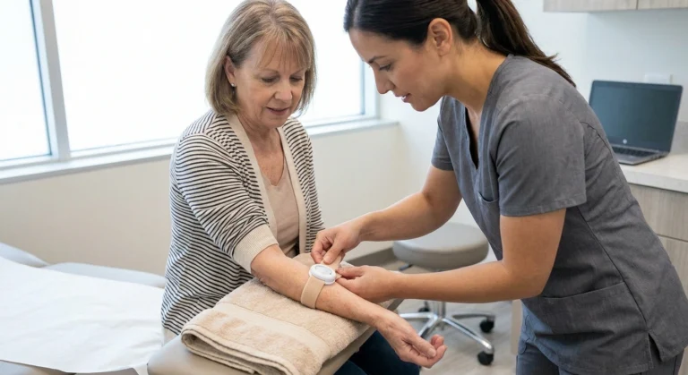
The first time my neurologist mentioned nerve ultrasound, I pictured the same wand-on-belly scan they used when I was pregnant with my daughter. I was not far off. It really is that simple — a bit of warm gel, a small probe, and pictures on a screen. What surprised me was that this old, gentle technology has quietly become one of the most useful tools in a modern neuropathy workup, and hardly anyone talks about it with patients.
If your doctor has mentioned “high-resolution nerve ultrasound,” or if you have been reading about it and wondering whether to ask for it, this guide walks through what it actually shows, what it cannot tell you, when it beats other tests, and what to expect if you go in for one.
What Nerve Ultrasound Actually Is
Nerve ultrasound (sometimes called high-resolution ultrasonography, or HRUS) uses very high-frequency sound waves — usually from a probe running between 15 and 24 megahertz — to build a picture of your peripheral nerves in real time. The probes are much higher frequency than the ones used for pregnancy or heart scans, which lets them see fine, superficial structures that would otherwise be too small to make out.
Nerve ultrasound is one of the few tests that shows you what a nerve looks like. EMG shows function, blood work shows metabolic causes, and skin biopsy shows the smallest fibers. Ultrasound answers the anatomical question: is the nerve swollen, being squeezed, or sitting under something it shouldn’t be?
What the sonographer or radiologist sees on the screen is a nerve in cross-section, appearing as a honeycomb-like bundle of fibers wrapped in a bright outer sheath. They can measure how big it is, how bright or dark it looks, whether it has extra blood flow, and whether it slides normally when you move a joint. From those four measurements — size, echogenicity, vascularity, mobility — a lot can be worked out about what is happening inside a nerve without ever putting a needle in it.
What a Nerve Ultrasound Can Show
The single most important thing nerve ultrasound gives you that no other test does is a picture of the actual nerve. Nerve conduction studies and EMG tell you how well nerves are firing electrically. Blood work tells you about metabolic causes. Skin biopsy tells you about the smallest fibers. Ultrasound tells you what the nerve looks like.
Practically, that means it can spot:
- Enlargement of a nerve. A nerve that is fatter than normal at a specific spot is one of the strongest signs of compression (like carpal tunnel), inflammation, or an inherited condition like Charcot-Marie-Tooth.
- A pinch point. The ultrasound can watch the nerve swell just before it enters a tight passage and become thin as it squeezes through — a very direct visualization of entrapment.
- A mass or tumor. Schwannomas, neurofibromas, ganglion cysts, and other growths sitting on or in a nerve show up clearly.
- Structural damage. After trauma, ultrasound can see whether a nerve has been fully cut, partially torn, or is just bruised.
- Multifocal thickening. Conditions like chronic inflammatory demyelinating polyneuropathy (CIDP) and multifocal motor neuropathy often show patches of enlargement in specific patterns.
- Nerve mobility problems. Because it is real-time, the technician can move your joint while scanning and see whether the nerve glides smoothly or gets stuck.
For a lot of patients, this is what finally answers the “why” question that standard neuropathy diagnostic testing could not.
Where Nerve Ultrasound Genuinely Shines
Not every nerve problem needs an ultrasound. But there are situations where it is quietly one of the best tools we have.
Published reviews of nerve ultrasound consistently support its usefulness in:
- Carpal and cubital tunnel syndrome — nerve enlargement at the entrapment site is a sensitive signal
- CIDP and multifocal motor neuropathy — multifocal nerve thickening in characteristic patterns
- Nerve trauma and post-surgical injury — visualizing continuity, neuromas, and hardware impingement
- Hereditary neuropathies like CMT1A — diffuse nerve enlargement supports the diagnosis
Entrapment Neuropathies
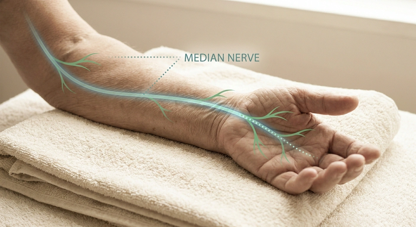
This is the sweet spot. Carpal tunnel syndrome, cubital tunnel (ulnar nerve at the elbow), tarsal tunnel at the ankle, peroneal nerve at the fibular head, radial nerve at the spiral groove — all can be diagnosed with ultrasound, often with more anatomical detail than EMG alone. A swollen median nerve at the wrist with a cross-sectional area above about 10 to 12 square millimeters is a strong signal for carpal tunnel, for example.
Inflammatory Neuropathies
CIDP and multifocal motor neuropathy often produce nerve enlargement in patterns that ultrasound can see clearly. This matters because these two conditions are treatable — sometimes dramatically so — but they can be missed for years if they are only chased with routine blood work.
Trauma, Masses, and Post-Surgical Problems
If you were fine before an injury or surgery and then something changed in a specific limb, ultrasound is often the first imaging step. It can find nerve tumors, scar tissue wrapping around a nerve, hardware pressing on a nerve after orthopedic surgery, or a neuroma (a painful bundle of nerve endings that formed after a cut).
Hereditary Neuropathies
In Charcot-Marie-Tooth type 1A, the classic finding is diffuse enlargement of many peripheral nerves. Ultrasound can support the diagnosis and, in some cases, help sort out which subtype is most likely before genetic testing is ordered.
Where Nerve Ultrasound Does Not Help
Nothing in medicine is a universal answer, and being honest about limits is part of what makes a test trustworthy. Here is where nerve ultrasound is not the right tool.
A normal nerve ultrasound does not rule out neuropathy. Small-fiber neuropathy, early metabolic neuropathies, and problems in deep nerves (roots, brachial plexus) can all look normal on ultrasound while causing real symptoms. If your ultrasound comes back clean but you still have symptoms, ask what test the anatomy might have missed — the answer is often a skin biopsy or a targeted MRI.
- Small-fiber neuropathy. The tiny unmyelinated C-fibers and thin A-delta fibers that carry burning, temperature, and autonomic signals are too small to see. Small-fiber problems still need skin biopsy or QSART.
- Deep-lying nerves. Nerve roots inside the spinal canal, the brachial plexus behind the clavicle, and the sciatic nerve deep in the buttock are difficult or impossible to see well. MRI wins for those.
- Pure functional questions. Ultrasound cannot tell you whether a nerve fires slowly, whether the signal is dropping out midway, or whether muscle is receiving instructions. That is what nerve conduction studies and EMG do.
- Metabolic screening. If your neuropathy is from diabetes, B12 deficiency, kidney disease, or alcohol, ultrasound will usually be normal or nonspecific. Blood work is the tool for those.
The takeaway: ultrasound is a beautiful anatomical test but a limited functional and microscopic one. It works best alongside other tests, not instead of them.
Nerve Ultrasound vs. EMG and Nerve Conduction Studies
This is the most common question I get, and the answer is not “one is better.” They tell you different things.
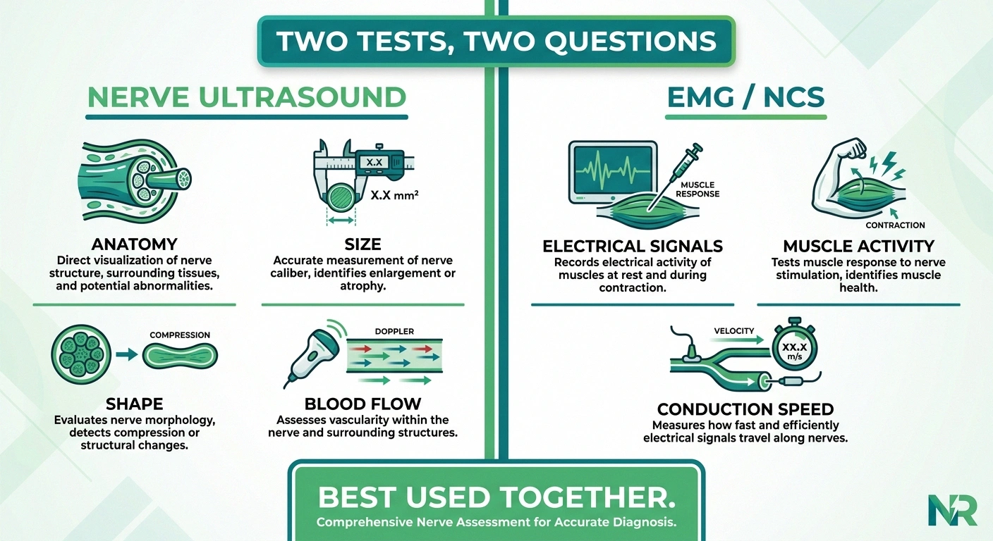
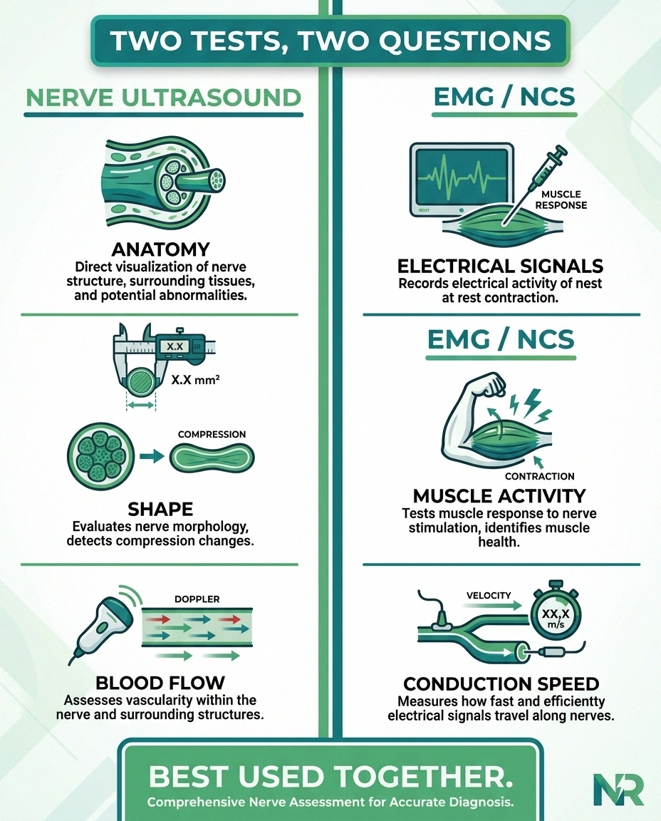
Two Tests, Two Different Questions
Size · Shape · Blood flow · Mobility
Speed · Signal strength · Muscle response
In most workups, they are complementary — not competing.
Nerve conduction studies and EMG measure electrical function. They send small pulses along a nerve and time how fast and how strong the signal travels, and they listen to how muscle behaves at rest and during contraction. That gives you information about whether the nerve is losing its insulation (demyelination), losing fibers (axonal loss), or whether the muscle it feeds has been chronically starved of nerve input.
Nerve ultrasound measures anatomy. It shows the size, shape, blood flow, and position of the nerve itself.
Put together, they answer two different questions: “Is this nerve working?” (EMG/NCS) and “What does this nerve look like?” (ultrasound). In carpal tunnel, for example, EMG can prove the nerve is being squeezed hard enough to slow signals down, and ultrasound can prove the nerve is swollen and get an eye on where exactly the pinch is. In CIDP, EMG can show the classic slow, block-y pattern, and ultrasound can show the enlarged, thickened nerve segments.
If a doctor forces you to pick one, EMG/NCS is still the workhorse for most peripheral neuropathy workups. But for entrapment, inflammatory conditions, or when EMG results are ambiguous, adding ultrasound often turns “we think it’s this” into “we can see it.”
Nerve Ultrasound vs. MRI
MRI of the peripheral nerves (sometimes called MR neurography) can also image nerves, and in some cases it is the better choice. Here is a quick way to think about it:
- Ultrasound is better for: superficial nerves in the arms and legs, dynamic testing (looking at the nerve during joint movement), quick and inexpensive answers, patients with pacemakers or metal implants, and patients who cannot tolerate the tube.
- MRI is better for: nerve roots inside the spine, the brachial plexus (behind the collarbone), pelvic and lumbosacral plexus, deep sciatic nerve problems, and when you need a very wide field of view.
Ultrasound is faster, cheaper, and does not require you to lie still for 45 minutes in a loud tunnel. MRI sees deeper and gives your neurologist a wider anatomical map. Increasingly, good neuromuscular centers use them as complementary tools.
What to Expect at the Appointment
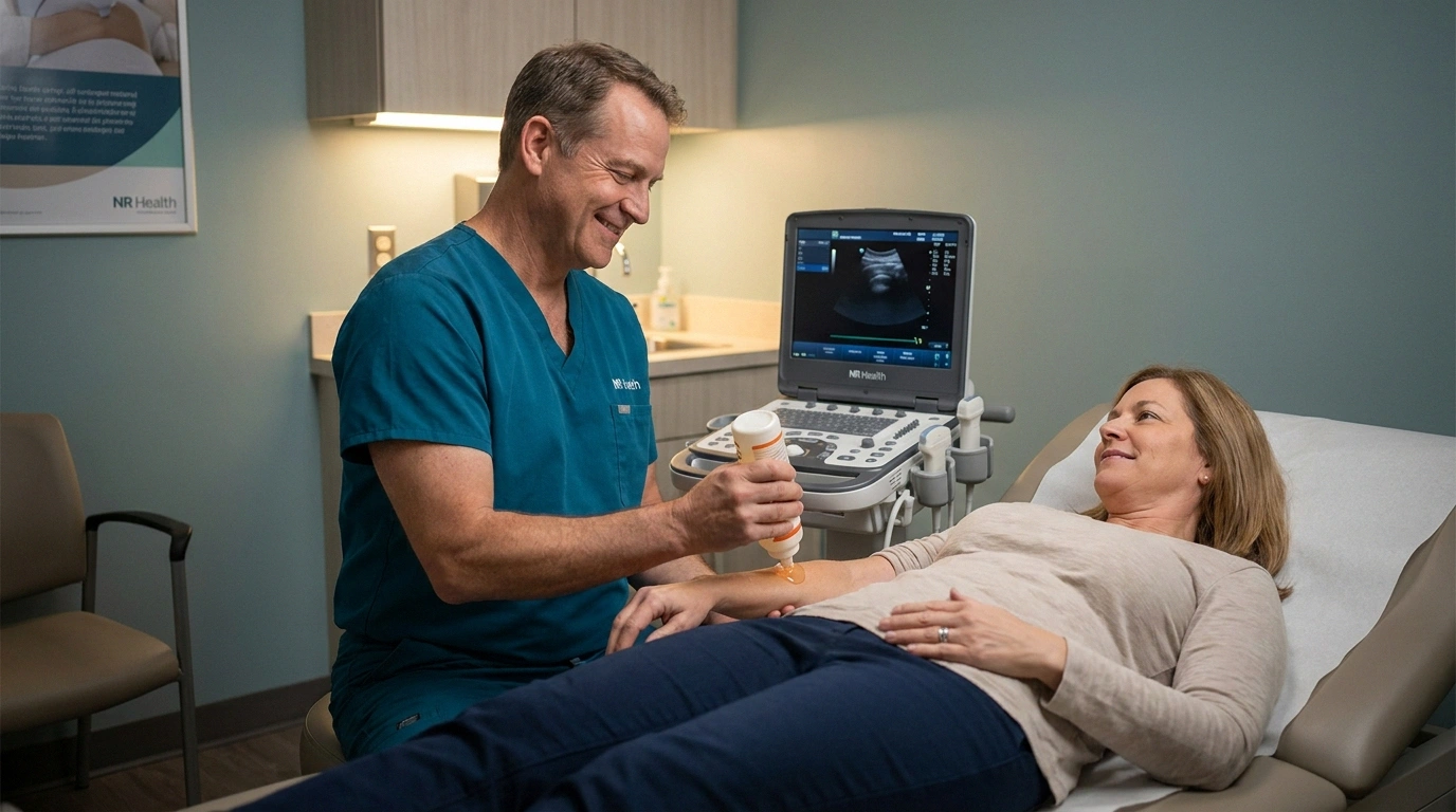
If your doctor orders a nerve ultrasound, here is how the day usually unfolds. It is one of the easiest tests you will ever have.
Your Nerve Ultrasound, Step by Step
You do not need to fast. You do not need to stop any medications. Wear comfortable clothes that give easy access to the arm or leg being scanned — a short-sleeve shirt for an upper-limb study, loose pants that roll up for lower-limb. Expect the exam itself to take between 15 and 45 minutes depending on how many nerves need to be checked and whether they are looking at one focused problem or doing a broader survey.
You will lie on an exam table. The sonographer will apply warm ultrasound gel to your skin, then slide the small probe along the path of the nerve. There is no pain, no needle, no radiation, no injection, and no dye. Sometimes they will ask you to bend or straighten a joint so they can see how the nerve moves. If they are looking for a compression, they may compare the “bad” side to the “good” side.
Some centers will give you preliminary impressions on the spot. Others send the images to a radiologist or neuromuscular specialist to read, with a formal report to your ordering doctor within a few days.
What the Report Will Actually Say
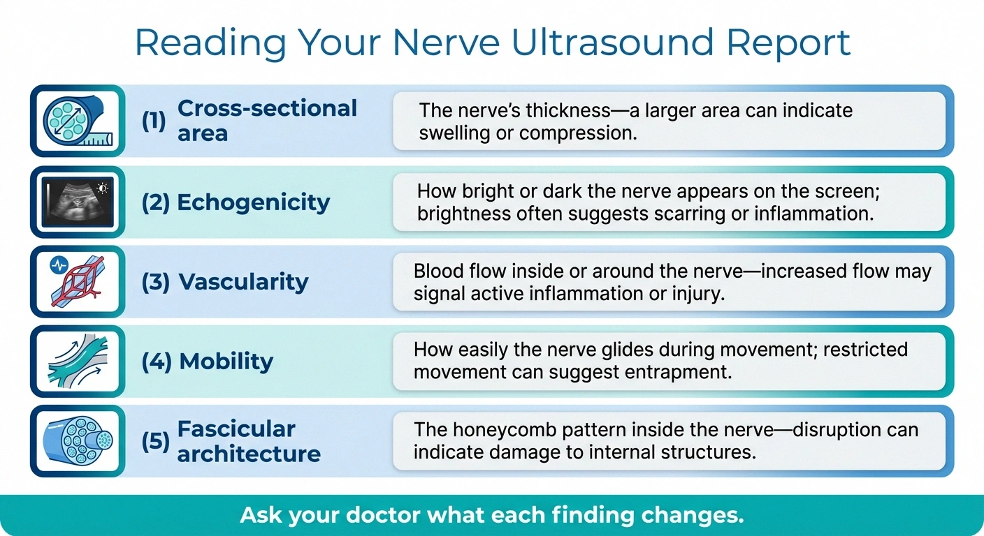
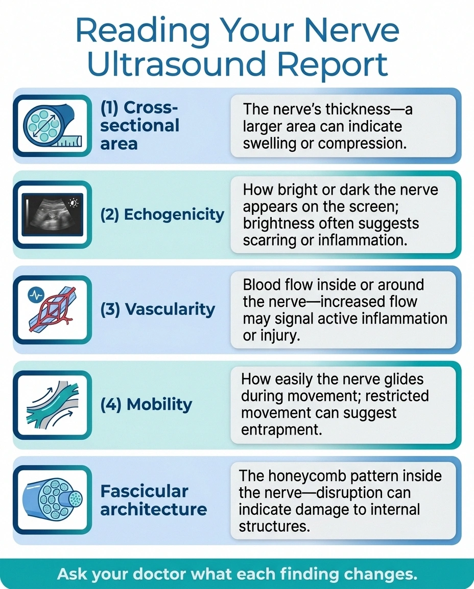
Ultrasound reports use vocabulary that can look intimidating on paper. A few translations will help.
- Cross-sectional area (CSA): the thickness of the nerve at a specific spot, measured in square millimeters. Normal ranges depend on which nerve; larger than expected suggests swelling, compression, or inflammation.
- Echogenicity: how bright or dark the nerve appears. A nerve that is more “hypoechoic” (darker) than usual can suggest edema or an active inflammatory process.
- Vascularity / hyperemia: increased blood flow inside the nerve, seen with the power Doppler setting. Usually a sign of active inflammation.
- Mobility / gliding: whether the nerve slides smoothly under the skin when you move. Reduced gliding can suggest tethering by scar or adhesions.
- Fascicular architecture: the internal honeycomb pattern. Loss of the normal pattern can point to more serious injury.
If your report mentions a specific enlarged spot with a measurement, ask your neurologist to walk you through where that is anatomically and what it changes about the plan. Numbers on a page are only useful in the context of what the doctor is doing about them.
How Much It Costs and Whether Insurance Covers It
This is the practical question most people forget to ask until the bill lands. In the United States, nerve ultrasound is generally billed under CPT codes for extremity or musculoskeletal ultrasound. Rough out-of-pocket ranges I have seen and heard from readers:
Nerve Ultrasound: What It Costs
| Payment Scenario | Typical Out-of-Pocket |
|---|---|
| Insurance + prior authorization | Standard specialist copay |
| Hospital radiology, self-pay | ~$200 – $600 |
| Outpatient neuromuscular clinic, self-pay | ~$150 – $400 |
| Medicare / most commercial plans | Usually covered for specific indication |
Denials are often reversed on appeal when the ordering doctor clarifies the specific question the ultrasound will answer.
- With insurance and prior authorization: often a standard specialist copay, similar to any diagnostic imaging.
- Self-pay at a hospital radiology department: commonly $200 to $600.
- Self-pay at an outpatient neuromuscular clinic: often $150 to $400.
- Medicare and most commercial plans: usually cover the study when ordered for a specific indication (suspected entrapment, mass, inflammatory neuropathy, post-traumatic nerve injury).
Coverage tends to be smoother when the ordering doctor documents a specific question the ultrasound will answer — “rule out median nerve entrapment at the wrist” or “assess for nerve enlargement suspicious for CIDP” — rather than a vague “neuropathy workup.” If you get denied, that is often the first thing to fix in the appeal.
Who Should Perform Your Nerve Ultrasound
This is where I have to be direct: nerve ultrasound is heavily operator-dependent. The images are only as good as the person holding the probe. A radiologist or neuromuscular specialist who scans nerves every week will see things that a general ultrasound technician will miss.
When possible, ask for:
- A neuromuscular ultrasound clinic at an academic center or large neurology practice — the person scanning also reads the study and often knows the clinical question.
- A radiologist fellowship-trained in musculoskeletal or neuroradiology — comfortable with peripheral nerve anatomy, not just tendons and joints.
- A center that scans a decent volume of nerve cases — not the same place doing three of these a year.
If your only option is a general imaging center, it can still be useful — but interpret a “normal” result with a grain of salt, and don’t rule out a real problem based only on that scan.
Practical Situations Where I’d Ask for a Nerve Ultrasound

Speaking as someone who has spent years learning to advocate for herself in the exam room, these are the moments where I would gently but firmly ask my neurologist, “would nerve ultrasound help here?”
A gentle way to ask your doctor:
Framing the request as a specific question — not a shopping list — usually gets a specific answer back.
- Symptoms are one-sided or focal. A single arm, a single leg, or “only on the pinky side” pattern suggests something anatomical — an entrapment, a mass — rather than a diffuse metabolic cause. Ultrasound thrives here.
- EMG is normal but symptoms are real. Sometimes an entrapment is early or partial, and EMG missed it. Sometimes there is a structural lesion the EMG cannot detect.
- EMG suggests demyelination. If your report mentions “conduction block,” “prolonged latencies,” or “features suggestive of CIDP or MMN,” ultrasound looking for nerve enlargement can support the diagnosis and speed up the decision to treat.
- Post-surgical or post-injection pain. When new nerve pain shows up after an operation, a shot, or a fracture, ultrasound can look for nerve injury, hardware impingement, or a neuroma.
- Persistent, well-localized pain with a normal MRI. A perfectly normal spine MRI does not rule out a peripheral nerve problem downstream. Ultrasound covers the anatomy the MRI does not.
How Nerve Ultrasound Fits Into a Complete Workup
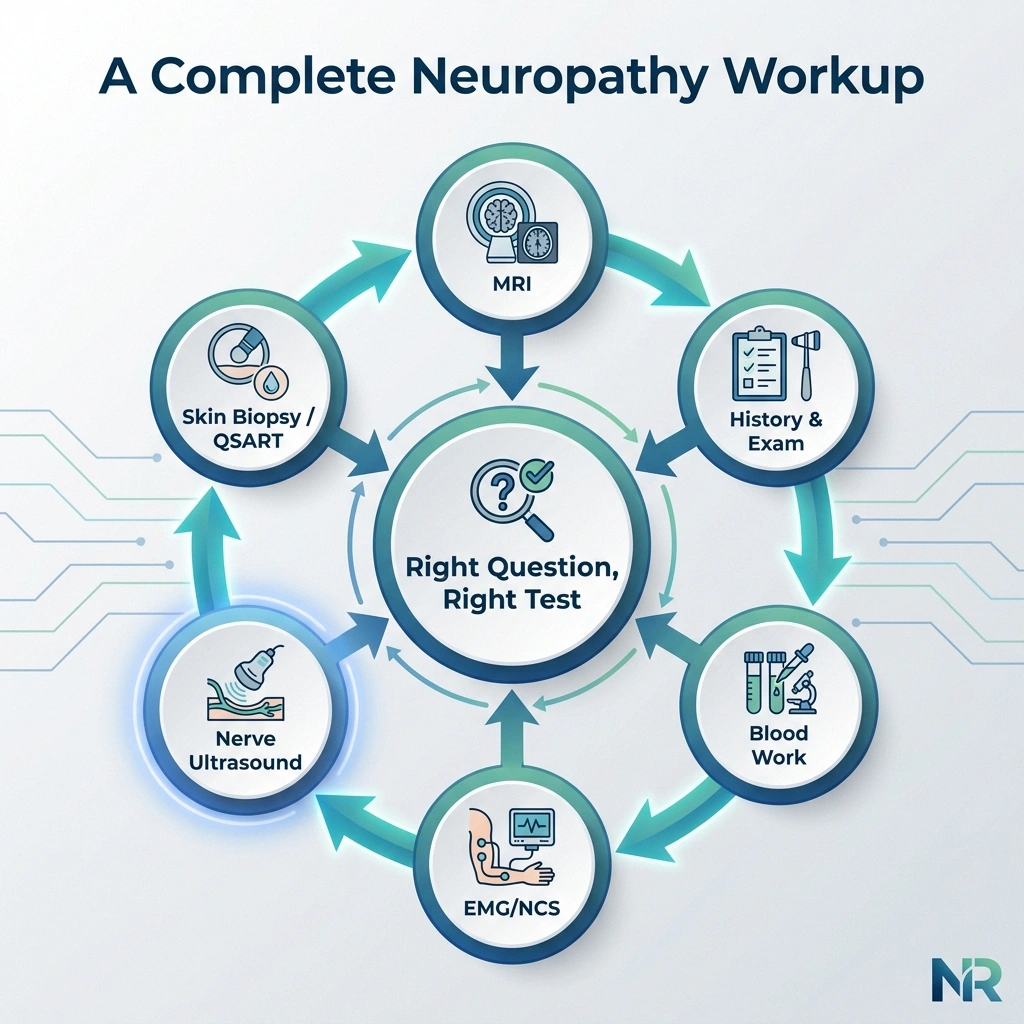
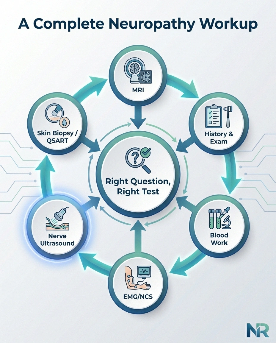
The best neuropathy workups I have seen use tests as answers to specific questions, not as a shotgun blast. Here is a rough map of how the pieces fit together:
- Story and physical exam answer: “what pattern of nerve involvement are we dealing with, and what is the most likely cause?”
- Blood work answers: “is there a metabolic, autoimmune, or nutritional cause driving this?”
- EMG and nerve conduction studies answer: “how well are the medium and large nerves functioning, and is the damage in the axon or the myelin?”
- Nerve ultrasound answers: “what do these nerves look like, and is there structural cause I can see?”
- Skin biopsy or QSART answer: “are the smallest fibers involved?”
- MRI answers: “is the problem deeper — the spine, plexus, or a hard-to-reach nerve — and is there a mass?”
Your job as the patient is not to master any of this. Your job is to know that these tests each have a specific role, and to gently push back when someone tells you “the EMG was normal so there is nothing more to look for.” Sometimes the next question is a scan, not a shrug.
Frequently Asked Questions
Does nerve ultrasound hurt?
No. It uses sound waves and a small handheld probe on the skin with gel. There are no needles, no electrical pulses, and no radiation. Occasionally the sonographer will apply pressure to see how a nerve responds, but nothing that would count as painful. Most people describe it as the easiest test in their whole workup.
Can nerve ultrasound detect small fiber neuropathy?
No. Small fibers are the tiniest unmyelinated nerve endings that carry burning pain, temperature, and autonomic signals. They are simply too small to see on ultrasound. If small fiber neuropathy is what your doctor is chasing, the appropriate tests are a punch skin biopsy for epidermal nerve fiber density, QSART for sweat function, or corneal confocal microscopy — not ultrasound.
How is nerve ultrasound different from a Doppler ultrasound of the legs?
They use the same underlying technology but look at very different structures. A venous Doppler evaluates blood vessels and blood flow, usually to rule out a clot. Nerve ultrasound uses a much higher-frequency probe to visualize the nerves themselves and adds power Doppler mainly to check for inflammatory blood flow inside a nerve. If you have had a Doppler before, the setup will feel very familiar — same table, same gel, same wand — but the pictures and interpretation are completely different.
How long does the appointment take?
A focused single-nerve study — for suspected carpal tunnel, for example — often takes 15 to 20 minutes. A broader survey looking at multiple nerves in the arm or leg can take 30 to 45 minutes. Studies that check both sides for comparison run longer. Most centers will have your images to a reader within a few days and a report back to your ordering doctor within about a week.
Is nerve ultrasound covered by Medicare?
Generally yes, when it is ordered for a specific medical indication such as suspected nerve entrapment, evaluation of a mass, follow-up of a nerve injury, or an inflammatory neuropathy. Coverage improves when the ordering doctor documents the clinical question the ultrasound is meant to answer. If a claim is denied, the appeal often succeeds when the documentation is clarified.
Can nerve ultrasound replace an EMG?
Not for most workups. EMG and nerve conduction studies measure how well nerves are conducting electrical signals, and how the muscles they feed are behaving. Ultrasound measures anatomy — size, shape, and blood flow. Both are useful and answer different questions. In a few specific entrapment situations, an experienced neuromuscular team may reasonably diagnose the problem with ultrasound alone, but in most peripheral neuropathy cases the two studies complement each other.
My EMG was normal. Should I still ask for a nerve ultrasound?
If your symptoms are focal, unilateral, or worse on one side of a specific joint, yes — it is a reasonable next question. EMG can be normal early in entrapment or when the compression is intermittent. Ultrasound can catch swelling or a mass that the EMG has no way of showing. If your symptoms are diffuse, symmetric, and burning (a classic small-fiber pattern), then skin biopsy or QSART is likely a better next step than ultrasound.
Are there any risks or side effects?
None significant. There is no radiation, no injected contrast, no electrical current, and no needle. The ultrasound gel can occasionally cause skin irritation, but that is rare. Some people find lying still on a hard exam table uncomfortable if they are already in pain, so bring a small pillow if you know you will need positioning support.
How do I find a center that does nerve ultrasound well?
Look for one of three things: a dedicated neuromuscular ultrasound clinic (usually at an academic medical center), a musculoskeletal or neuroradiology-trained radiologist at your hospital, or a neurologist who trained in EMG and ultrasound together. Volume matters. When you call to schedule, it is fair to ask, “how often do you scan peripheral nerves here?” A center that does them regularly will answer without hesitation.
Nerve ultrasound is not the answer to every neuropathy question, and it will never replace the careful conversation with a doctor who is willing to actually look at your whole story. But when the question is anatomic — is a nerve being squeezed, swollen, or sitting under something it shouldn’t be — it is one of the kindest, safest, most informative tests we have. If your workup has stalled and no one has mentioned it, it may be worth asking the question yourself.
About the Author