There is a specific kind of appointment that a lot of people reading this have already survived. The nerve conduction study was normal. The bloodwork was unremarkable. Maybe the skin biopsy came back with a fiber count somewhere in the acceptable range, or somewhere just below it, and either way nobody could tell you why your feet feel like they are being held against a radiator at two in the morning.
The tests measured what they were built to measure. They were not built to measure the thing that is actually wrong.
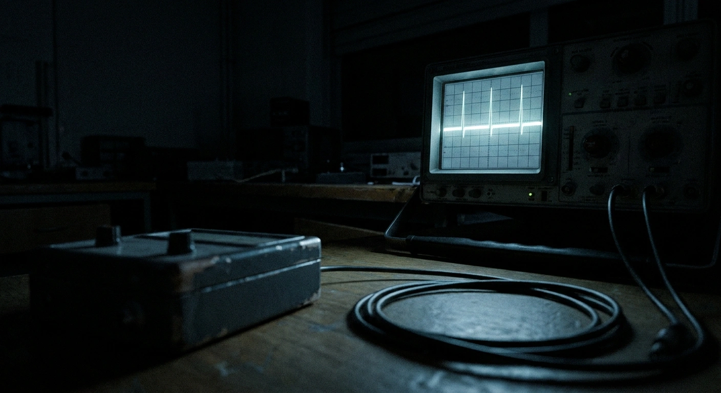
There is a technique that can measure it. It involves sliding a hair-thin electrode into a living nerve in an awake person and listening to individual nerve fibers, one at a time, while they fire. It is called microneurography, and it has produced the clearest evidence anyone has ever gathered that the burning is not in your head.
You almost certainly cannot get it. That sounds like a cruel setup, and I want to be honest about it up front rather than lead you through 2,000 words toward a disappointment. But the reason to understand this test anyway is that what it found changes how you should think about every other test you have already had.
Listening to One Nerve Fiber at a Time
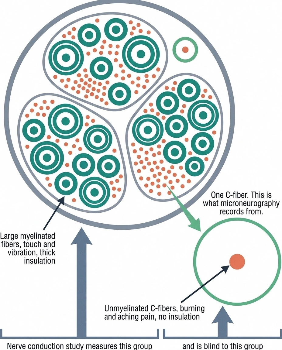
Start with the scale of the thing.
The scale problem, in numbers
Why one class of fiber is easy to measure and the other is not.
| Fiber type | Diameter | Signal speed | Carries |
|---|---|---|---|
| A-beta, myelinated | 6 to 12 micrometers | 30 to 70 m/s | Touch, vibration, position |
| A-delta, thinly myelinated | 1 to 5 micrometers | 5 to 30 m/s | Sharp first pain, cold |
| C-fiber, unmyelinated | 0.2 to 1.5 micrometers | Under 2 m/s | Burning, aching, itch, warmth |
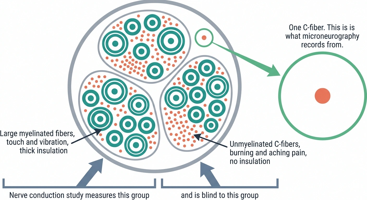
A standard nerve conduction study reports the fastest signal it can detect. C-fibers conduct roughly thirty times slower than the fibers being measured, so their contribution never surfaces in the result at all. The test is not failing. It is reporting on a population your symptoms are not coming from.
A peripheral nerve is a bundle. Inside the sheath there are thousands of individual fibers packed together, some thick and insulated in myelin, some bare and impossibly thin. The C-fibers, the ones that carry burning and aching pain, are among the thinnest structures in your body. A standard nerve conduction study cannot see them at all. It sends a shock along the nerve and times the response, which works beautifully for the big myelinated fibers and is completely blind to the small ones. This is why so many people with real small fiber damage get told their nerve testing was normal.
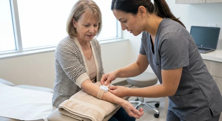
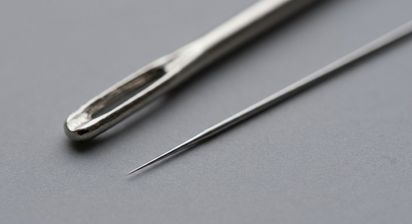
Microneurography goes a different route entirely. A tungsten microelectrode, tapering to a tip measured in fractions of a micrometer, is inserted through the skin and advanced into a nerve fascicle. A second electrode sits just under the skin nearby as a reference point. The person is awake the entire time, usually lying still with their leg supported, often for hours.
Then the researcher hunts. The electrode is advanced in tiny increments while they listen to the amplified signal, searching for the moment the tip lands close enough to a single fiber to pick up its individual action potentials. That hunting phase is the hard part, and it is why this remains a specialist craft rather than a routine procedure.
When it works, you get something no other method in human medicine can give you. Not an average across a whole nerve. Not a count of fibers in a piece of skin. The actual electrical traffic of one identified nerve fiber, in real time, in a conscious human being who can tell you what they are feeling while it fires.
The Fibers That Were Not Supposed to Be Talking
Here is where the research gets genuinely important for anyone living with unexplained burning.
Reading the 34 percent figure correctly
The number counts fibers, not patients. It means that within the small fiber neuropathy group, roughly a third of the individual silent nociceptors sampled were firing without a stimulus. It does not mean a third of patients had the finding and two thirds did not.
Sample sizes in this work are small by necessity, because each recording session yields a handful of fibers after hours of effort. The findings have been reproduced across groups, which matters more here than any single study’s headcount.
And spontaneous firing has been documented in fibromyalgia at a nearly identical rate. Whatever this measures, it is not specific to nerve damage from a known cause, which is one reason it has not become a diagnostic test.
Not all C-fibers behave the same way. A large subgroup are called mechano-insensitive nociceptors, and they have an unglamorous nickname in the literature: silent nociceptors. Under normal conditions they do almost nothing. Poke the skin above one and it will not respond. They sit quiet, waiting for inflammation or injury to wake them up, at which point they become part of the alarm system.
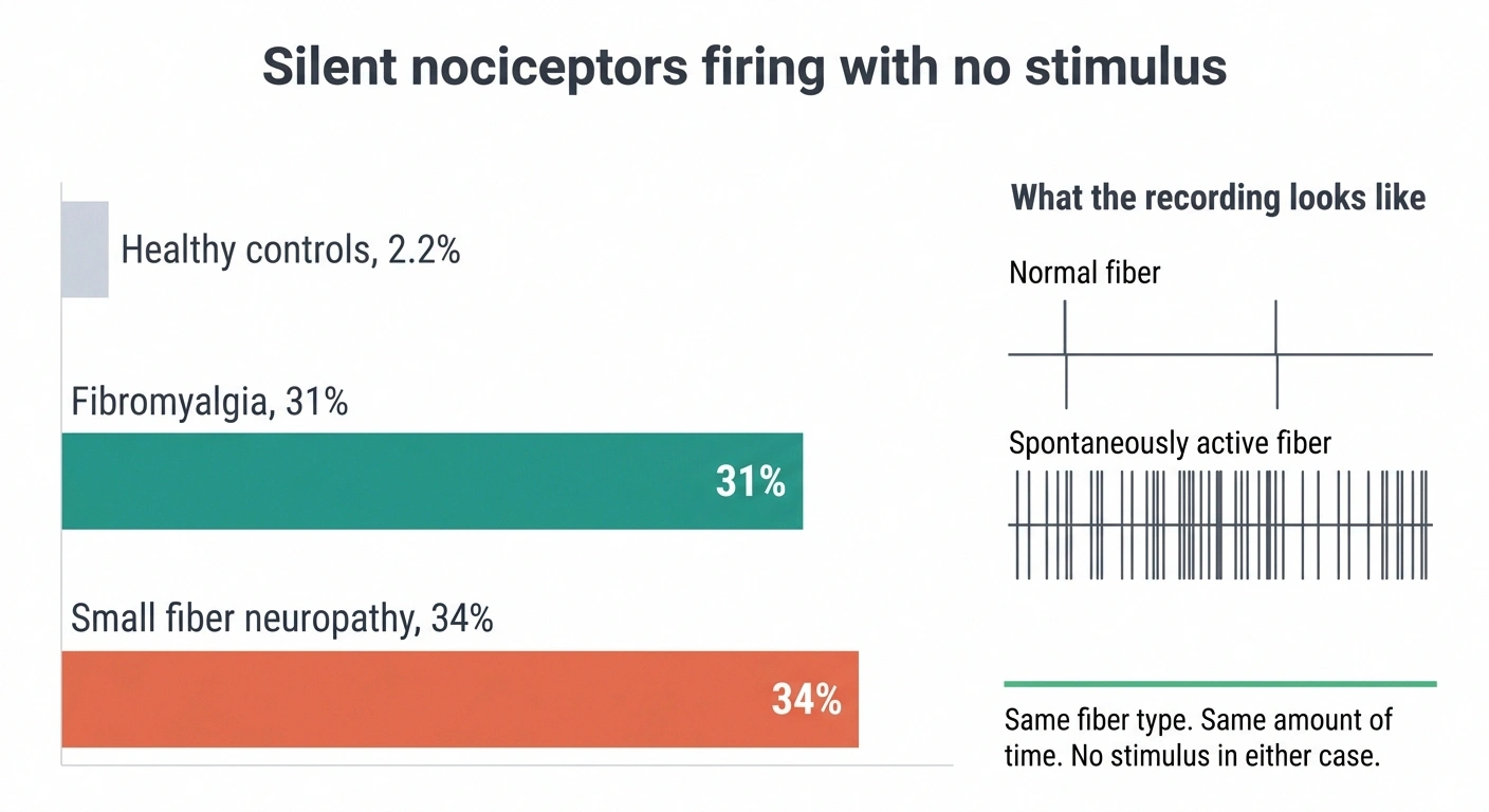
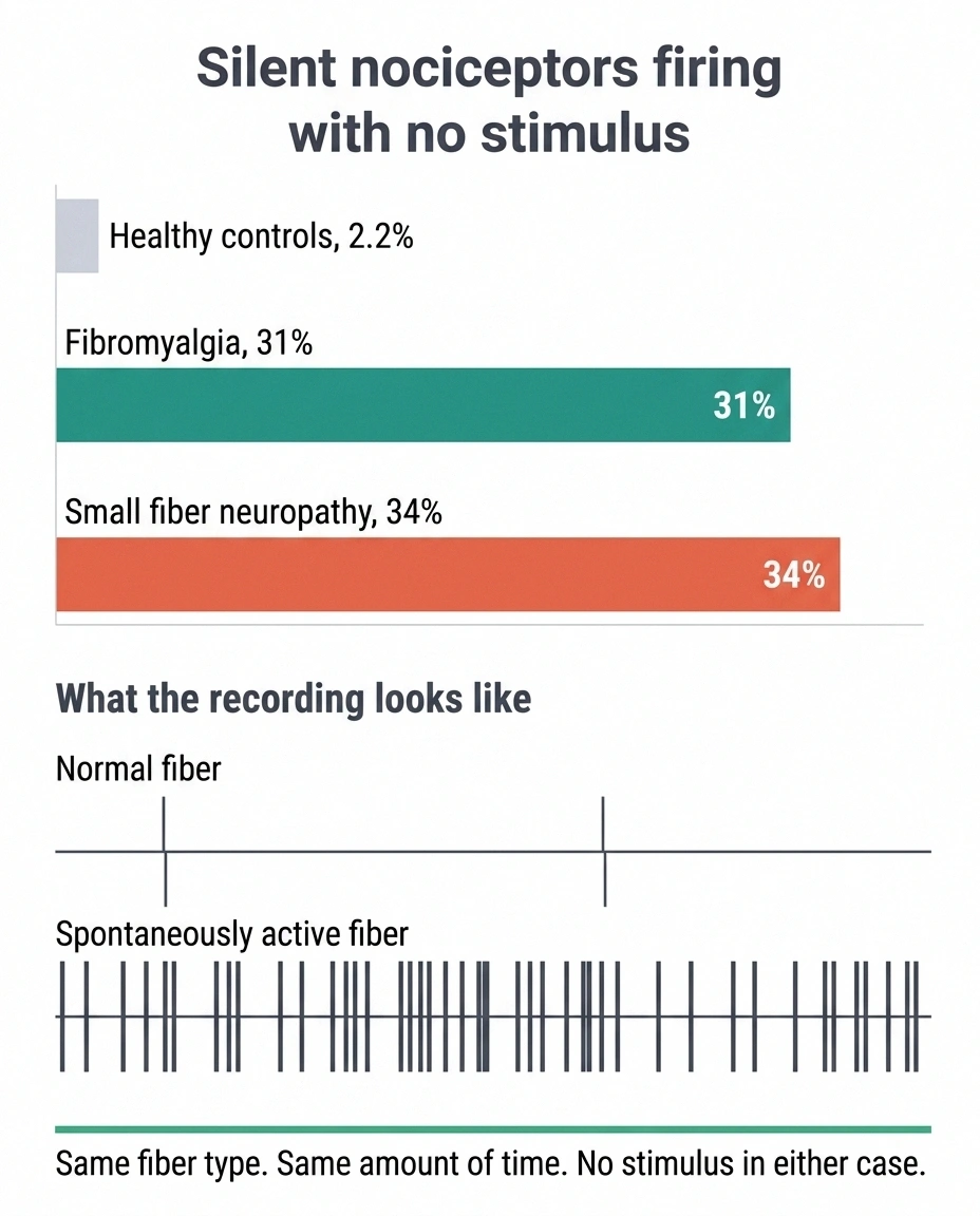
In healthy people, these fibers stay quiet. Researchers using microneurography found spontaneous activity in roughly 2.2 percent of silent nociceptors in control subjects. That is the background noise level of a normal nervous system.
In people with small fiber neuropathy, the same measurement came back at 34 percent.
Roughly a third of the silent fibers were not silent. They were firing on their own, with no stimulus, no injury happening in that moment, nothing touching the skin. Just firing. Sending pain signals up toward the spinal cord about an event that was not occurring.
The same research group found a nearly identical pattern in fibromyalgia, at 31 percent, which is its own conversation and one of the more interesting findings in modern pain research.
And within the neuropathy group, the divide held. Patients who reported ongoing pain and patients who did not were separated by exactly this: whether their C-nociceptors showed spontaneous activity and mechanical sensitization. The fibers of people in pain behaved differently from the fibers of people with nerve damage who were not in pain.
Why a Normal Fiber Count Does Not Mean Nothing Is Wrong
This is the part that should change how you read your own test results, so I want to slow down here.
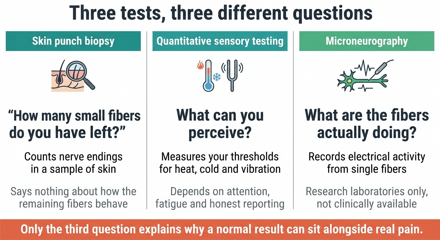
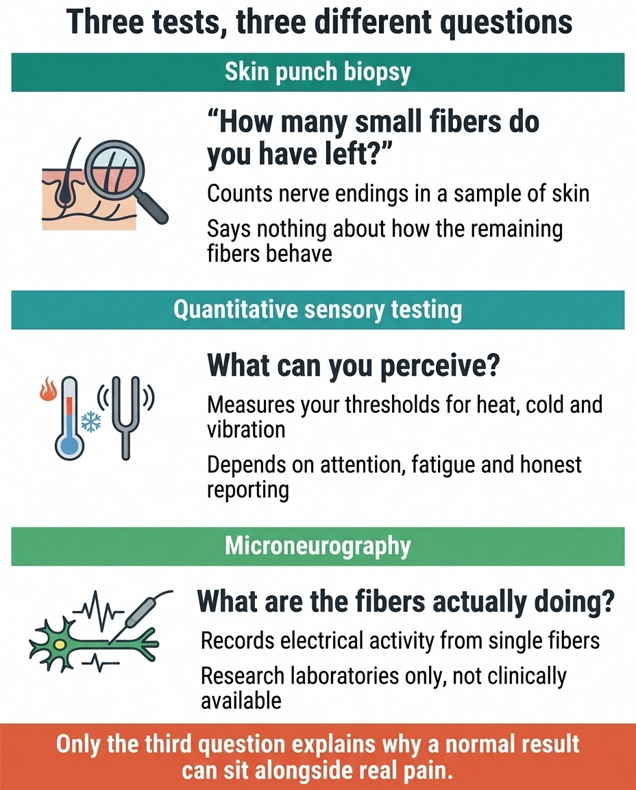
Skin punch biopsy, the current standard for diagnosing small fiber neuropathy, counts nerve endings. A pathologist stains a small cylinder of skin, counts the fibers crossing into the epidermis, and compares that number against normal values for your age and sex. It is a good test. It answers one question well: how many small fibers do you have left?
Quantitative sensory testing asks a different question: what can you perceive? It measures your thresholds for heat, cold, and vibration, which depends on your fibers, your attention, your fatigue, and your honest reporting.
Neither one asks the question microneurography asks, which is: what are the remaining fibers doing?
Those are three genuinely different questions, and a person can fail one while passing another. You can have a fiber count in the normal range and have a substantial fraction of those normal-count fibers firing spontaneously. Your biopsy looks fine. Your nerves are screaming. Both statements are true at the same time, and only one of them has ever been measured in your case.
This is a large part of why idiopathic neuropathy stays idiopathic, and why people whose symptoms follow a non-length-dependent pattern so often spend years being told the workup was unremarkable. The workup was unremarkable. The workup also did not look at fiber behavior, because outside a handful of research labs, nothing does.
What the Procedure Is Actually Like
People imagine something medieval. The reality is stranger and less dramatic.
What a session actually involves
| Anesthetic used | Usually none, because numbing the area would interfere with the sensory reporting the session depends on |
| Nerve search phase | Capped at 45 to 60 minutes by protocol, often stopped earlier |
| Recording phase | Commonly around 30 minutes lying still |
| Typical after-effects | Tenderness, soreness or a patch of numbness near the insertion site for a few days |
| Uncommon after-effect | Tingling along the outer calf for a few hours, rarely up to about two weeks |
| Track record | Over 3,000 published studies since 1979; some labs report 300 or more recordings without major complications |
The realistic difficulty is not risk. It is holding a leg motionless for hours, which is a genuine ask for someone whose legs burn worst when they stop moving.
You lie down. The site is usually the peroneal nerve near the knee or the median nerve at the wrist, depending on what is being studied. There is no general anesthetic and typically no local anesthetic either, because numbing the area would interfere with the sensory reporting the whole session depends on.
The insertion itself is described by participants as a needle stick followed by odd referred sensations while the electrode is being positioned. Pins and needles. Cramping. A dull ache that comes and goes as the tip moves. Those sensations are actually useful information, because they tell the researcher which part of the nerve the electrode is near.
Once the electrode settles into position, most people report that discomfort largely stops. If the nerve is being electrically stimulated as part of the protocol, you may feel involuntary twitching or tingling in the foot or lower leg, and it stops when the stimulation stops.
The searching phase is deliberately time-limited. Protocols commonly cap the nerve search at 45 to 60 minutes to reduce the chance of irritating the nerve, and many labs stop earlier. The recording itself often runs about 30 minutes with the participant lying still.
Safety record: this method has been used in more than 3,000 published studies since 1979. Individual laboratories have reported hundreds of recordings without major complications. The realistic after-effects are tenderness, soreness, or a patch of numbness near the insertion site that resolves on its own within a few days. Some participants notice tingling along the outer calf for a few hours. In rare reports it lasted around two weeks. Longer than that has not been described in the published series.
The genuine burden is not danger. It is time and stillness. Holding a leg motionless for several hours while someone hunts for a single fiber is difficult for anyone, and for a person whose legs burn when they stay still, it is a real ask.
Why Your Neurologist Cannot Order This
The 2025 review literature says it plainly: microneurography is not readily applicable to clinical practice. It remains a research technique.
Several things stand in the way at once, and none of them are close to being solved.
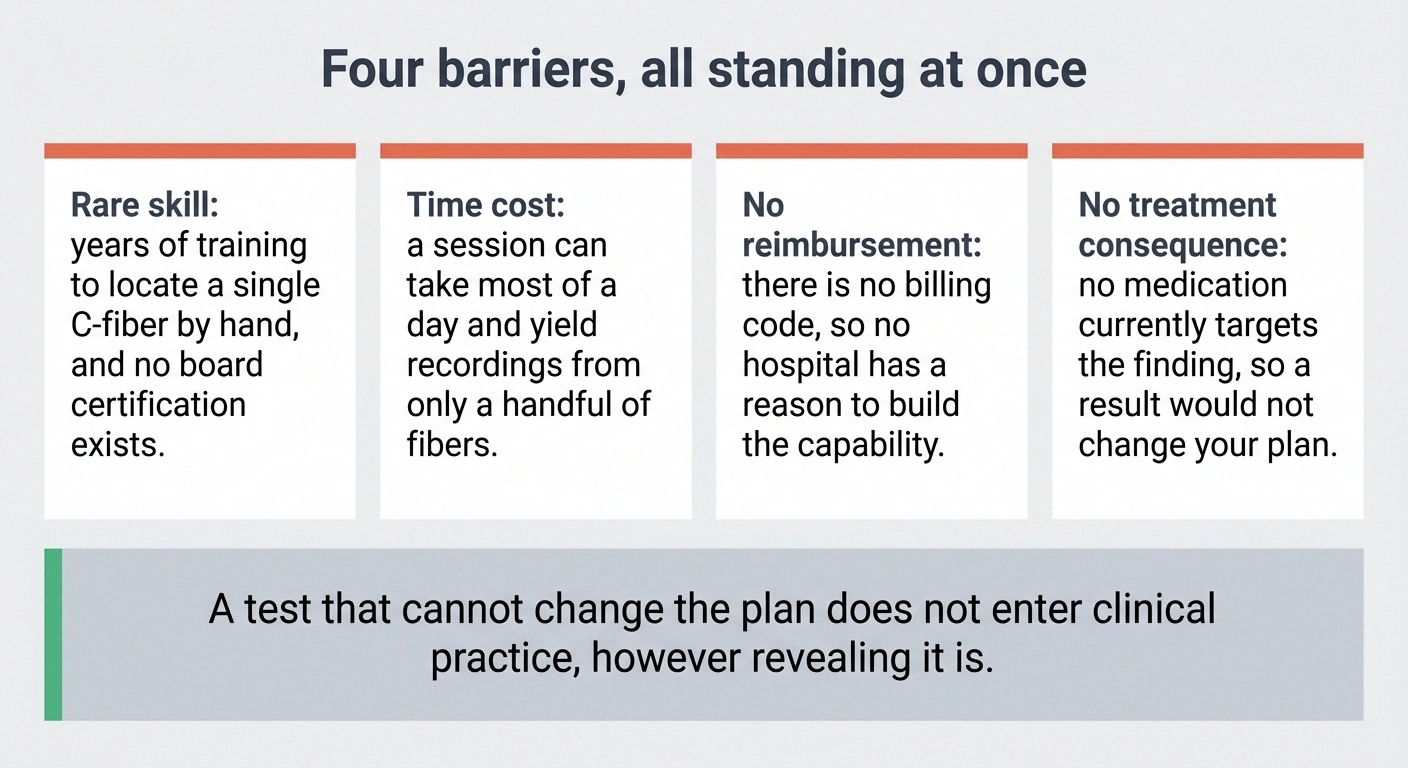
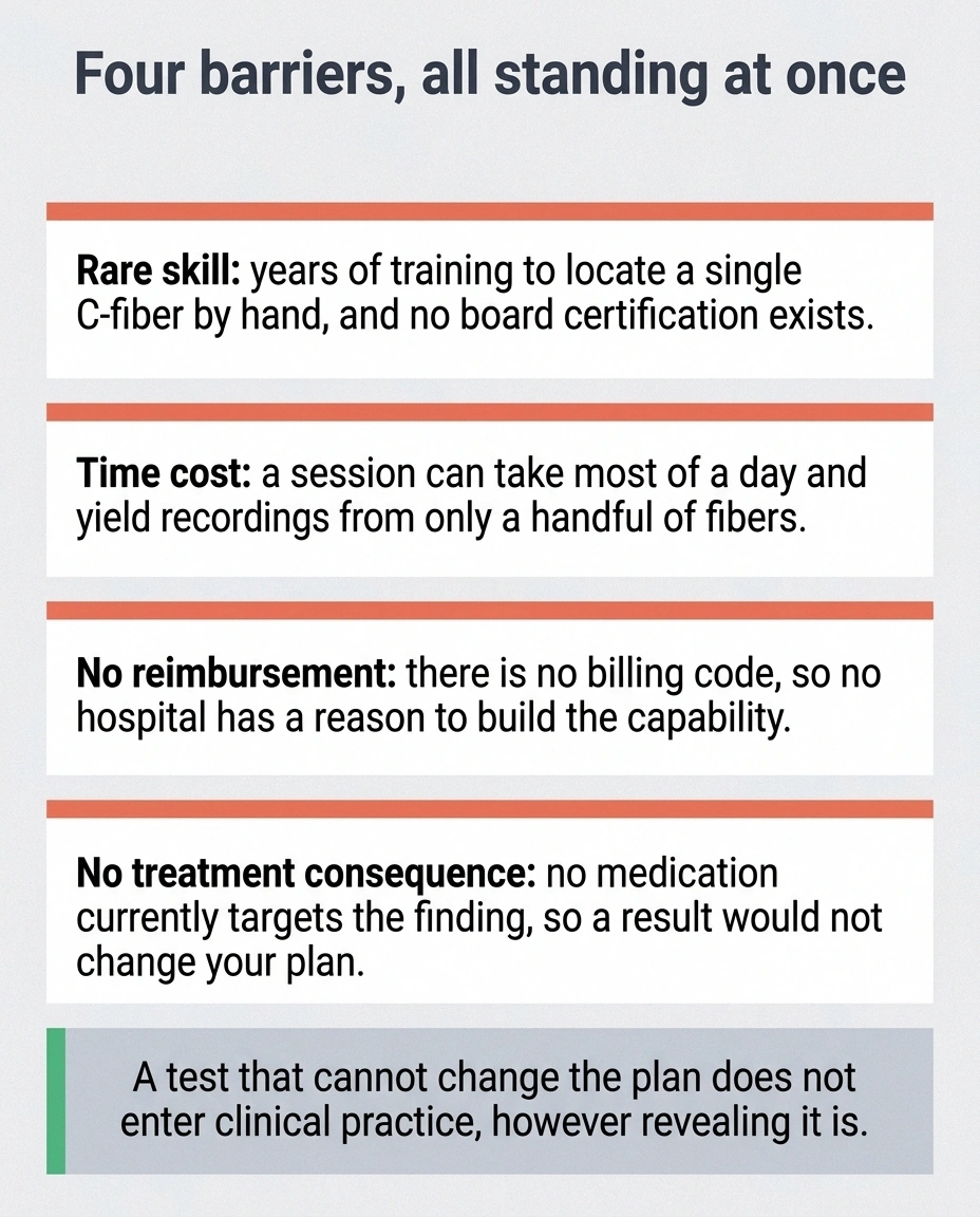
The skill is rare. Locating a single C-fiber with a handheld electrode takes years of training, and the number of people worldwide who do it well is small enough that the field runs on personal collaborations. There is no board certification for this.
The time cost is enormous. A single session can consume most of a day for a technician and a researcher, and it may yield recordings from only a handful of fibers.
There is no billing code and no reimbursement pathway, which means no hospital has a financial reason to build the capability.
And critically, the results do not yet change treatment. If a session confirmed that 30 percent of your silent nociceptors were firing spontaneously, there is no medication that specifically targets that finding today. A test that cannot alter the plan does not enter clinical practice, no matter how revealing it is.
In the United States, established microneurography laboratories operate at institutions including Virginia Commonwealth University, the University of Iowa, Mayo Clinic, and the University of Colorado at Boulder. Strong groups exist in Germany at RWTH Aachen and Friedrich-Alexander University Erlangen-Nurnberg, and in Canada at Western University. Most of that capacity is aimed at cardiovascular and sympathetic nerve research rather than pain, and the pain-focused labs are fewer still.
If you ever do encounter this procedure, it will be as a participant in a study, not as a patient receiving a test. That is a meaningful distinction. A research participant is contributing to a question, and the answer comes back to the field rather than to their chart. Anyone genuinely interested in that path should look at how neuropathy clinical trials work before getting attached to the idea.
What This Research Is Actually Building Toward
The near-term value of microneurography is not diagnostic. It is that the technique gives drug developers something they have never had, which is a direct physical readout of spontaneous pain.
The measurement problem this could solve
Placebo response rates in neuropathic pain trials commonly land in the 20 to 30 percent range, and have reached higher. A drug producing a real but moderate effect has to clear that noise to register as working, which is why several plausible compounds have failed trials that a better endpoint might have passed.
A count of hyperactive fibers does not respond to expectation, sleep quality, or which arm a participant believes they were assigned to. That is the entire argument for developing this into a biomarker, and it is a drug-development argument rather than a diagnostic one.
Consider the problem a company faces when testing a new nerve pain drug. The primary endpoint is almost always a pain scale, where a patient rates their pain from zero to ten. That number is genuinely meaningful to the person living it and genuinely terrible as a scientific measurement. It moves with sleep, mood, weather, expectation, and the powerful placebo response that pain trials are notorious for. Drugs that work have failed trials on that noise. Drugs that do nothing have passed on it.
Now imagine measuring whether hyperactive C-nociceptors actually quieted down. That is a physical event in a nerve fiber. It does not care how the participant slept or whether they believed they got the real drug.
This is why the phrase “biomarker of spontaneous pain” keeps appearing in the literature around this work. If hyperactive mechano-insensitive fibers can be validated as that biomarker, early-stage pain trials get dramatically more efficient, and the failure rate that has kept the neuropathy pipeline so thin for so long starts to come down. The broader search for nerve repair has the same underlying problem, and better measurement helps there too.
The technology side is moving as well. Work on multi-channel percutaneous probes aims to record from many fibers at once rather than hunting for them one by one, which is the single biggest bottleneck. If that succeeds, the calculation around clinical feasibility changes.
None of that is a treatment. It is the instrumentation that treatments get built on top of, and instrumentation tends to arrive quietly and then matter enormously a decade later.
What to Take From This If You Are the Patient

Three things worth carrying out of this article.
Normal test results describe the test, not your experience. A normal nerve conduction study means the large fibers conduct normally. A normal skin biopsy means you have an ordinary number of small fibers. Neither one measured whether those fibers are behaving correctly, and in small fiber neuropathy, behavior is where the pain lives. This is worth understanding before your next appointment, alongside what your other neuropathy lab tests can and cannot show.
The burning has a documented physical mechanism. Somebody put an electrode in a human nerve and recorded pain fibers firing at nothing, in people who described exactly what you describe. Whatever else is uncertain, that is not. If you have ever been made to feel that unexplained meant imagined, the recordings say otherwise.
Do not chase this test. If you find a clinic advertising microneurography as a diagnostic service for neuropathy patients, treat that as a warning sign rather than an opportunity. The technique lives in research laboratories, and the labs that have it are not marketing it to patients.
The most useful version of this knowledge is the sentence you can now say to a clinician who tells you your workup was clean. Not an argument, just a clarification: the tests I have had count fibers and measure conduction speed, and my symptom pattern is consistent with abnormal fiber activity, which those tests do not assess. That is a factually accurate statement, and it changes the tone of the conversation. It moves you from a person with no findings to a person describing a specific gap in the available testing.
That reframe will not fix your feet tonight. It does put you on more solid ground, which over the long course of this condition turns out to matter more than most people expect. Understanding where you sit in the overall progression of neuropathy works the same way, and both are worth the effort of learning properly.
Frequently Asked Questions
What is microneurography?
Microneurography is a research technique in which a very fine tungsten electrode is inserted through the skin into a peripheral nerve in an awake person, allowing researchers to record the electrical activity of individual nerve fibers. It is the only method that can directly record C-fiber activity in living humans.
How is microneurography different from a nerve conduction study?
A nerve conduction study measures how fast a signal travels along a whole nerve, and it only assesses large myelinated fibers. Microneurography records from single fibers, including the thin unmyelinated C-fibers that carry burning pain and that standard nerve conduction testing cannot detect at all. This is why nerve conduction studies are frequently normal in small fiber neuropathy.
Can I get microneurography for my neuropathy?
Almost certainly not as a clinical test. It is performed at a small number of research laboratories, requires specialized training, has no billing code or insurance pathway, and does not currently change treatment decisions. The only realistic route to undergoing it is enrolling in a research study that uses it.
Does microneurography hurt?
Participants typically describe the needle insertion as a stick followed by pins and needles, cramping, or dull aching while the electrode is positioned. Once the electrode is in place, significant discomfort is uncommon. Electrical stimulation during the recording can cause twitching or tingling that stops when the stimulation stops. Sessions require lying still for a long period, which many people find harder than the sensations themselves.
Is microneurography safe?
It has been used in more than 3,000 studies since 1979 with a strong safety record, and some individual laboratories report hundreds of recordings without major complications. The most common after-effects are soreness, tenderness, or numbness near the insertion site lasting a few days. Tingling along the outer calf for a few hours is occasionally reported, and in rare cases has lasted about two weeks.
What did microneurography find in small fiber neuropathy?
Researchers found spontaneous activity in about 34 percent of silent nociceptors in people with small fiber neuropathy, compared with about 2.2 percent in healthy controls. Within the patient group, those experiencing ongoing pain were distinguished from those who were not by the presence of spontaneous firing and mechanical sensitization in their C-nociceptors.
What are silent nociceptors?
Silent nociceptors, also called mechano-insensitive C-fibers, are pain-sensing nerve fibers that normally do not respond to mechanical stimulation and stay largely inactive until inflammation or injury sensitizes them. In small fiber neuropathy, a substantial fraction of them fire spontaneously without any triggering stimulus.
If my skin biopsy was normal, can I still have small fiber neuropathy pain?
Yes. A skin biopsy counts nerve fiber density, which is a measure of how many fibers remain rather than how they are behaving. Microneurography research shows that fiber hyperactivity can be present in ways a fiber count does not capture. A normal or borderline biopsy does not rule out genuine small fiber pain, and this is worth raising directly with a neurologist familiar with small fiber disorders.
Will microneurography ever become a standard clinical test?
Not in its current form. The barriers are the rarity of the skill, the hours required per session, the absence of reimbursement, and the fact that results do not yet alter treatment. Development of multi-channel probes that record many fibers simultaneously could change the feasibility picture, but that work is early and no timeline should be assumed.
About the Author