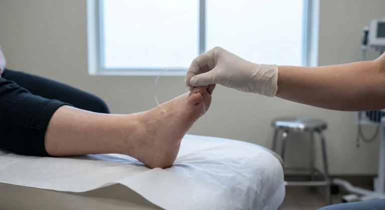
Roughly four out of ten people carrying a diagnosis of idiopathic small fiber neuropathy have an antibody in their blood that nobody looked for.
That statistic comes out of a series of studies on cryptogenic small fiber neuropathy, which is the clinical term for the version where every standard cause has been ruled out and the file gets closed. Among those patients, 40 percent tested positive for at least one of three specific autoantibodies.
It is a genuinely important finding and it is also easy to oversell, which is what most of the internet does with it. So this article does both halves. What the antibodies are, who should be tested, what a positive result actually means, and the uncomfortable part that most coverage skips, which is that finding one does not reliably get you treated.
Three Antibodies With Unwieldy Names
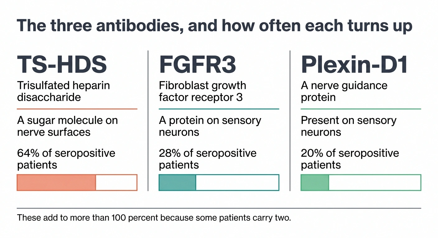
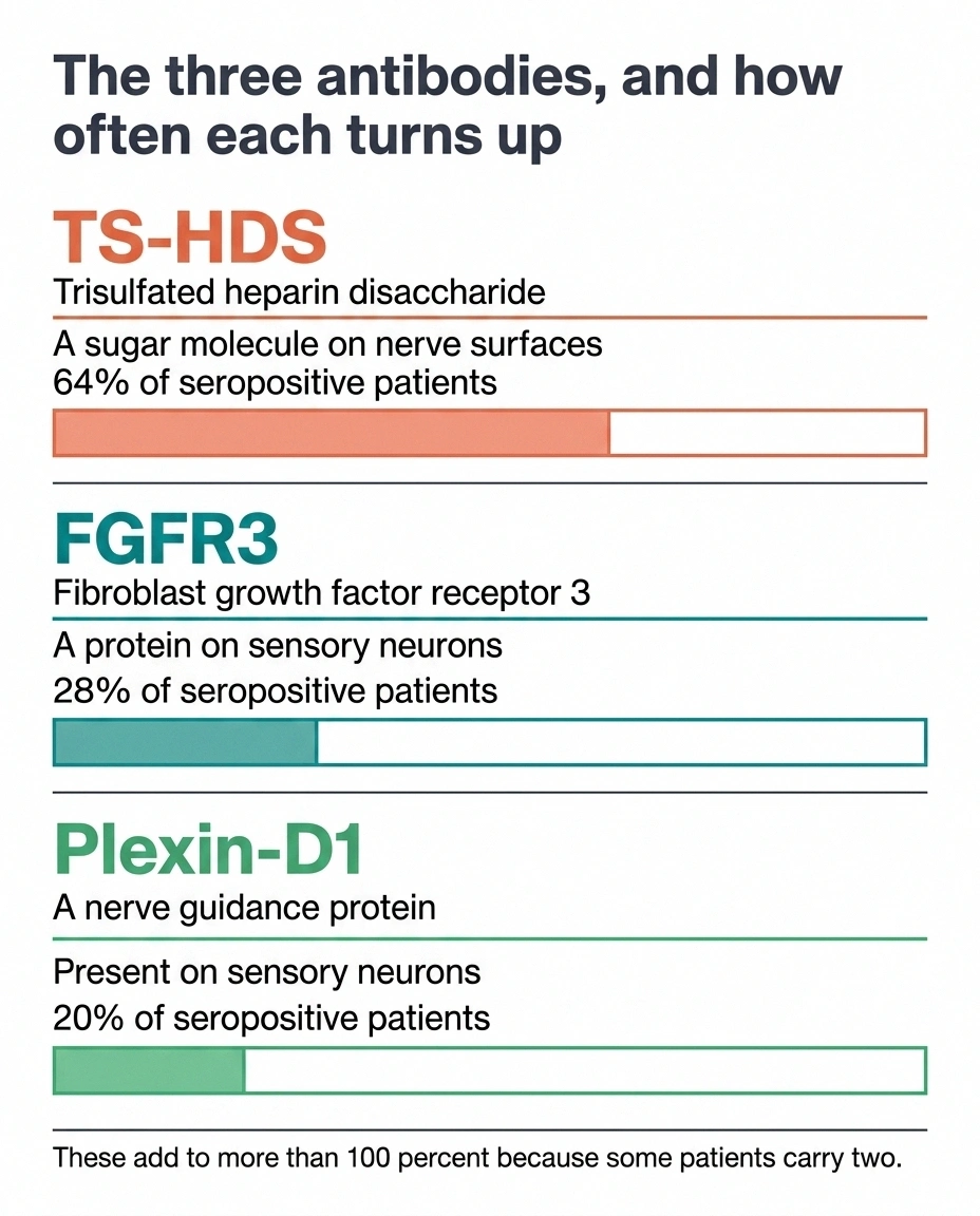
The three that dominate this conversation:
Where the attack lands, and why it changes the map
- Dorsal root ganglion
- A cluster of sensory nerve cell bodies sitting just outside the spinal cord, one pair at nearly every vertebral level. Every sensory fiber in your body grows outward from a cell body housed in one of these.
- Why its blood supply matters
- The ganglia sit outside the blood-nerve barrier that protects nerve fibers further along their length. Circulating antibodies reach them more easily than they reach the nerve trunks, which is a structural reason an immune process would concentrate there.
- The consequence for your symptoms
- Damage at a cell body kills that neuron’s entire fiber regardless of how long it is. So the pattern follows which ganglia were hit rather than which fibers are longest. A ganglion serving your face and one serving your thigh can be affected while the ones serving your feet are spared, which is precisely the patchy distribution that confuses a length-dependent workup.
TS-HDS, short for trisulfated heparin disaccharide. A sugar-based molecule found on nerve surfaces. This is the most common of the three by a wide margin.
FGFR3, fibroblast growth factor receptor 3. A protein expressed on sensory neurons in the dorsal root ganglion, the cluster of nerve cell bodies sitting just outside the spinal cord.
Plexin-D1, a guidance protein that helps nerve fibers find their targets during development and remains present on sensory neurons afterward.
Within the seropositive group in the largest series, the split ran roughly 64 percent TS-HDS, 28 percent FGFR-3, and 20 percent Plexin-D1. Those add to more than 100 because some people carry two.
What they share is a target: not the long nerve fibers running down to your feet, but the cell bodies those fibers grow out of. If the immune attack is happening at the ganglion, the damage does not have to follow the usual pattern. That single fact explains most of what makes this form of neuropathy look different.
The Pattern That Should Make Someone Ask
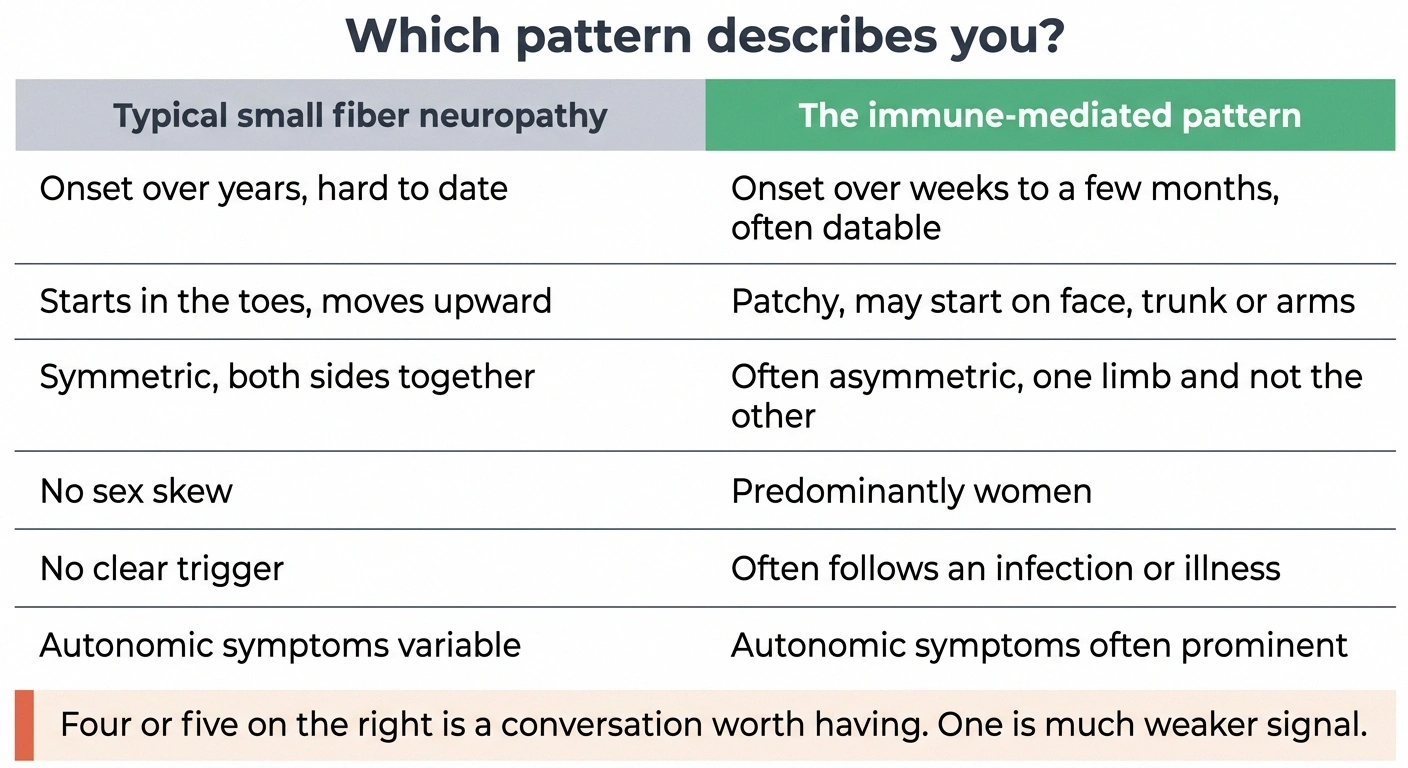
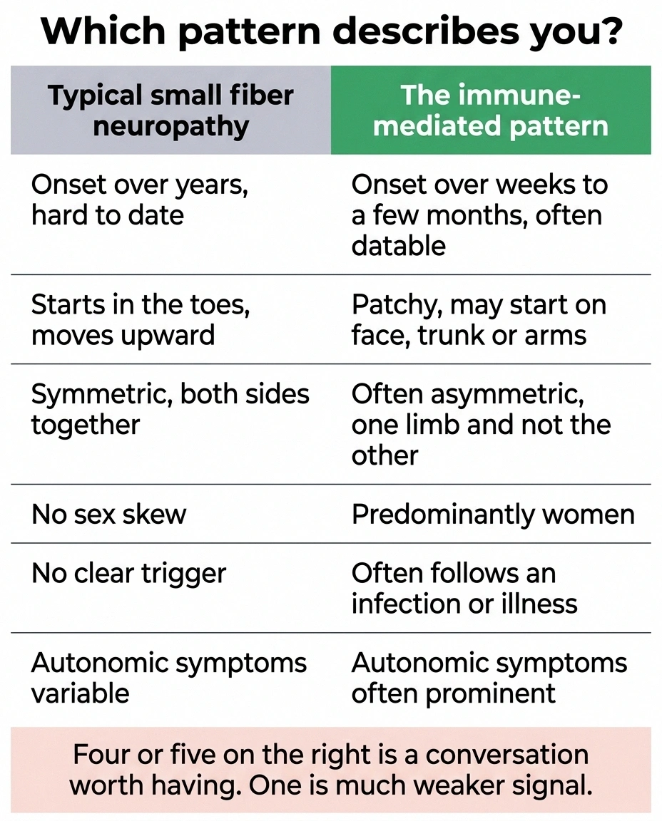
Immune-mediated small fiber neuropathy has a clinical fingerprint. Not every patient shows every feature, but the more of these that describe you, the more the question is worth raising.
It came on fast. Classic small fiber neuropathy creeps. Tingling in the toes one year, up to the ankles two years later, a slow forward march you can barely date. The immune-mediated form frequently arrives acutely or subacutely, over weeks to a few months. People can often name the season it started. Some can name the week.
It is not following the stocking-glove map. Length-dependent neuropathy hits the longest nerves first, so it starts at the toes and works upward, and hands join in only once the legs are well involved. Immune-mediated cases are frequently non-length-dependent, meaning patchy and asymmetric. Burning on the face. A band across the trunk. One thigh but not the other. Arms involved as early as the feet, or before them. This shows up on biopsy too, which is why the site of the punch matters.
You are a woman. The seropositive population skews strongly female.
Something preceded it. A viral illness, an infection, occasionally another immune event. Post-infectious onset is common enough to be part of the described pattern.
The autonomic symptoms are prominent. Lightheadedness on standing, heart rate that jumps out of proportion to what you are doing, altered sweating, digestive changes. TS-HDS and FGFR3 antibodies have been documented in patients with clear autonomic involvement, and autonomic neuropathy riding alongside sensory symptoms is a recognized part of this picture.
Autoimmunity already runs in your history. Personal or family history of thyroid disease, rheumatoid arthritis, lupus, celiac, or Sjogren’s. Small fiber involvement is well described in Sjogren’s syndrome and in lupus, and those conditions are checked in most standard workups. The three antibodies here are not.
If you read that list and recognized yourself in four or five items, that is the conversation to bring to a neurologist. If you recognized yourself in one, it is much weaker signal.
How the Testing Actually Works

These are blood tests, drawn like any other. In the United States they are most commonly ordered as a panel through Washington University’s neuromuscular laboratory, and several commercial reference labs now offer them as well. The turnaround is typically a few weeks.
Two practical things worth knowing before you ask.
First, they are not part of any standard neuropathy panel. Ordering them is a deliberate decision, and a general neurologist who does not focus on peripheral nerve disease may not have them on their radar. This is a reasonable thing to name specifically rather than hint at.
Second, coverage varies and denials happen. Ask what the self-pay price is before the blood is drawn, because finding out afterward is a bad surprise. The usual sequence is that the standard neuropathy lab workup comes back clean first, and these get added afterward as the second tier.
Before you spend money on this panel
These antibodies are not validated diagnostic criteria. They appear in some healthy people and in people with other conditions, and no professional society has issued a guideline stating that a positive result establishes a diagnosis or mandates treatment. A neurologist with peripheral nerve expertise is the right person to decide whether the test is worth ordering in your specific case and what to do with the answer.
What a Positive Result Does and Does Not Mean
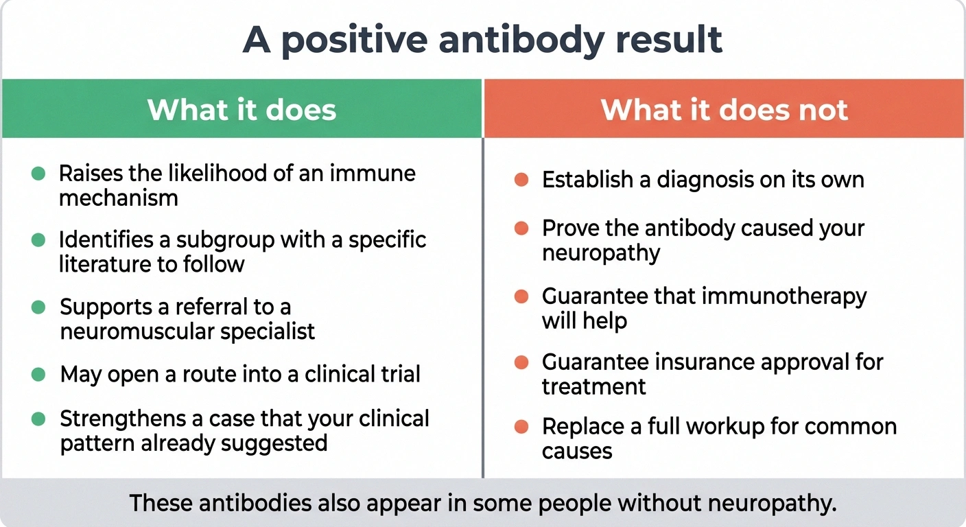
This is the section where honest coverage separates from marketing, so here is the state of the evidence in plain terms.
The case for these antibodies mattering: They cluster in a specific clinical population rather than appearing at random. Inflammation has been observed in a portion of skin biopsies from seropositive patients. Small-vessel vasculitis has been reported in seropositive small fiber neuropathy, which is a structural finding, not a statistical one. A 2025 laboratory study argued that FGFR3 autoantibodies are directly pathogenic rather than incidental. And treatment response, where it has been reported, has clustered in seropositive patients.
The case for caution: None of these antibodies has an established sensitivity and specificity you could hang a diagnosis on. They turn up in people without neuropathy. The pathogenicity work is early and not yet replicated across independent groups. And the treatment evidence, which is the part that actually determines whether the test changes your life, is thinner than the enthusiasm around it suggests.
A useful way to hold it: a positive result is a meaningful clue that shifts the probability toward an immune mechanism. It is not a diagnosis, and it is not a prescription. A neurologist weighs it against your clinical pattern, your biopsy, and your other results. In someone with rapid onset, patchy distribution, and autonomic symptoms, a positive antibody strengthens a case that was already forming. In someone with a textbook slow length-dependent pattern and a positive result, it means considerably less.
The Treatment Question
The therapy that gets discussed is intravenous immunoglobulin, and readers who want the general background should start with how IVIG treatment for neuropathy works before wading into the specifics here.
The controlled evidence, such as it is
Everything else in this section is retrospective or uncontrolled. These are the two studies designed to actually settle the question.
| NCT03401073 | NCT04153422 | |
|---|---|---|
| Design | Double-blind, placebo-controlled pilot | Phase II, double-blind, placebo-controlled |
| Antibodies included | TS-HDS or FGFR-3 | TS-HDS, FGFR-3 or Plexin-D1 |
| Duration | 6 months | Reported in progress through 2025 |
| Primary endpoints | Change in pain score and change in nerve fiber density on skin biopsy | Pain and nerve fiber density measures |
Both use nerve fiber density as an endpoint alongside pain, which is the right design choice. A pain score alone could not distinguish an immune effect from a placebo effect, and fiber density is the harder thing to move.
What has been reported in this population:
A retrospective series described significant improvement in both nerve fiber density and pain in seropositive patients treated with IVIG. One small report followed three patients through a six-month course and found an average increase in epidermal nerve fiber density of 721 percent on repeat punch biopsy. That number deserves a second look before it does any work on you. Three patients is three patients, and a percentage increase calculated from a very low starting fiber count produces a spectacular-looking figure from a modest absolute change. It is encouraging. It is not evidence of efficacy.
A separate analysis reported that IVIG was effective specifically in patients whose biopsies showed a non-length-dependent pattern along with Plexin-D1, TS-HDS, or FGFR-3 antibodies, which fits the idea that the immune subgroup is the one that responds.
Two controlled trials have been run at this question. NCT03401073 was a double-blind, placebo-controlled pilot of IVIG in TS-HDS or FGFR-3 positive small fiber neuropathy, running six months, with co-primary endpoints of change in pain score and change in intraepidermal nerve fiber density on skin biopsy. NCT04153422 is a phase II double-blind placebo-controlled trial covering TS-HDS, FGFR-3, and Plexin-D1, reported as in progress through 2025.
Rituximab has also been tried. The published experience is a series of five cases reporting benefit in antibody-positive patients. Five cases is a signal worth following, not a basis for a treatment decision.
Where that leaves things: no immunotherapy is approved for this indication. The supporting evidence is largely retrospective, from single centers, in small numbers, and the placebo-controlled work that would settle it is either recently completed or still running. IVIG is also expensive, time-consuming, and carries real risks, and insurers routinely deny it for small fiber neuropathy on exactly the grounds that the indication is not established. People do get it approved. It generally takes a neurologist willing to document the case carefully and appeal.
For patients who want to be part of resolving this rather than waiting on it, the relevant studies are open and enrolling, and understanding how neuropathy clinical trials select participants is worth doing before you inquire.
How This Differs From CIDP
People encountering immune neuropathy for the first time often collapse these into one category, and the distinction is worth holding clearly.
If you are pursuing immunotherapy coverage
Denials for IVIG in small fiber neuropathy are usually issued on the grounds that the indication is not established, not on the grounds that your case is weak. That distinction matters, because it tells you what an appeal has to contain.
Approvals in this population have generally required documentation of the specific antibody result, objective evidence of nerve involvement such as skin biopsy or autonomic testing, a record of what has already been tried and failed for symptom control, and a letter of medical necessity that cites the published series directly.
Ask your neurologist’s office whether they have a staff member who handles prior authorizations for infusion therapies. Practices that treat CIDP routinely usually do, and that person is worth more to this process than any argument you make yourself.
CIDP is an immune attack on myelin, the insulation around large nerve fibers. It produces weakness, loss of reflexes, and abnormal nerve conduction studies. It has established diagnostic criteria and approved treatments, including IVIG.
Immune-mediated small fiber neuropathy involves the small unmyelinated fibers. Strength stays normal. Reflexes stay normal. Nerve conduction studies come back normal, because the fibers involved are invisible to that test. Diagnosis rests on skin biopsy, autonomic testing, and the antibody panel, and there are no consensus criteria.
The practical consequence is a difference in institutional footing. CIDP has a name that insurers recognize and guidelines that support treatment. The immune small fiber form has neither, and people living with it are frequently caught in the gap between having something real and having something officially recognized.
Where the Biopsy Punch Goes Changes the Answer
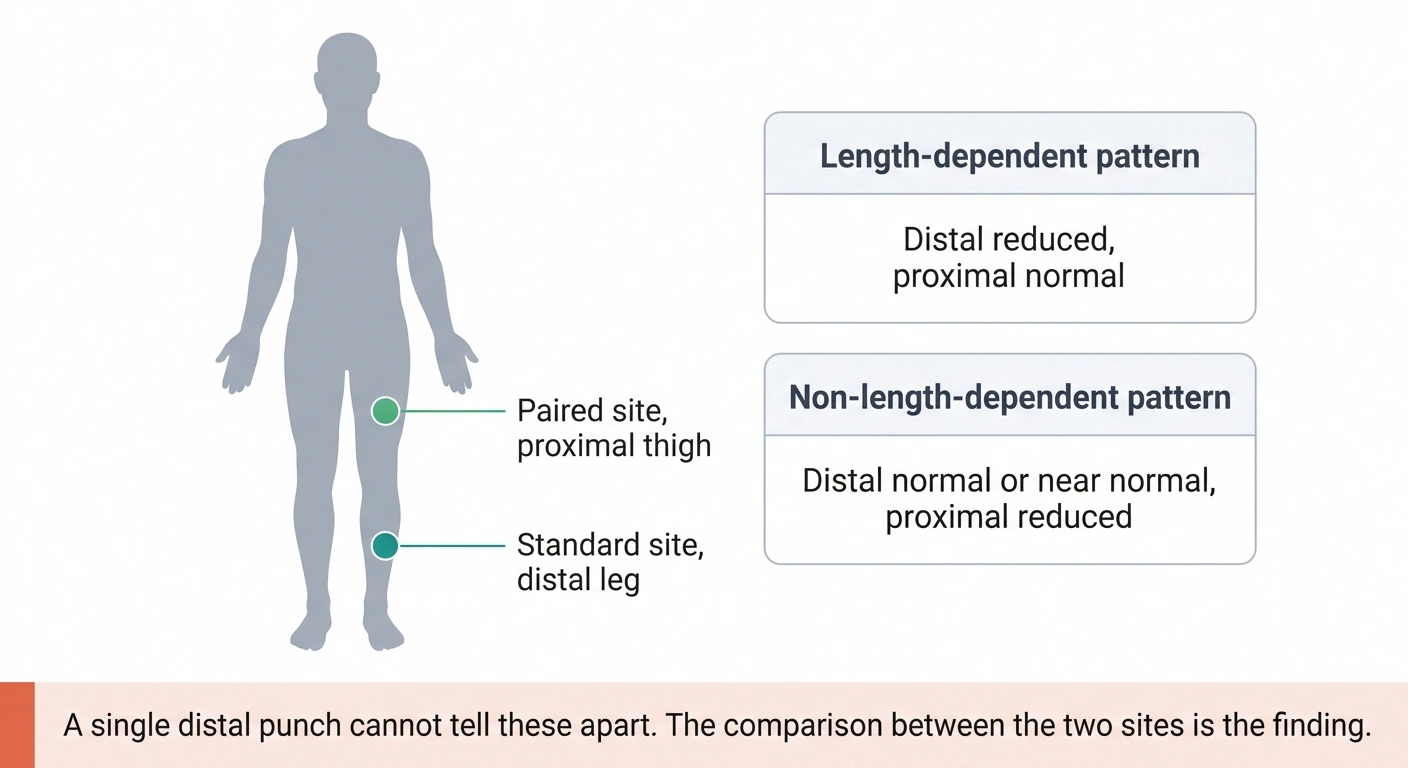
One practical detail deserves its own section, because it decides whether a test finds anything at all.
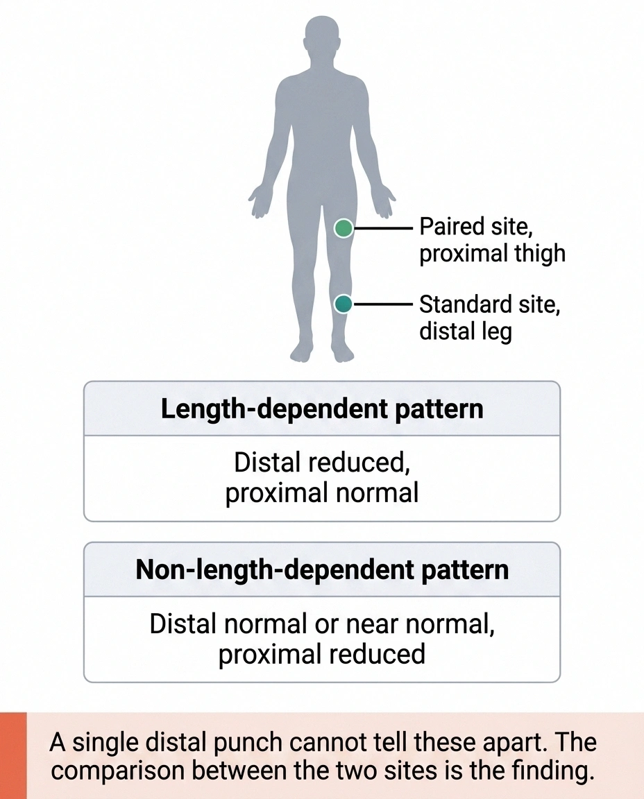
Standard skin punch biopsy for small fiber neuropathy takes a sample from the distal leg, about 10 centimeters above the outer ankle. That site was chosen deliberately. It sits at the far end of the longest sensory nerves, which is exactly where length-dependent damage shows up first and most severely. For the common form of the condition, it is the right place to look.
Now apply that to a patchy, ganglion-level process. If your immune system is attacking sensory neuron cell bodies, the resulting fiber loss does not concentrate at the ankle. It can be scattered, and it can spare the distal leg entirely while showing up clearly on the thigh, the trunk, or the arm.
This is why the protocol for suspected non-length-dependent disease uses paired sites, typically distal leg and proximal thigh, and compares them. A normal distal count alongside a reduced proximal count is itself the finding. That comparison is diagnostic information you simply cannot obtain from a single ankle punch, and a report showing a normal distal biopsy tells you very little about a patchy process.
So if a single distal biopsy came back normal and your symptoms have never followed a stocking-glove pattern, that result did not rule out small fiber neuropathy. It sampled the wrong territory. Bring the actual pathology report to the appointment rather than the summary sentence you were given, and ask which sites were taken.
What to Do With This

If the clinical fingerprint above describes you, the actionable sequence is short.
The workup that comes first
Every one of these is more common than the three antibodies, and several are treatable in ways that change the disease rather than the symptoms. Confirm each was actually run before spending money further down the list.
- Fasting glucose and HbA1c, plus a 2-hour glucose tolerance test, since prediabetes is missed by fasting glucose alone
- Vitamin B12 with methylmalonic acid, because B12 can read normal while functional deficiency is present
- Vitamin B6, which causes neuropathy at both deficient and excessive levels
- Thyroid function
- Celiac serology
- Serum protein electrophoresis with immunofixation, looking for a monoclonal protein
- Kidney and liver function
- HIV and hepatitis C
- ANA, SS-A and SS-B for Sjogren’s, which is a frequent and frequently missed cause
Ask for the actual result values rather than the word normal. Borderline B12 and impaired glucose tolerance both get reported as unremarkable and both matter here.
Confirm the standard workup was genuinely complete first. Diabetes and prediabetes, B12 and B6, thyroid, celiac, kidney and liver function, monoclonal protein studies, HIV and hepatitis, Sjogren’s antibodies. These account for far more small fiber neuropathy than the three antibodies discussed here, and several are treatable in ways that matter more. Chasing an exotic answer past an unexamined common one is the most expensive mistake in this whole area, and reviewing what your existing blood work already showed is the cheap first step.
Then, if that is clean and the clinical pattern fits, ask specifically. Name the three antibodies. Ask whether a neuromuscular specialist rather than a general neurologist should be involved, because the people who order these tests routinely are the people who know what to do with the results.
And go in knowing what a positive result buys you: a better explanation, a specific subgroup to follow in the literature, a possible route to a trial, and an argument for immunotherapy that you would still have to make. That is real value. It is not a cure, and being clear-eyed about the difference is what keeps hope from turning into disappointment later.
The word idiopathic has always meant we do not know. For a meaningful fraction of the people carrying it, it turns out to have quietly meant we did not check.
Frequently Asked Questions
What is immune-mediated small fiber neuropathy?
It is small fiber neuropathy in which the immune system appears to be attacking sensory nerve tissue, often at the dorsal root ganglion. It is associated with autoantibodies against TS-HDS, FGFR3, or Plexin-D1, and tends to present more acutely and in a more patchy, non-length-dependent pattern than typical small fiber neuropathy.
How common are these antibodies in idiopathic small fiber neuropathy?
In studies of cryptogenic small fiber neuropathy, about 40 percent of patients tested positive for at least one of the three. Among those who were positive, roughly 64 percent had TS-HDS, 28 percent had FGFR-3, and 20 percent had Plexin-D1, with some patients carrying more than one.
What are TS-HDS, FGFR3, and Plexin-D1?
TS-HDS stands for trisulfated heparin disaccharide, a sugar molecule found on nerve surfaces. FGFR3 is fibroblast growth factor receptor 3, a protein expressed on sensory neurons. Plexin-D1 is a guidance protein present on sensory neurons. Antibodies against each have been associated with small fiber neuropathy.
Does a positive antibody test mean I have autoimmune neuropathy?
Not by itself. These antibodies are not validated diagnostic criteria and can appear in people without neuropathy. A positive result raises the likelihood of an immune mechanism, particularly when the clinical pattern also fits, but it must be interpreted alongside your symptoms, examination, skin biopsy, and other testing by a clinician experienced with peripheral nerve disorders.
How do I get tested for these antibodies?
They are ordered as a blood panel, most commonly through a specialized neuromuscular laboratory, and are not included in standard neuropathy panels. A neurologist has to order them deliberately. Ask about cost and insurance coverage before the draw, since denials are common.
Does IVIG work for autoimmune small fiber neuropathy?
The evidence is preliminary. Retrospective series and small reports have described improvement in pain and nerve fiber density, particularly in patients with non-length-dependent biopsy findings. Controlled placebo-blinded trials have been conducted or are ongoing. No immunotherapy is approved for this indication, and insurance denials are frequent.
What does non-length-dependent mean?
Typical neuropathy affects the longest nerves first, producing symptoms that start in the toes and move upward. Non-length-dependent means the damage is patchy rather than following nerve length, so symptoms may appear on the face, trunk, or arms early, or affect one limb and not the other. It is a recognized clue toward an immune or ganglion-level process.
How is this different from CIDP?
CIDP is an immune attack on the myelin of large nerve fibers and causes weakness, reduced reflexes, and abnormal nerve conduction studies. Immune-mediated small fiber neuropathy affects small unmyelinated fibers, so strength, reflexes, and nerve conduction studies are typically normal, and diagnosis relies on skin biopsy, autonomic testing, and antibody results.
Should I ask for antibody testing if my neuropathy is called idiopathic?
It is a reasonable question to raise if your presentation was rapid or subacute, your symptoms are patchy rather than starting in the toes, you have prominent autonomic symptoms, you are female, or you have a personal or family history of autoimmune disease. Make sure the standard causes have been fully excluded first, since those are more common and several are treatable.
About the Author