Somewhere out there right now, there's a person sitting in a neurologist's office being told, gently, that the IVIG infusions haven't been working the way everyone hoped. Their CIDP diagnosis has been the working theory for months, maybe years, and the standard first-line treatment isn't putting a dent in the weakness, the tingling, the tremor, the fatigue. The conversation is turning to “let's try a different immunotherapy” or “let's tweak the dose.” What I want that person and their family to know, and what more neurologists are only recently starting to fold into their routine workup, is this: sometimes what looks like stubborn, treatment-resistant CIDP is actually a completely different disease. It has its own name, its own antibody signature, and its own treatment path. It's called autoimmune nodopathy.
I'm not a doctor. I'm a patient advocate who developed neuropathy in my mid-fifties and, in the process of clawing my way through the medical literature to understand my own condition, ended up learning enough to help other people navigate theirs. When I first stumbled across the term “autoimmune nodopathy” a few years back, I remember thinking, “How did I never hear about this?” The answer, honestly, is that this diagnostic category didn't formally exist until 2021, when a group of European neuromuscular experts published a new set of guidelines that carved it out from CIDP for the first time. If your neurologist trained more than a few years ago, they may still be catching up.
This article is my attempt to translate what the research actually says about autoimmune nodopathies — the antibodies involved, why the presentations look so different from one another, and, most importantly, why the standard CIDP treatment plan often fails these patients and what actually works instead. If someone you love has a CIDP diagnosis that isn't responding the way it should, this is the piece of the puzzle worth understanding.
What Autoimmune Nodopathy Actually Is
Let's start with the piece of anatomy the name is built around.
How Autoimmune Nodopathy Disrupts Nerve Signals
Key Takeaway
If a CIDP diagnosis isn't responding to IVIG, ask your neurologist about paranodal antibody testing. A different antibody signature usually means a different — and often more effective — treatment plan.
Your peripheral nerves are essentially long insulated wires. The “insulation” is a fatty wrapping called myelin, produced by cells called Schwann cells. Between each segment of myelin, there's a small gap in the coating — a bare patch of nerve fiber called the node of Ranvier. Those nodes matter enormously, because electrical signals in your nerves don't crawl smoothly down the wire the way they would in a copper cable. They jump from node to node, at high speed. If the nodes get disrupted, the whole signal-jumping system breaks down.
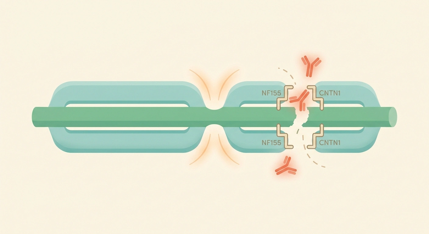
Right at each node, and in the tightly bound paranodal regions on either side of it, there's a scaffolding made of specialized proteins that hold everything together. Think of them as the rivets and brackets that keep the insulation snugged up against the wire in exactly the right way. The main proteins in this scaffolding include neurofascin-155 (which sits on the myelin side, at the paranode), contactin-1 and Caspr1 (which sit on the axon side and bind to neurofascin-155 like a molecular Velcro), and neurofascin-186 and neurofascin-140 (which sit at the node itself, on the axon).
In autoimmune nodopathy, the immune system, for reasons still not fully understood, starts producing antibodies against one of these scaffolding proteins. The antibodies bind to the target, disrupt the tight junctions between axon and myelin, and the whole node-of-Ranvier structure comes apart. Signals stop jumping cleanly. Muscles get weaker, sensation gets muddled, balance deteriorates. What's distinctive here is that the myelin itself isn't being stripped away the way it is in classic CIDP. The insulation is largely intact — it's the connection between the insulation and the wire that's failing. That anatomical difference turns out to matter a lot for treatment.
How Nodopathies Got Separated From CIDP
For decades, if you had a slowly progressive, immune-mediated peripheral neuropathy that wasn't Guillain-Barré, you got the label CIDP — chronic inflammatory demyelinating polyradiculoneuropathy. It was a big umbrella diagnosis that covered a lot of clinical territory, and the standard treatment ladder was IVIG, corticosteroids, or plasma exchange, tried in some order.
Research Says
The 2021 EAN/PNS guideline was the first to formally reclassify autoimmune nodopathies as distinct from CIDP — not just a “variant,” but a mechanistically different disease with a different antibody target, a different tissue signature, and a different treatment response.
The problem was that a subset of patients — maybe 10 to 20 percent of people with what looked like CIDP — didn't fit the mold. Their symptoms often came on more aggressively. They frequently had prominent sensory ataxia and hand tremor. Some had kidney trouble. And a striking number of them just didn't respond to IVIG at all, no matter how many rounds they were given.
Starting around 2012, researchers began identifying specific antibodies in the blood of some of these atypical patients — first against contactin-1, then against neurofascin-155, then Caspr1, then the pan-neurofascin group. As the case reports piled up, a pattern emerged. These weren't just “CIDP variants.” They were mechanistically different diseases that happened to share some surface features with CIDP.
In 2021, the European Academy of Neurology and the Peripheral Nerve Society (EAN/PNS) published a revised guideline on CIDP that formally recognized autoimmune nodopathies as a separate diagnostic category. The distinction wasn't cosmetic. It was made because the immune mechanism is different, the antibody target is different, the tissue damage is different, and — this is the load-bearing point — the treatment response is different. A patient with autoimmune nodopathy who is managed as if they have classical CIDP is likely to spend a long time on the wrong therapy.
The Four Main Antibodies and How They Present Differently
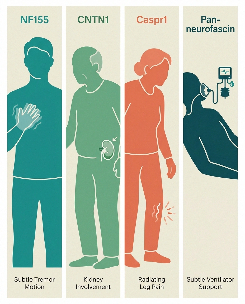
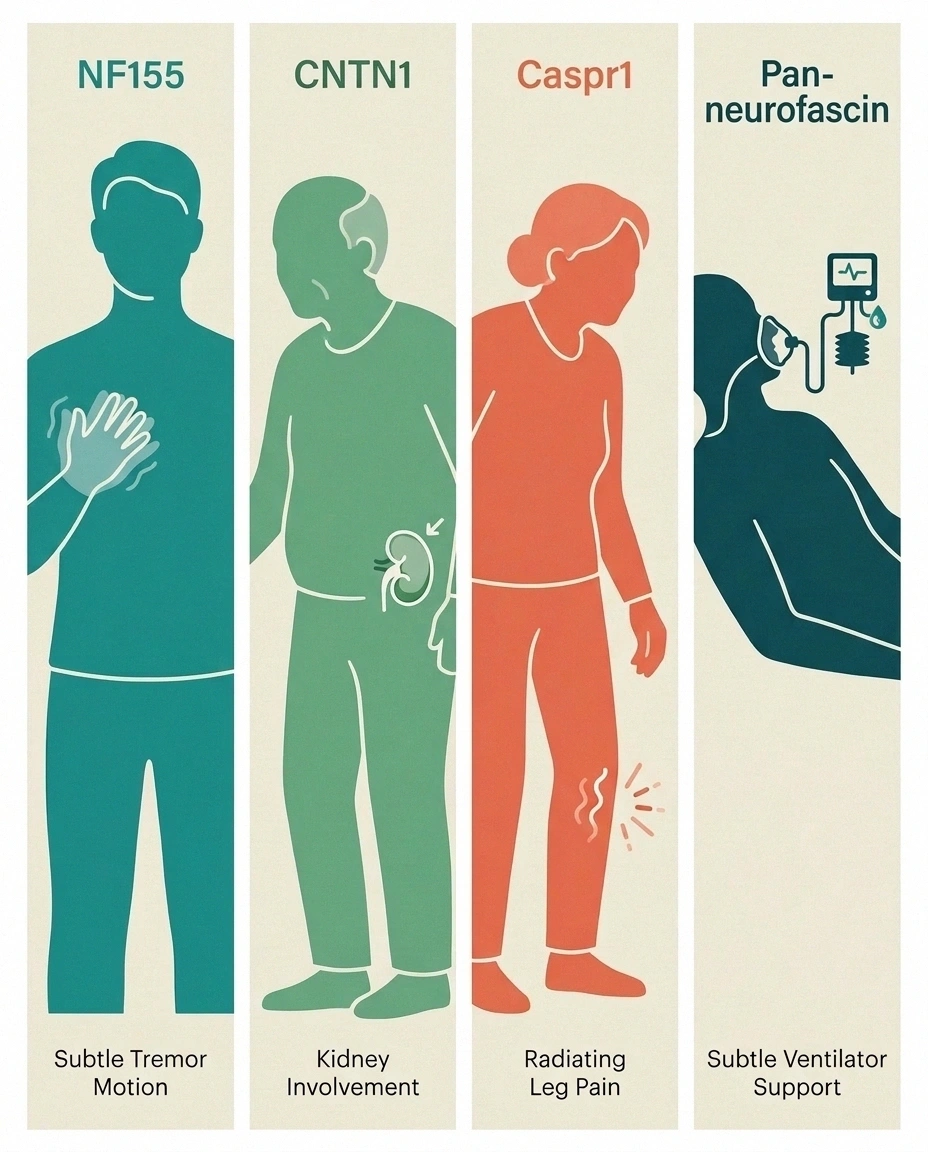
Autoimmune nodopathies aren't a single disease with a single face. Each antibody paints its own clinical portrait. Recognizing the pattern is what leads to the right test being ordered in the first place.
Autoimmune Nodopathy Antibodies at a Glance
| Antibody | Typical Patient | Distinguishing Features | Treatment Response |
|---|---|---|---|
| Anti-NF155 | Younger adults, often 30s-40s | Prominent hand tremor, sensory ataxia, possible CNS involvement | IVIG usually fails; rituximab often works well |
| Anti-CNTN1 | Older adults, often 60s-70s | Aggressive onset, early axonal loss, kidney (membranous nephropathy) link | IVIG usually fails; rituximab strong response |
| Anti-Caspr1 | Older adults | Severe painful neuropathy, gait disturbance, high CSF protein | IVIG usually fails; rituximab preferred |
| Anti-Pan-Neurofascin (NF140/NF186) | Any age, rare | Fulminant tetraparesis, GBS-like, respiratory failure risk | Plasma exchange rescue + rituximab; recovery can be dramatic |
Anti-NF155 (Neurofascin-155)
This is the most-studied and probably the most-encountered of the paranodal antibodies. It tends to hit younger patients — average age of onset in the late 30s to early 40s, though I've read reports of teenagers being diagnosed. Men are affected somewhat more often than women. The picture usually includes distal-dominant weakness (feet and hands more than the trunk), significant sensory ataxia, and — very characteristically — a prominent hand tremor that shows up when the person is holding a cup or trying to write. Cerebrospinal fluid protein tends to be quite high. There's also mounting evidence that NF155 nodopathy can involve the central nervous system in some patients, because NF155 isn't only made by Schwann cells in the periphery; it's also made by oligodendrocytes in the brain and spinal cord. That's why some folks with anti-NF155 nodopathy have imaging changes or symptoms suggestive of demyelinating disease alongside the peripheral picture.
Anti-CNTN1 (Contactin-1)
This is the antibody that tends to hit older patients hard and fast. Onset is usually in the 60s or 70s. The presentation can look almost Guillain-Barré-like — rapid progression to significant weakness over weeks, with sensory ataxia and disability building quickly. Early axonal damage is common, meaning that even after the antibody is controlled, some of the nerve fiber loss doesn't fully reverse. The signature association here is with membranous nephropathy, a form of kidney disease in which protein leaks into the urine and can produce nephrotic syndrome. In case series, this kidney involvement shows up in a striking minority of anti-CNTN1 patients, and the same antibody appears to be responsible for both the nerve and the kidney trouble. Any older man with progressive neuropathy plus new protein in the urine deserves an anti-CNTN1 workup.
Anti-Caspr1 (Contactin-Associated Protein 1)
Caspr1 nodopathy is rarer and, when it hits, tends to be severe. Patients skew older. The picture often includes severe distal weakness, gait disturbance with sensory ataxia and tremor, high CSF protein, and — like CNTN1 — a poor response to IVIG. One of its distinguishing features that neurologists talk about is prominent neuropathic pain, which stands out because paranodal antibodies as a group are more associated with sensory ataxia than with burning pain. Kidney involvement can occur here too.
Anti-Pan-Neurofascin (NF140 and NF186)
This is the smallest and most dramatic group. Pan-neurofascin antibodies bind to a shared portion of the neurofascin proteins that sit at the node itself (rather than the paranode), and the resulting disease can be fulminant — severe tetraparesis over days, cranial nerve involvement, dysautonomia, and respiratory failure requiring ventilation. In its acute form, it can look and act exactly like severe Guillain-Barré syndrome. Mortality has been reported to be high without recognition and aggressive B-cell-directed therapy. The extraordinary thing about pan-neurofascin nodopathy, though, is that once the antibody is depleted, recovery can be remarkable — even after months of near-tetraplegia. The underlying nerve fibers are largely intact; it's the node scaffolding that failed, and the body can reassemble it.
Why Standard CIDP Treatment (IVIG) Often Doesn't Work Here
Here's the part I think every reader with a “CIDP not responding to IVIG” story needs to sit with. It's a mechanistic reason, not a “your CIDP is just harder to treat” reason.
Most of the paranodal antibodies driving autoimmune nodopathy — anti-NF155, anti-CNTN1, anti-Caspr1 — are of the IgG4 subclass. IgG4 is a strange member of the antibody family. Unlike its cousins IgG1 and IgG3, it doesn't do a good job of activating the classical complement cascade, and it doesn't do a good job of recruiting immune cells to attack. What it does very effectively is bind to its target and physically get in the way — in this case, disrupting the molecular Velcro between contactin-1 and neurofascin-155 at the paranode.
Intravenous immunoglobulin (IVIG) works, when it works in autoimmune neuropathies, through a bunch of mechanisms that mostly boil down to interfering with complement activation, blocking Fc receptors on immune cells, and dampening antibody-driven inflammation. Every one of those mechanisms is aimed at how IgG1 and IgG3 antibodies do their damage. When the offending antibody is IgG4 — which uses none of those pathways — IVIG has very little to grab onto. That's why patient after patient with a paranodal-antibody-driven neuropathy sits through weeks or months of IVIG infusions with no meaningful improvement.
The right approach for IgG4-driven disease is to shut down the production of the antibody itself. That means targeting the B cells that are making it. And that's where rituximab comes in.
How Doctors Diagnose Autoimmune Nodopathy
Getting to the right diagnosis usually starts with clinical suspicion — a neurologist noticing that the case in front of them doesn't quite fit the CIDP mold. The clues that should prompt an antibody workup include:
Important Warning
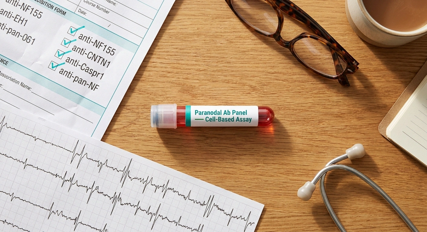
Standard CIDP antibody panels do NOT include paranodal antibodies. You (or your doctor) have to specifically request NF155, CNTN1, Caspr1, and pan-neurofascin testing — and it must be a cell-based assay, not a standard Western blot.
- Poor response to IVIG after an adequate trial (usually a couple of months at proper dosing).
- Prominent sensory ataxia and hand tremor in a supposed CIDP picture.
- Very high CSF protein on lumbar puncture, well above typical CIDP levels.
- Acute or subacute onset that looks Guillain-Barré-like but doesn't behave like GBS.
- Kidney involvement (nephrotic syndrome, membranous nephropathy) in a patient with neuropathy.
- Atypical age patterns — either strikingly young or strikingly aggressive for classical CIDP.
Research Says
Across published case series, IgG4 paranodal antibodies (NF155, CNTN1, Caspr1) are identified in roughly 10 to 20 percent of patients who carry a working diagnosis of CIDP. That means one in five to one in ten “CIDP” patients may actually have an autoimmune nodopathy that requires a different treatment plan. The 2021 EAN/PNS guideline was the first to formally split these patients out into a category of their own.
Once clinical suspicion is there, the confirmatory step is antibody testing using a cell-based assay. This is important: standard Western blot or ELISA tests, which is what most commercial labs default to, can miss the conformational form of the antibody that actually matters. A cell-based assay expresses the target protein on the surface of live cells and looks at whether the patient's serum binds it in its native shape. It's the gold standard for these antibodies. Not every commercial lab in the U.S. offers it. Academic neuromuscular centers, specialized reference labs, and a handful of dedicated providers like Athena Diagnostics are the usual referral points. Some patients end up sending samples to European or Chinese research labs when nothing closer is available.
Alongside the antibody test, the full neuropathy diagnostic workup — electromyography, nerve conduction studies, sometimes a lumbar puncture and MRI of the nerve roots — is still needed. The antibody result doesn't stand alone; it has to fit the clinical and electrical picture.
Rituximab and Other Treatments That Work Better

Rituximab is the treatment that has changed the story for autoimmune nodopathy. It's a monoclonal antibody that binds to CD20, a protein on the surface of B cells (the immune cells that eventually mature into antibody-producing plasma cells). Rituximab depletes most of those B cells for a period of months. With the cells that make the offending IgG4 antibody knocked back, antibody levels fall, and the paranodal damage begins to reverse.
Nodopathy Treatment Escalation
The clinical response is often dramatic — improvement in strength, in balance, in tremor, in reflexes — but it's not instantaneous. Because antibodies that are already made take time to clear from the body, and because the nerves need time to rebuild their damaged scaffolding, meaningful improvement usually unfolds over weeks to a few months after infusion. That patience piece is critical; families who expect an IVIG-like short-term response can be discouraged early.
Rituximab is typically given as either two infusions two weeks apart or four weekly infusions, sometimes repeated every six to twelve months based on how B cells and clinical status are trending. Side effects most people should know about: infusion reactions (usually mild and manageable), increased risk of infections during the period of B-cell depletion, and reactivation of certain viruses like hepatitis B in patients who have a history of exposure. Screening bloodwork before the first infusion catches most of the risk factors.
When rituximab isn't fast enough — for example, in a patient with fulminant pan-neurofascin nodopathy who's crashing toward respiratory failure — plasma exchange is often used as a bridge. Plasma exchange physically removes circulating antibodies from the blood over a series of sessions, buying time while the slower B-cell depletion takes hold. In some centers, plasma exchange is the initial rescue for the sickest patients, followed by rituximab for durable control.
Corticosteroids play a variable role. In some anti-NF155 cases, moderate-dose steroids appear to help, particularly in combination. For fulminant pan-neurofascin disease, pulse steroids are sometimes part of the initial rescue package. But steroids are generally not adequate as long-term monotherapy for nodopathies; B-cell-directed therapy is where the durable control lives.
What to Do If Your CIDP Isn't Responding to IVIG
This is the practical, deployable piece. If you or someone you love has been diagnosed with CIDP and the standard treatment plan just isn't working, here's a plain-language script for the next neurology visit.
Ask For Paranodal Antibody Testing If You Have…
- ✓ Poor response to IVIG after an adequate treatment trial.
- ✓ Prominent sensory ataxia — balance loss especially in the dark or with eyes closed.
- ✓ A hand tremor that shows up when holding, writing, or pouring.
- ✓ Kidney involvement — protein in urine or a nephrotic-syndrome workup alongside the neuropathy.
- ✓ Atypical age of onset — either strikingly young or rapidly progressive in an older adult.
First, ask your neurologist directly: “Given that I'm not responding to IVIG, should we be testing for paranodal antibodies — specifically NF155, contactin-1, Caspr1, and pan-neurofascin? Would you refer me to a lab that does cell-based assays for these?” The specific wording matters. “Autoimmune antibody panel” is too vague; most standard CIDP panels do not include these. You are asking for something specific.
Second, ask about referral to an academic neuromuscular center. Even if your local neurologist is excellent, the reality is that expertise in autoimmune nodopathies is concentrated in a relatively small number of dedicated centers. Places like Mayo, Johns Hopkins, Washington University in St. Louis, UCSF, Cleveland Clinic, and a handful of other academic hubs see enough of these cases that they can pattern-match quickly. A one-time consultation, with your local neurologist continuing routine care, is often the practical arrangement.
Third, understand up front that the antibody testing may take weeks and may not be cheap. Insurance coverage varies. Some patients pay out of pocket for send-out testing. Weigh that cost against the cost of another six or twelve months of an ineffective therapy — for most people the math is clear.
Fourth, if the antibody test does come back positive, ask specifically about the timeline for switching to a B-cell-directed therapy like rituximab. Insurance approval for rituximab in this off-label context can require a peer-to-peer review, letters of medical necessity, and sometimes documentation of IVIG failure. Your neurologist's office is usually the one to shepherd this, but knowing what's involved lets you keep the ball moving.
Living With Autoimmune Nodopathy: What Janet Has Learned From the Community
Beyond the medications and the antibody tests, there's a life to be lived with this condition, and the folks I've spoken to over the years have taught me a few things worth passing along.
The tremor is often the first quality-of-life battle. Especially in anti-NF155 nodopathy, the postural tremor of the hands can make coffee cups, forks, and small buttons genuinely difficult. Weighted utensils, two-handled mugs, and voice-to-text software are simple accommodations that make daily life more workable. Occupational therapy referrals are often more useful than people expect.
Balance and proprioception need active work. When your nerve scaffolding at the node of Ranvier isn't cleanly conducting position-sense signals, your brain loses the map of where your feet are in space. Dark rooms and uneven surfaces become high-fall zones. The recovery of balance after treatment is real but takes months, and dedicated vestibular and proprioceptive physical therapy makes a meaningful difference. Our full article on proprioception loss and neuropathy goes deep on this piece — the fall-prevention playbook applies directly.
Fatigue is real and undervalued. The immune process, the treatment protocols, and the daily energy cost of moving a body whose nerves aren't cooperating add up. Nap permission, calendar buffers between commitments, and honest conversations with employers and family members are all part of the picture. This isn't laziness; it's biology.
The community piece matters. Autoimmune nodopathy is rare enough that most people diagnosed with it have never met anyone else with the same condition. Online patient communities — CIDP and GBS foundations, autoimmune neuropathy Facebook groups, the neuromuscular community more broadly — are where practical knowledge and emotional companionship live. Some readers will find kinship with the broader autoimmune neuropathy world that includes Sjögren's-related nerve disease and other overlaps.
The Coming Wave of Nodopathy Research

The pace at which the autoimmune nodopathy field is moving right now is honestly one of the more hopeful stories in neuromuscular medicine. A few threads to watch over the next several years.
Broader antibody panels. Beyond the four antibodies we've talked about, researchers are identifying additional paranodal and nodal targets, and commercial testing is slowly expanding to cover them. Expect the diagnostic yield to keep climbing.
Next-generation B-cell and antibody-clearing therapies. Newer anti-CD20 agents like obinutuzumab, plasma-cell-directed therapies like daratumumab, and the neonatal Fc receptor (FcRn) blockers currently being studied in CIDP proper (efgartigimod, rozanolixizumab) all have potential mechanisms of action in nodopathy. Case reports of FcRn blockers in refractory nodopathy are already appearing in the literature.
Better biomarkers. Serum neurofilament light chain is emerging as a general marker of nerve injury and treatment response. Combined with antibody titers, it may eventually let neurologists track disease activity in real time rather than relying only on clinical exams.
The general trajectory is: more antibodies identified, more targeted therapies, faster recognition. A patient diagnosed with autoimmune nodopathy in 2030 will almost certainly have a smoother path than one diagnosed today.
Frequently Asked Questions
Is autoimmune nodopathy the same as CIDP?
No. Autoimmune nodopathy used to be lumped together with CIDP because the clinical pictures can overlap and both involve immune attack on peripheral nerves. In 2021, the European Academy of Neurology and Peripheral Nerve Society formally separated autoimmune nodopathies out as their own diagnostic category. The distinction matters because the underlying mechanism is different (IgG4 antibodies against node-of-Ranvier scaffolding proteins, not classical demyelination), and the treatment response is different (rituximab often works well; IVIG often doesn't).
Why doesn't IVIG work as well for nodopathy?
Most nodopathy-driving antibodies are of the IgG4 subclass. IgG4 antibodies cause damage by physically blocking normal protein-to-protein interactions at the nerve node, rather than by activating the complement cascade or recruiting immune cells the way IgG1 and IgG3 do. IVIG works mostly by interfering with those complement and Fc receptor pathways. Because IgG4 doesn't use those pathways, there's very little for IVIG to interrupt. The fix is to shut down production of the offending antibody at its source, which is what rituximab does by depleting B cells.
What's the difference between NF155 and CNTN1 nodopathy?
Both are IgG4-driven paranodal nodopathies, but they tend to present in very different patient profiles. NF155 nodopathy usually affects younger adults, often in their 30s or 40s, and is characterized by prominent hand tremor, sensory ataxia, and sometimes central nervous system involvement. CNTN1 nodopathy tends to hit older adults, often in their 60s or 70s, with a more aggressive onset that can look Guillain-Barré-like, and a distinctive association with kidney disease (membranous nephropathy). Both respond to rituximab; both tend not to respond to IVIG.
How do I get tested for paranodal antibodies?
You have to specifically ask your neurologist to order the test, and it needs to be sent to a lab that uses a cell-based assay for these specific antibodies. Standard autoimmune neuropathy panels usually do not include NF155, CNTN1, Caspr1, or pan-neurofascin. In the United States, specialized reference labs like Athena Diagnostics offer testing, and academic neuromuscular centers can arrange it through research or clinical protocols. Coverage varies by insurance, and results can take several weeks. The specific request to make of your doctor is a cell-based assay for anti-NF155, anti-CNTN1, anti-Caspr1, and anti-pan-neurofascin IgG antibodies.
Can nodopathy go into remission?
Yes, and this is one of the more hopeful features of the condition. When the antibody producing the damage is successfully depleted through B-cell-directed therapy like rituximab, many patients experience substantial or even near-complete recovery of strength, sensation, and balance. Some patients enter durable remissions lasting years, particularly in the NF155 group. Others need periodic re-treatment when B cells repopulate and antibody levels rise again. Even patients who spent months on ventilators with pan-neurofascin nodopathy have had striking recoveries once the antibody was cleared. The nerve fibers themselves are usually still there; it's the connections that need to rebuild.
Is rituximab safe for nodopathy?
Rituximab has been used for decades in autoimmune disease and cancer, and its safety profile is well characterized. The main considerations are infusion reactions (usually mild and managed with pre-medication), increased infection risk during the months of B-cell depletion after each treatment cycle, and the potential for reactivation of hepatitis B in patients with prior exposure, which is why screening before the first infusion is standard. Vaccines, including COVID-19 boosters, are less effective while B cells are depleted, so timing matters. For most patients, the risks are far outweighed by the potential benefit of treating an otherwise progressive, disabling neuropathy. The specific risk-benefit conversation should happen with your treating neurologist, ideally one experienced with immunotherapy in neuromuscular disease.
The Bottom Line
Autoimmune nodopathy is the diagnosis that too many people with treatment-resistant CIDP have never had spelled out for them. It's a distinct disease with its own antibody signature, its own molecular mechanism, and its own treatment path. IVIG, the workhorse of classical CIDP care, is often not the right tool for the job here. Rituximab and other B-cell-directed therapies are, and when they're deployed against the right target, the response can be remarkable.
If your CIDP diagnosis has stopped making sense — if the infusions aren't working, if the disability keeps climbing, if the clinical picture just doesn't fit the textbook — you owe it to yourself to ask about paranodal antibody testing. The path from a wrong-tool treatment to a right-tool treatment sometimes runs through a single specialized blood test. That's the piece of the puzzle worth chasing down.
You're not alone in this, and the field is finally catching up to you. Ask the specific questions, seek the specific tests, and don't accept “let's just try a higher dose of IVIG” as the final answer. There's a better answer, and for a meaningful percentage of people who've been struggling with unresponsive CIDP, it's the one that will finally get them their life back.
About the Author