Minute four is fine. Minute twelve is manageable. Somewhere around minute twenty-two, the burning in your feet stops being background noise and becomes the only thing in the world, and the one thing you are not allowed to do about it is move.
That is the problem with an MRI when you have neuropathy, and almost nothing written about MRI preparation addresses it. The standard advice assumes your difficulty is claustrophobia or boredom. Yours is that lying perfectly still is exactly the condition under which nerve pain gets loudest.
Most of what makes this scan tolerable gets decided before you are on the table, and several of the decisions have to be made days ahead. Here is what to arrange, what to ask for, and what to say.
Why Neuropathy and MRI Work Against Each Other
Understanding the mechanics helps, because it tells you what to target.
Four forces working against you, and what each one costs
Only one of the four is fixed by the machine. The other three are negotiable.
Enforced stillness
The same mechanism that makes nerve pain worse at 2am. Fixable with positioning support.
Cold room, low to mid 60s F
A magnet requirement, not a thermostat setting. Fixable only with blankets and socks.
Hard, thinly padded table
A skin risk when you cannot feel a pressure point forming. Fixable with heel and sacral padding.
Motion, repeat, longer scan
Every comfort measure also shortens the scan, because comfort reduces repeats.
Stillness makes nerve pain worse. The same reason symptoms flare at bedtime applies here: with no movement, no walking, no shifting position, there is nothing competing for your attention and nothing changing the pressure and circulation in your feet. Anyone who has read about why neuropathy gets worse at night already knows this pattern. An MRI table is a bed you are forbidden to move in.
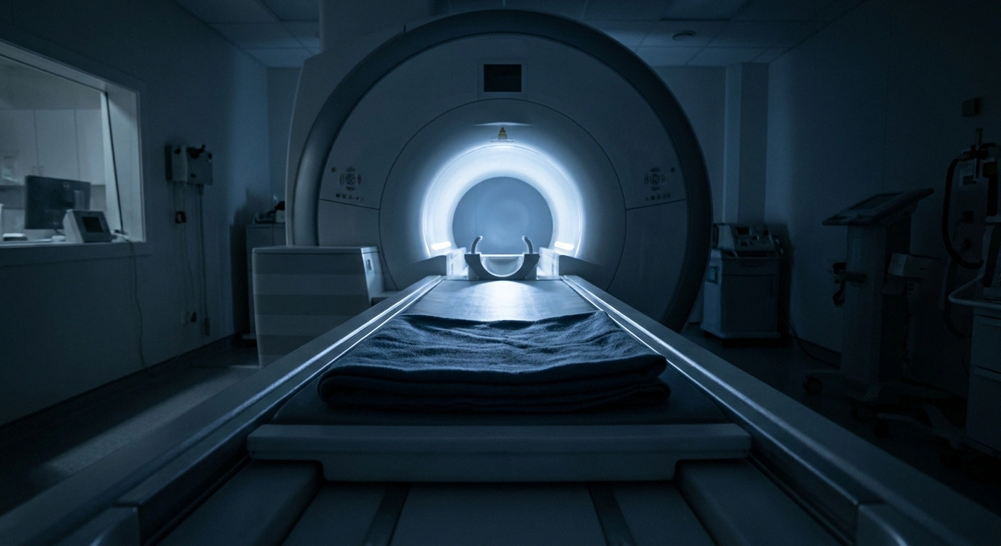
The room is cold. Scanner rooms are typically kept somewhere in the low to mid sixties Fahrenheit to protect the magnet and its electronics. Cold constricts blood vessels and makes burning, aching and cold-sensitivity symptoms sharper. This is not adjustable. The room temperature is a machine requirement, not a comfort setting.
The table is hard. It is a flat, thinly padded surface. If sensation in your feet, heels or sacrum is reduced, you may not register a pressure point developing until after the scan, which is a genuine skin risk on longer protocols.
And there is a feedback loop that turns discomfort into more discomfort. Movement creates image artifact. Artifact means the technologist repeats that sequence. Repeats extend the scan. A longer scan means more time in exactly the position that hurts. This is the loop worth attacking, because everything you do to stay comfortable also shortens the scan.
The Screening Conversation That Cannot Wait Until the Day
Several devices common in neuropathy care create real MRI complications, and every one of them is easier to sort out at booking than in the changing room.
Say these on the booking call
| What you have | What it needs | Bring |
|---|---|---|
| Spinal cord stimulator | Model-specific conditions: charged battery, checked impedances, set mode, transmitter removed | Device ID card |
| TENS unit | Off and out of the magnet room for the whole appointment | A plan for pain without it |
| Insulin pump or CGM | Removed before entry; sensors are generally not MR safe | Replacement sensor, glucose plan |
| Medicated patches | Checked individually; some backings contain metal and can heat | The box or name of the patch |
| Contrast ordered | Kidney function screening before the appointment | Recent bloodwork date |
A spinal cord stimulator is the big one. Modern systems are generally described as MR conditional, meaning scanning is possible only under specific conditions, and those conditions differ by manufacturer and by model. Common requirements include a fully charged battery, lead impedance values within an expected range, particular device settings, and removal of any external transmitter. Some older systems restrict which body regions can be scanned at all. Bring your device identification card, and tell the scheduler about the stimulator when you book, not when you arrive.
A TENS unit comes off and stays out of the magnet room. Beyond the obvious issue of metal and wires, surface stimulation can drive current through an implanted device. If you normally rely on a TENS unit for pain control, plan for the fact that you will be without it for the whole appointment, including waiting time.
An insulin pump or continuous glucose monitor must be removed before entering the room. Sensors and transmitters are generally not MR safe, and a sensor can be damaged or can cause a burn. Ask when you book how long you will be disconnected and plan glucose management around it, including whether to bring a replacement sensor.
Then the standard metal screening: pacemakers and defibrillators, aneurysm clips, cochlear implants, joint replacements, surgical hardware, and any history of metal fragments from an injury or from grinding and welding work.
Finally, contrast. If the scan uses gadolinium, kidney function gets checked first. That matters more in this population than most, since diabetes and several other causes of neuropathy commonly come with reduced kidney function. If nobody has mentioned recent bloodwork, ask.
Positioning: The Requests Worth Making
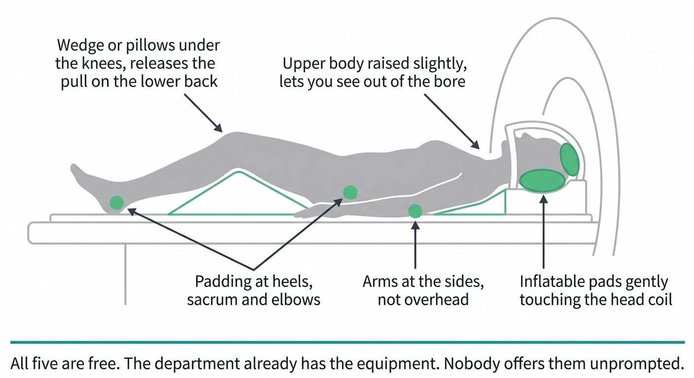
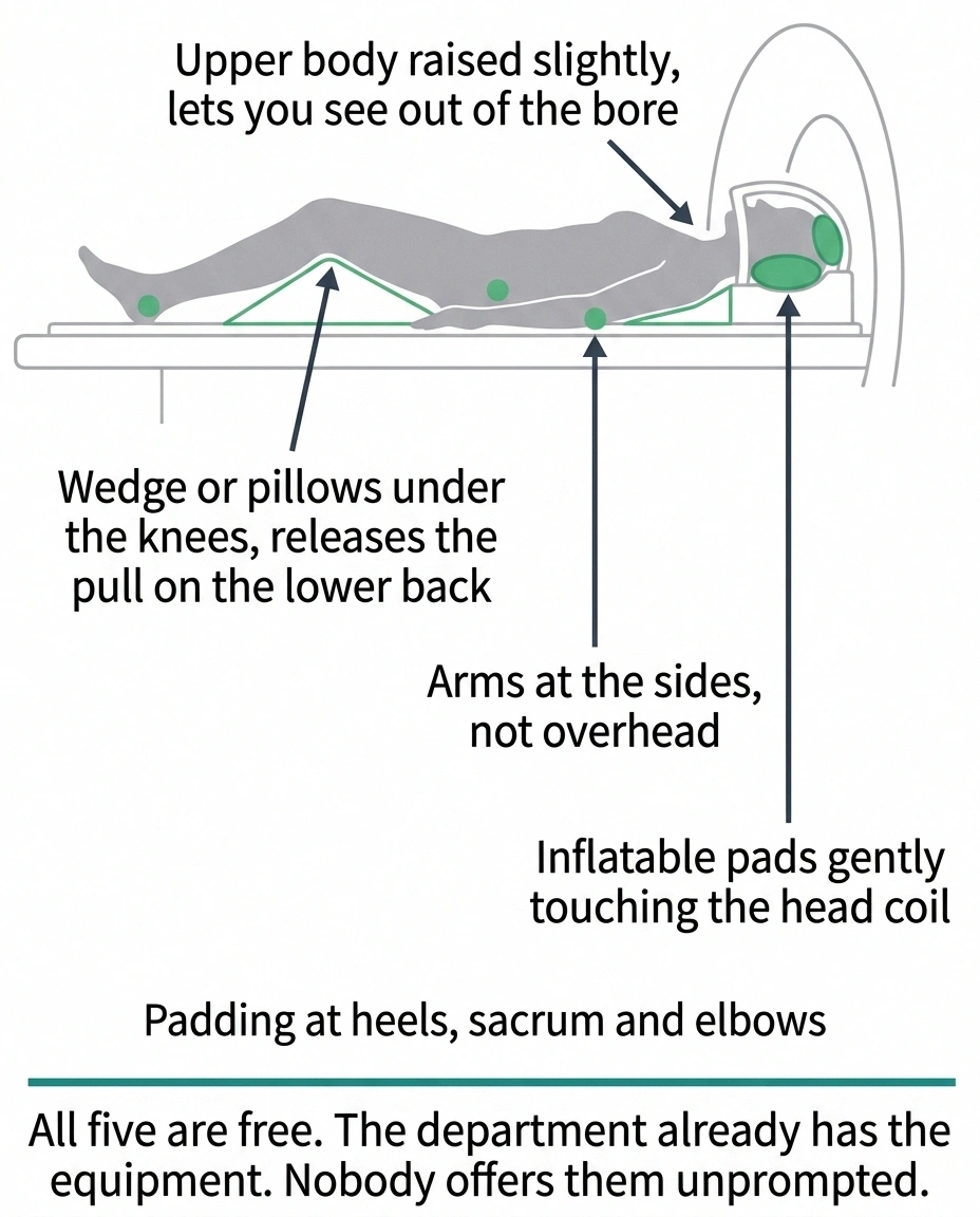
Positioning support is the highest-value thing you can ask for, it costs the department nothing, and technologists generally have the equipment on hand. Most people never ask because nobody tells them they can.
Five requests, all free, most never asked for
Technologists usually have this equipment within arm's reach. Nobody offers it unprompted.
- Wedge or pillows under the knees. Releases the pull on the lower back that flat legs create. The single highest-value ask.
- Upper body raised slightly on a wedge. Changes your sight line so you can see out of the bore instead of into it.
- Arms at your sides, not overhead. Ask whether your specific protocol actually requires overhead positioning. Often it does not.
- Padding at heels, sacrum and elbows. These are the pressure points you are least likely to feel forming.
- Inflatable head pads touching the coil. Reduces head motion, and most people find it feels like being held rather than confined.
A wedge or pillows under the knees is the single best request for anyone lying on their back. Flattening the legs pulls on the lower back and makes the whole position harder to hold. Bending the knees slightly releases that, and it is the difference between fidgeting and lying still for many people.
For claustrophobia, elevating the upper body on a wedge changes what you can see. With the head and shoulders raised slightly, many people can see out of the opening of the bore rather than staring at the wall of it a few inches from their face. That single change of sight line does more for anxiety than most breathing advice.
Arms at your sides or resting on your abdomen, rather than overhead, matters more than it sounds. Overhead positioning is uncomfortable within minutes for anyone and is often unnecessary depending on what is being scanned. Ask what your protocol requires.
Ask for padding under the heels, the sacrum and the elbows specifically. These are the pressure points, and they are the ones you are least likely to feel developing if sensation is reduced.
Inflatable pads placed alongside the head and gently inflated until they just touch reduce head motion and, for a lot of people, produce a feeling of being held rather than being confined.
Handling the Cold Room
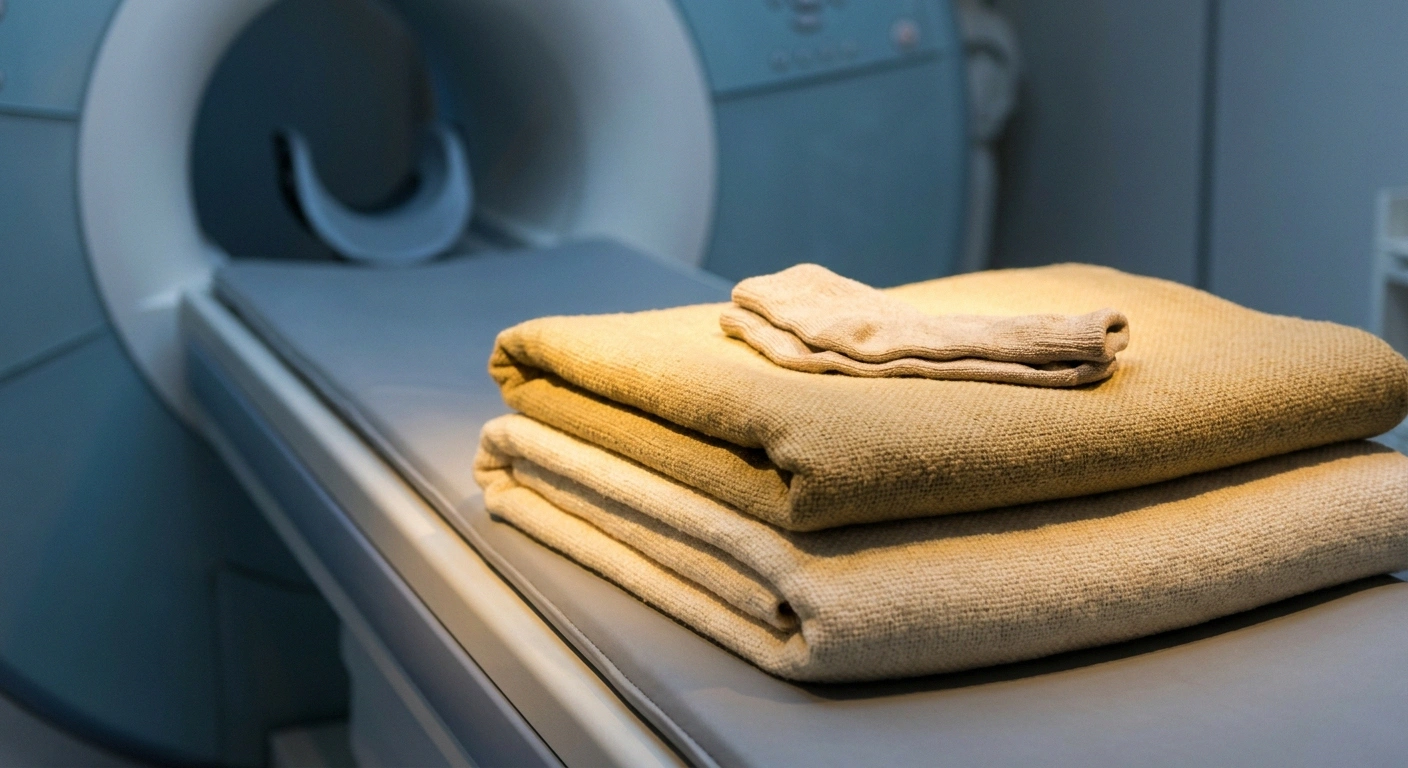
Ask for warmed blankets before the scan begins, not after you have already gotten cold. Rewarming while lying still takes far longer than staying warm in the first place, and by the time you notice you are cold, your symptoms have usually already escalated.
Socks are typically allowed, with one caveat that catches people out. Silver-infused or metallic-thread socks, including some diabetic and antimicrobial styles, are not appropriate in the scanner. If you normally wear specialized neuropathy socks, bring a plain cotton pair for the appointment and check them with the technologist.
Everything else metallic comes off: jewelry, hearing aids, wigs with clips, hairpins, magnetic clasps. Medicated patches deserve a specific check, since some have metallic backing layers that can heat. Mention any patch you are wearing rather than assuming it is fine.
Timing the Appointment and the Medication
Book for the time of day your symptoms are quietest. For most people whose pain peaks in the evening and overnight, that means mid-morning. A first-thing appointment can be worse if mornings are stiff for you. You know your own pattern; use it, because scheduling is one of the few variables entirely under your control.
Ask how long the protocol takes when you book. There is a wide range, from about twenty minutes for a simple sequence to an hour or more for multi-region imaging with contrast. Knowing the number in advance changes how you prepare and removes the worst part of the experience, which is not knowing when it ends.
Ask whether the study can be split into shorter blocks with a brief break in between. Many protocols can accommodate this. Some cannot, particularly contrast-timed sequences. It is worth one question.
Ask whether the facility has a wide-bore or open scanner. Wide-bore units have a larger opening and help considerably with both claustrophobia and body size. Availability varies, and image quality on open units is sometimes lower, so this is a trade-off to raise with whoever ordered the scan.
On medication, the goal is to have your routine nerve-pain medication at peak coverage during the scan window rather than wearing off. That is a timing conversation with whoever prescribes it, not a dose you adjust yourself. If anxiety rather than pain is the limiting factor, a short-acting anxiolytic taken before the appointment is a prescriber decision and requires someone else to drive you home. Neither of these can be arranged on the morning of the scan, which is why they belong in the booking call. Our guide to getting a useful answer from your doctor covers how to frame that request.
The Squeeze Ball Problem
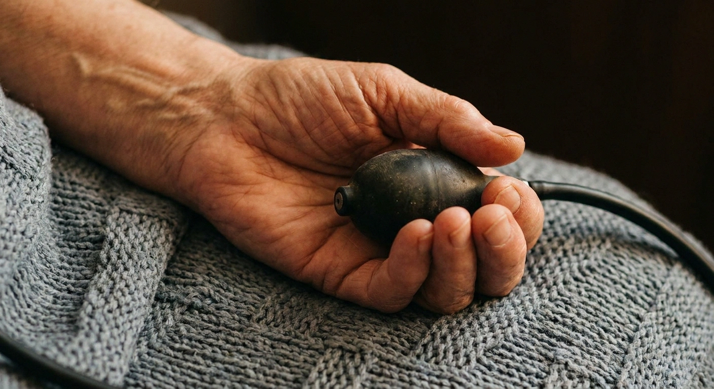
You will be given a rubber bulb or ball to squeeze if you need the scan stopped. It is the entire communication system, and it quietly assumes two things about your hands that neuropathy may have made untrue.
Your entire communication system assumes two things about your hands
It assumes you can feel the bulb if it shifts, and that you can squeeze it hard enough to register. Reduced sensation and reduced grip strength both break that assumption quietly.
Before the table moves: name your better hand out loud, squeeze the bulb, and confirm the technologist registered it.
Then ask what happens when you signal. People who know the answer use the ball early, when a thirty-second pause is enough, instead of holding on until they have to come all the way out.
It assumes you can feel it. If it slips slightly in a numb hand, you may not notice until you try to use it.
It assumes you can squeeze it hard enough. Hand weakness and reduced grip are common, and the bulb requires a definite squeeze.
Two things fix this. First, put it in your better hand, and say out loud which hand that is rather than letting it be handed to whichever side is nearer. Second, test it before the table moves. Squeeze it, confirm the technologist registered it, and confirm you felt yourself doing it.
Also agree on what happens next. Ask what the technologist will do when you signal: pause the sequence, talk to you, or bring you out. Knowing the answer in advance makes people far less likely to panic and far more likely to use the ball early, when a thirty-second pause is enough, instead of enduring until they have to come out entirely.
What an MRI Can and Cannot Tell You
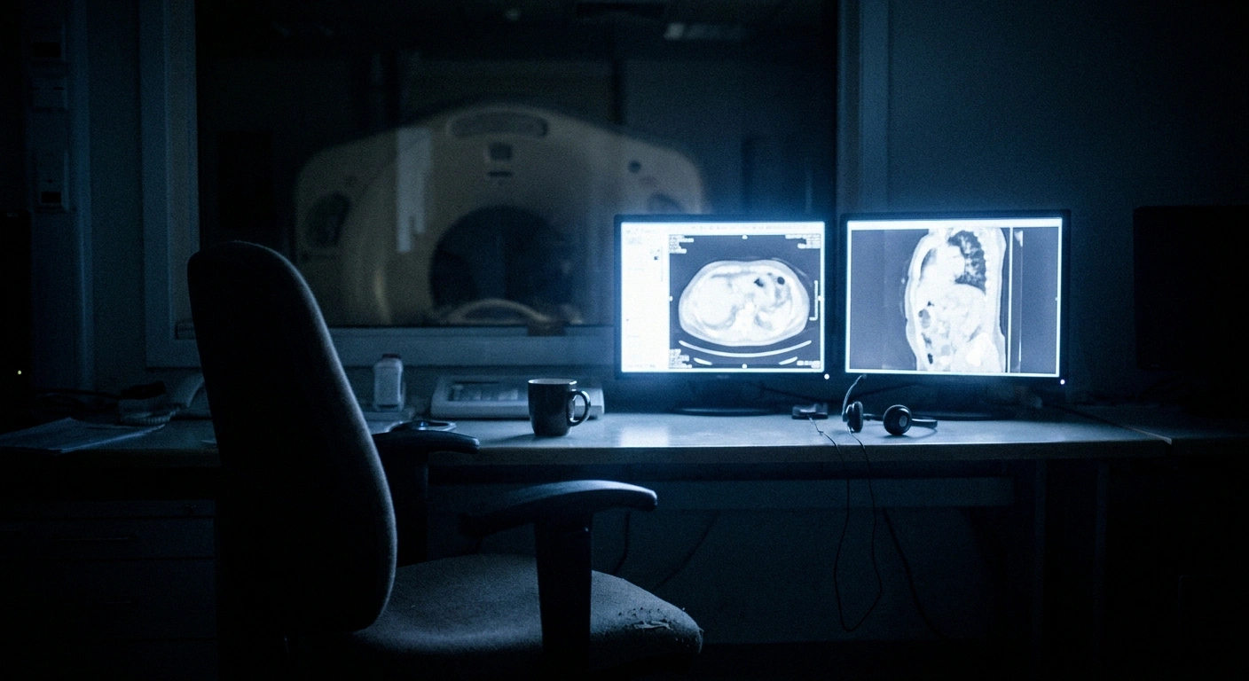
This is worth understanding before the results conversation, because it prevents a specific and common disappointment.
Two different questions, two different tests
What MRI answers
Is something structural causing or mimicking this? Disc or stenosis compressing a nerve root, tumor, MS lesion, plexus injury. MR neurography images larger nerves directly at some centers.
What answers the neuropathy question
Nerve conduction studies and EMG, skin biopsy for small fiber density, and blood work for causes. MRI cannot see the small fibers in your feet at all.
A normal MRI means the structural causes it was looking for were not found. That is useful, and it is not a clean bill of nerve health.
An MRI does not diagnose peripheral neuropathy. It cannot see the small nerve fibers in your feet at all. The tests that establish neuropathy are nerve conduction studies and EMG, skin biopsy, and blood work, which is covered in our overview of how neuropathy is diagnosed.
What an MRI is for is finding a cause or a mimic. Spinal stenosis or a disc compressing a nerve root can produce symptoms that resemble neuropathy. So can a tumor pressing on a nerve, multiple sclerosis lesions, or a plexus injury. Some centers offer MR neurography, which images the larger peripheral nerves directly, though it is not widely available.
So a normal MRI does not mean nothing is wrong with your nerves. It means the specific structural causes the scan was looking for were not found, which is genuinely useful information and is not the same as a clean bill of nerve health.
If You Have to Stop
Some people cannot complete the scan. That is a known outcome, not a failure, and there are established alternatives.
Ask about splitting the study across two appointments. Ask about sedation, which requires an anesthesia or radiology consultation and an escort home. Ask whether a different imaging method, such as CT or ultrasound, could answer the clinical question, which sometimes it can. And ask whether another facility in the area has a wide-bore or open scanner.
The important thing is to raise it rather than absorb it. A scan abandoned at minute thirty with nothing said is a wasted appointment. A scan stopped at minute thirty with a plan attached is a rescheduled one.
A Short Checklist for the Day
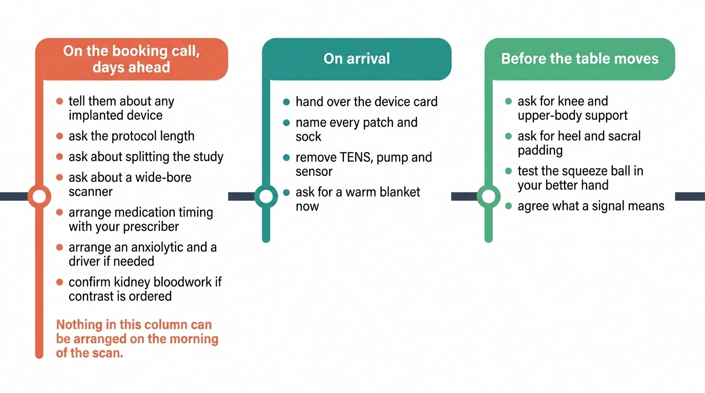
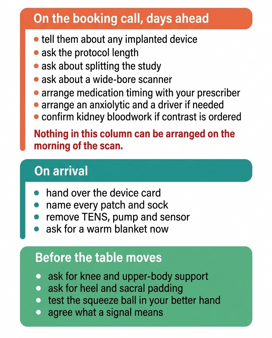
When you book, tell them about any implanted device and ask the protocol length. When you arrive, hand over your device card, name every patch and sock you are wearing, and ask for knee support, a head or upper-body wedge, heel and sacral padding, and a warm blanket before you lie down. Test the squeeze ball in your better hand and agree on what a signal means. Then, once the scan starts, the only job left is breathing slowly and letting the noise happen.
If you are also facing an overnight admission or a procedure around the same time, our guide to hospital stays with neuropathy covers the longer-stay version of these same problems. And if anxiety is the dominant issue rather than pain, our page on neuropathy and anxiety may be more useful than anything on this one.
Frequently Asked Questions
Can I have an MRI if I have a spinal cord stimulator?
Usually yes, but only under specific conditions that vary by manufacturer and model. Common requirements include a fully charged battery, lead impedances within an expected range, particular device settings, and removal of any external transmitter. Some older systems limit which body regions can be scanned. Tell the scheduler when you book, and bring your device identification card to the appointment.
Does an MRI show nerve damage or neuropathy?
Not peripheral neuropathy itself. MRI cannot see the small nerve fibers in the feet and hands. It is ordered to look for structural causes or mimics, such as spinal cord or nerve root compression, tumors, multiple sclerosis lesions, or plexus injuries. Neuropathy itself is diagnosed with nerve conduction studies and EMG, skin biopsy, and blood work.
How long does an MRI take?
It ranges widely, from roughly twenty minutes for a single straightforward sequence to an hour or more for multi-region imaging with contrast. Ask for the specific number when you book, because knowing the endpoint is one of the most effective things you can do for tolerance.
Can I take something for anxiety before an MRI?
A short-acting anxiolytic before the scan is common and is prescribed in advance rather than given on the day. It requires arranging a ride home. Raise it during the booking call, since the prescription and the transport both need to be sorted out beforehand.
Can I wear socks during an MRI?
Plain cotton socks are generally fine and help with the cold room. Silver-infused, metallic-thread and some antimicrobial diabetic socks are not appropriate in the scanner. Bring a plain pair and check them with the technologist.
What happens if I move during the scan?
Movement blurs the images for that sequence, and the technologist repeats it. Nothing is damaged and nobody is upset, but the scan takes longer. Signaling with the squeeze ball for a short pause usually costs less time than a repeated sequence does.
Is an open MRI as good as a closed one?
Not always. Open and wide-bore scanners are much easier to tolerate for claustrophobia and larger body size, but some open units use a lower magnetic field strength, which can reduce image detail. Wide-bore closed scanners are often a good middle ground. Whether the trade-off is acceptable depends on what is being looked for, so ask the doctor who ordered the scan rather than deciding alone.
Can I keep my insulin pump or glucose monitor on?
No. Insulin pumps, sensors and transmitters are generally not MR safe and must be removed before entering the magnet room. Ask when you book how long you will be disconnected, and plan glucose management and any replacement sensor around that window.
About the Author