
For most people who end up with this diagnosis, the order of events runs backward from what you would expect. The feet go first. Tingling, then numbness creeping up from the toes, then a growing sense that the floor is further away than it used to be. Months pass. A neurologist orders blood work, and buried in it is a protein electrophoresis showing a spike of IgM that nobody was looking for. A hematologist enters the picture. And somewhere in that appointment a word appears that you have never heard spoken aloud.
Waldenström macroglobulinemia is a rare blood cancer, and roughly a quarter of people diagnosed with it already have nerve symptoms at the time of diagnosis. In a series of 182 patients, 47 of them reported neuropathic symptoms at initial presentation. For a meaningful share of those people, the nerve trouble is what started the whole investigation.
That sequence matters, because it changes what you are actually dealing with. You are not a cancer patient who later developed a side effect. You are someone whose nerves detected a blood problem before any blood count did.
What follows is how one disease reaches the nerves through four separate routes, why the pattern in your hands and feet is specific enough to have its own name, and the part that frustrates patients more than anything else: why the number your hematologist tracks so carefully has such a loose relationship with how your feet feel.
Why the Nerves Usually Notice First
Waldenström macroglobulinemia is a lymphoplasmacytic lymphoma. A population of abnormal B cells sets up in the bone marrow and produces one antibody, over and over, in enormous quantity. That antibody is immunoglobulin M, IgM, the largest of the antibody classes.
How rare, in numbers you can actually hold
4.2
new cases per million people per year
~1,500
diagnoses a year across the United States
25%
had nerve symptoms already at diagnosis, in a 182-patient series
5
units bound together in one IgM molecule, which is why its size matters so much
Rarity has a practical cost. Most neurologists see this pattern a handful of times in a career, which is part of why an unexplained slowly progressive neuropathy should prompt a serum protein electrophoresis rather than a second year of watching.
It is genuinely rare. Incidence runs at about 4.2 cases per million people per year, which in the United States works out to somewhere near 1,500 new diagnoses annually. Many people live with it for years without treatment, monitored rather than medicated, because the disease is often slow and the blood counts hold.
The nerves are a different story. IgM is a physically enormous molecule, and unlike most antibodies it circulates as a pentamer, five units bound together. That size has consequences. It makes blood thicker. It makes the protein prone to depositing where it should not. And when the abnormal clone happens to produce an IgM that recognizes something in nerve tissue, the size means it hits hard.
So the nerves become an early warning system for a disease that is otherwise quiet. Not because nerves are especially fragile, but because several distinct properties of this one protein all happen to converge on peripheral nerve.
What the Protein Does to a Nerve
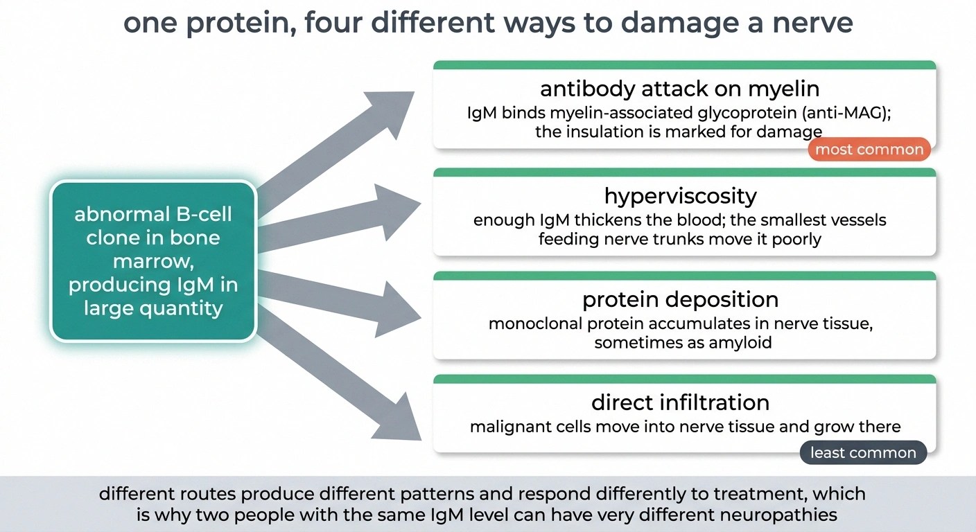
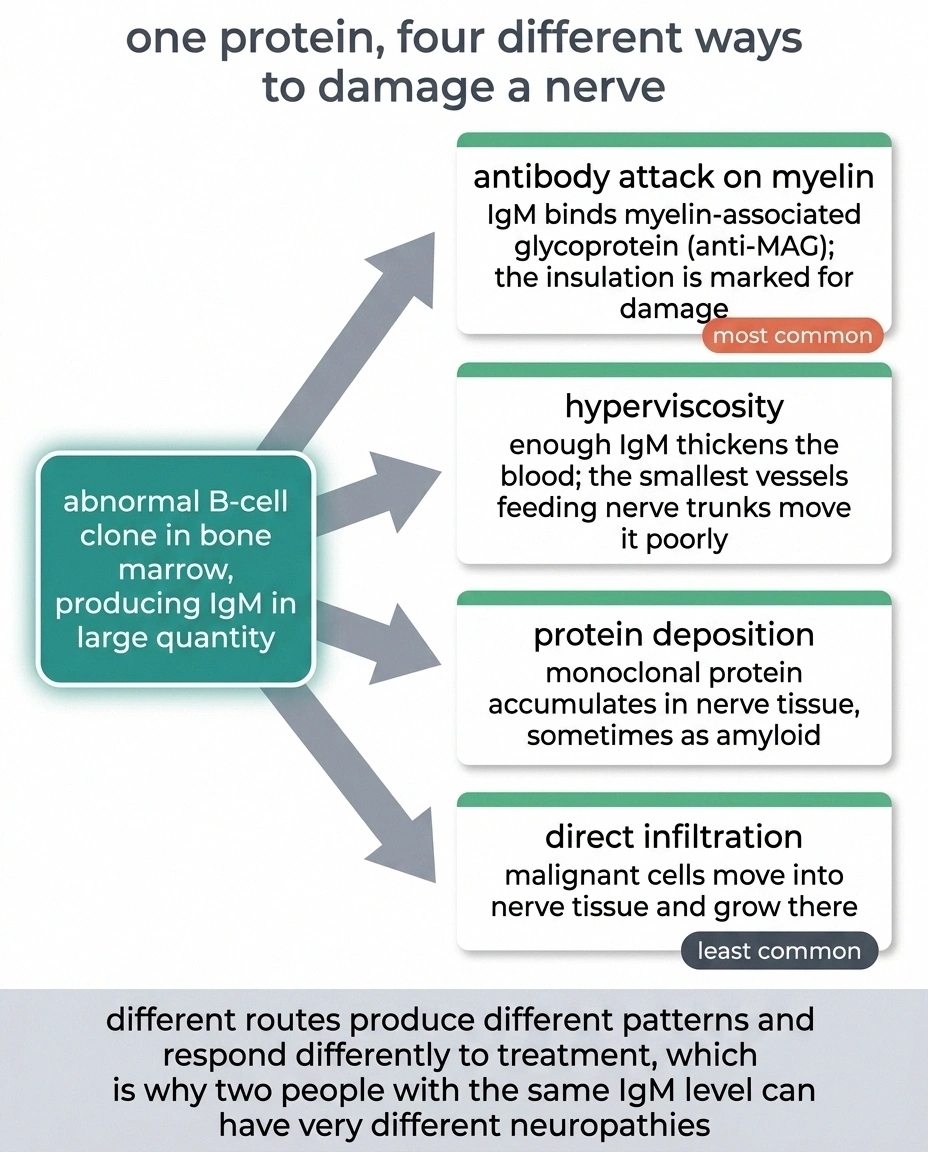
There is no single mechanism here, and that is the most useful thing to understand early. Four separate processes can damage nerve in this disease, they produce different patterns, and they respond differently to treatment. Two people with the same diagnosis and similar IgM levels can have completely different neuropathies.
The first and most common is an autoimmune attack. The IgM the clone produces sometimes binds to myelin-associated glycoprotein, a structural protein in the insulating sheath around nerve fibers. When that happens the antibody is not just floating in the blood. It is docking onto your myelin and marking it for damage. Antibodies against GM1 and other nerve targets show up less often but do the same kind of thing.
The second is hyperviscosity. Enough IgM makes blood measurably thicker, and thick blood moves poorly through the smallest vessels. The tiny arteries feeding nerve trunks are among the smallest in the body.
The third is deposition. The monoclonal protein can precipitate out and accumulate in nerve tissue directly, sometimes as amyloid, which is a folding problem rather than a targeting problem and behaves more like amyloid neuropathy than like an autoimmune attack.
The fourth is infiltration. The malignant cells themselves can move into nerve tissue and grow there. This is the least common route and the most aggressive.
These are worth separating because the first one has a name, a specific test, and a specific look on nerve conduction studies. It is also, by a wide margin, the one most people reading this page have.
Anti-MAG: The Pattern With a Name
When IgM targets myelin-associated glycoprotein, the resulting neuropathy is distinctive enough that neurologists recognize it on description alone.
Three findings that carry more weight together than apart
- Distal sensory loss without much weakness
- Numbness that outruns any measurable loss of strength. Grip and ankle power often test close to normal while the numbness is severe.
- A large, slow tremor when reaching or holding
- High amplitude and low frequency, appearing on action rather than at rest. Routinely written off as essential tremor and therefore left out of the history entirely.
- Balance that fails specifically with eyes closed
- Showering with eyes shut, walking to the bathroom at night, or turning in the dark. Vision had been compensating, and removing it exposes the deficit.
Worth saying out loud at the appointment: “My hands shake when I reach for things, and I lose my balance in the shower when my eyes are closed.” Both halves of that sentence are diagnostic information, and most people never think to mention either one.
It is a demyelinating neuropathy, meaning the insulation is damaged rather than the fiber itself, at least initially. That distinction matters for recovery, and it is worth understanding the difference between demyelinating and axonal nerve damage before any conversation about prognosis, because they heal on completely different timescales.
What makes anti-MAG unusual is that it preferentially strips myelin from the most distal fibers. Damage concentrates at the far ends. Clinically that produces a picture doctors call DADS, for distal acquired demyelinating symmetric neuropathy, and it looks like this:
Sensory symptoms dominate. Numbness and unsteadiness rather than weakness. Some people have almost no measurable weakness at all despite significant disability. It is symmetric, affecting both sides similarly, and it involves all four limbs, though the legs are usually well ahead of the arms. It progresses slowly, over years rather than months, which is part of why the diagnosis so often arrives late.
And it comes with two features that are worth naming separately, because patients rarely connect them to the diagnosis on their own.
The Tremor and the Ataxia

People with anti-MAG neuropathy frequently develop a tremor in the hands and arms. It is characteristically high-amplitude and low-frequency, a large slow shake rather than a fine buzz, and it shows up when reaching for something or holding a position. It is common enough in this specific neuropathy that its presence alongside distal numbness should prompt a check for IgM.
Many people spend a long time attributing it to age, or to anxiety, or to a separate essential tremor. It is neither. It comes from the same demyelination that is causing the numbness, and it is a documented feature of the disease rather than a coincidence.
The second feature is gait ataxia. Not weakness, and not dizziness, but a specific kind of unsteadiness that comes from losing position sense. Your legs are strong. Your brain has simply stopped receiving reliable reports about where they are. The tell is that balance collapses in the dark or with eyes closed, because vision was silently compensating the whole time. This is sensory ataxia, and it is the reason so many people with this condition describe walking as an act of concentration.
Both features come as a package with the sensory loss. If you have all three, that combination is doing real diagnostic work.
Why Your IgM Number Does Not Track How You Feel
Here is the part that causes the most frustration, and the part that almost no patient-facing source explains.
Your hematologist will track your IgM level closely. It is the number on the chart, the thing that goes up and down, the thing that decisions get made around. It is entirely reasonable that they track it. And it will often have very little to do with how your feet feel.
Discordance between symptoms and IgM level is common and well described in the literature. People with modest IgM elevations can have severe, disabling neuropathy. People with dramatic IgM levels can have minimal nerve symptoms. Bringing the level down does not reliably bring the symptoms down on any predictable schedule.
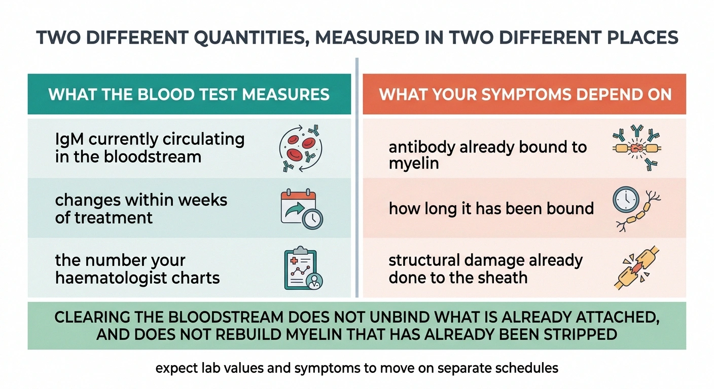
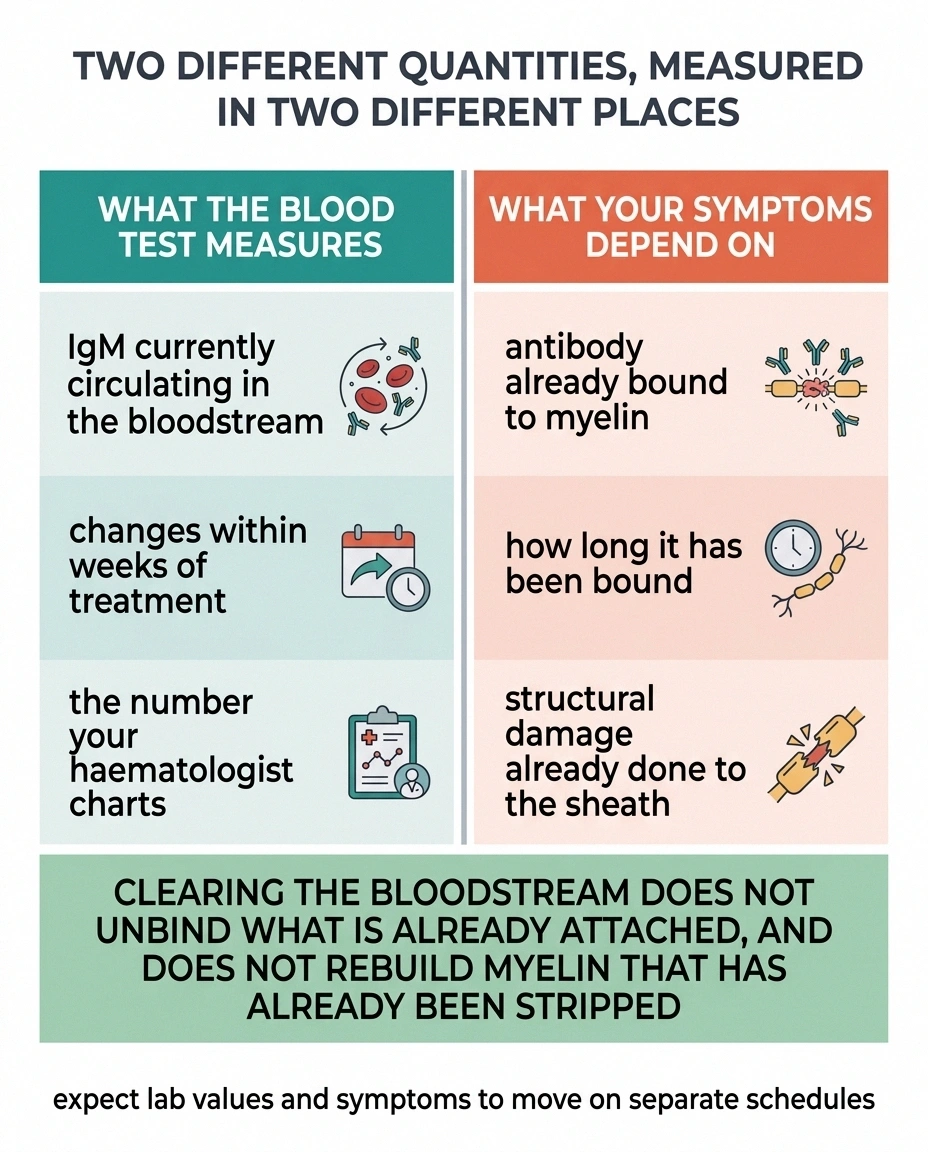
There is a mechanical reason for this, and once you see it the frustration eases somewhat. The blood level measures how much protein is circulating. The neuropathy depends on how much antibody has already bound to your myelin, how long it has been there, and how much structural damage it has done. Those are different quantities. Clearing the bloodstream does not un-bind what is already attached, and it certainly does not rebuild myelin that has already been stripped.
Think of it as the difference between shutting off a leaking pipe and drying the floor. Necessary, and not the same job.
The practical consequence is that you should expect your nerve symptoms and your lab values to move on separate schedules, and you should not read a rising IgM as proof that your feet are about to get worse, or a falling one as a promise that they will improve.
Getting to the Diagnosis

Three pieces of evidence usually converge.
The first is the blood work. Serum protein electrophoresis with immunofixation identifies the monoclonal IgM and quantifies it. An anti-MAG antibody titer tests specifically for the myelin-targeting antibody, and high titers are strongly supportive. A bone marrow biopsy establishes the underlying lymphoma and distinguishes Waldenström from IgM MGUS, which produces an identical-looking neuropathy from a much smaller clone that does not meet the threshold for cancer.
That distinction is worth pausing on. If your marrow shows a small clone and your blood counts are normal, you may have MGUS rather than Waldenström, and the neuropathy can be just as significant. The nerve disease does not care much which side of the line the clone falls on. The treatment decisions differ, though, so the labelling is not merely academic.
The second piece is electrodiagnostic testing. Nerve conduction studies and EMG show demyelination that is disproportionately concentrated at the distal ends of nerves. The specific measurement is a prolonged distal motor latency out of proportion to the slowing along the rest of the nerve, expressed as an abnormal terminal latency index. That pattern is close to a fingerprint. If you want to make sense of the report yourself, our guide to reading EMG and nerve conduction results walks through what the columns mean.
The third is simply the clinical picture: slow, distal, symmetric, sensory-dominant, with tremor and gait ataxia. On its own it is suggestive. Combined with the other two it is close to definitive.
A nerve biopsy is occasionally done, mostly when amyloid deposition or infiltration is suspected rather than antibody attack. It is not routine here.
Treatment Aims at the Clone
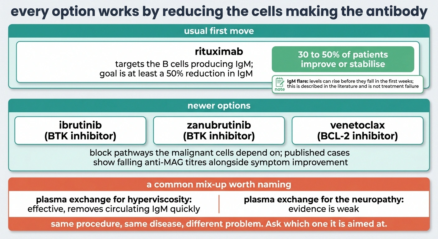
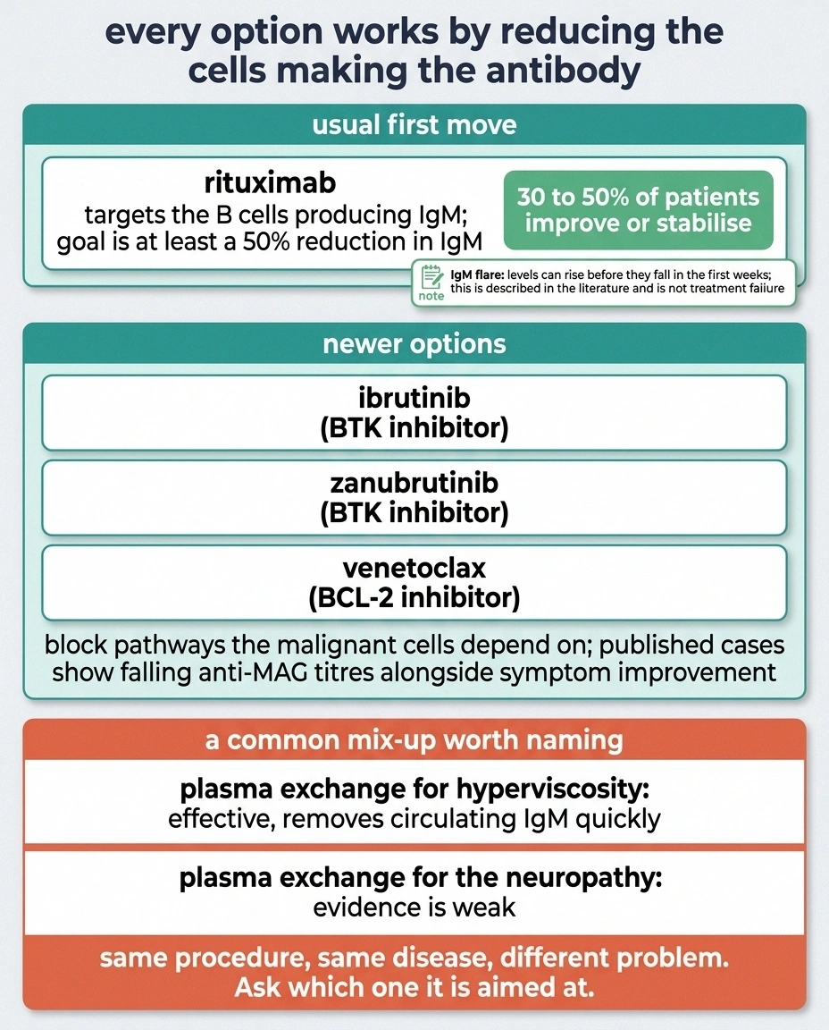
There is no drug that repairs anti-MAG damage directly. Every available treatment works by reducing the population of cells making the antibody, on the reasoning that less antibody eventually means less ongoing injury.
Five questions that change what you are told
- Which of the four mechanisms do you think is driving my neuropathy, and what is that based on?
- Has an anti-MAG titre been run, and what was it?
- If we start rituximab, what would a flare look like, and at what point should I call rather than wait?
- Are we treating to protect nerve function, to control blood counts, or both? The answer changes what success looks like.
- What objective measure will we repeat, and how often, so we can tell progression from a bad week?
Question four does the most work. A haematologist optimising blood counts and a patient optimising foot sensation can both be satisfied with the same appointment and mean entirely different things by it.
Rituximab is the usual first move. It targets the B cells producing the IgM, and the treatment goal is typically a reduction of at least half in the IgM level. Honest numbers matter here: roughly 30 to 50 percent of patients improve or stabilize. That is a real benefit and it is also a coin flip, and you deserve to hear it framed that way before you start.
Rituximab also has a documented quirk in this specific setting. A subset of patients experience an IgM flare in the weeks after starting, where the level rises before it falls, occasionally with a temporary worsening of symptoms. It has been described in a systematic review of the literature. It is a recognized phenomenon rather than a sign that treatment has failed, and knowing it exists in advance saves an enormous amount of alarm. Rarely, a more serious post-rituximab worsening resembling CIDP has been reported.
BTK inhibitors are the newer option. Ibrutinib and zanubrutinib block a signaling pathway the malignant cells depend on. Published cases document falling anti-MAG titers alongside symptom improvement, and these agents are now a standard part of the Waldenström armamentarium generally.
Venetoclax, a BCL-2 inhibitor, also lowers IgM and is used in some settings.
Combination chemoimmunotherapy is used when the disease itself needs treating for other reasons.
One point deserves emphasis because it is a frequent misunderstanding. Plasma exchange is genuinely used in Waldenström, and it works well, for hyperviscosity. Filtering the blood removes circulating IgM quickly and relieves the thick-blood emergency. The evidence that it helps the antibody-mediated neuropathy is weak. Same disease, same procedure, different problem. If plasma exchange is offered to you, it is worth asking which of the two it is aimed at.
The Decision Nobody Prepares You For
Waldenström is frequently managed with observation. If blood counts are fine and nothing is causing trouble, treating a slow lymphoma can do more harm than leaving it alone. That is a legitimate and well-supported approach.
Progressive neuropathy is one of the recognized reasons to break from it and start treatment anyway.
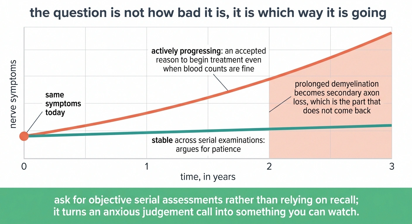
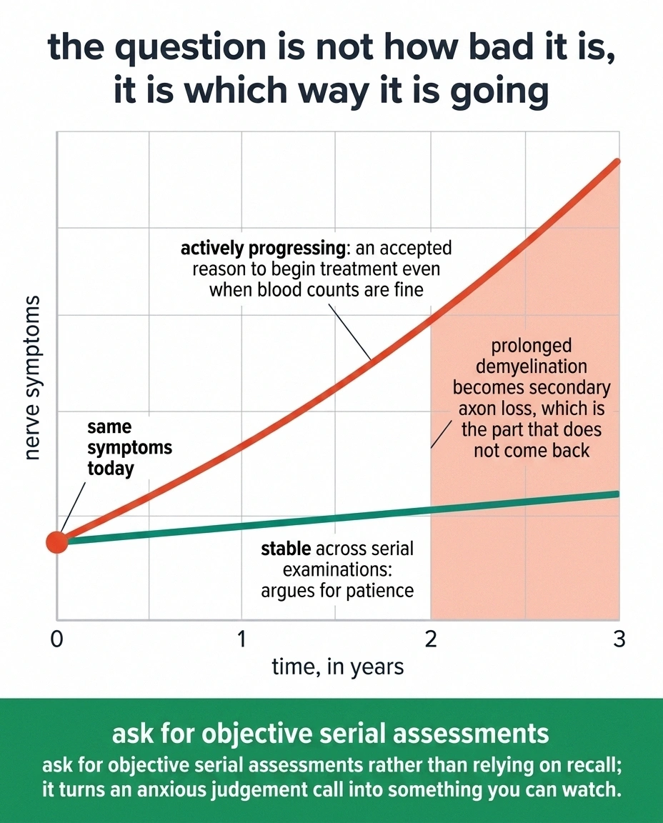
This puts some people in a genuinely uncomfortable position: being asked to accept treatment for a cancer that is not otherwise bothering them, in order to protect nerves that may or may not respond. There is no formula that resolves it. What tips the balance in practice is trajectory. Nerve damage that is actively progressing is worth intervening against early, because demyelination that persists long enough becomes secondary axon loss, and axon loss is the part that does not come back. Stable symptoms that have not changed in two years argue for patience.
If you are facing this decision, the useful question is not whether your neuropathy is bad. It is whether it is getting worse, how fast, and whether that pace is measured on serial examinations rather than on memory. Ask for objective tracking. It converts an anxious judgment call into something you can watch.
What Recovery Realistically Looks Like

Demyelinating damage can remyelinate. That is the good news, and it is the reason this neuropathy has better recovery potential than most.
What repairs, and on what clock
| What was damaged | Realistic timescale | What you notice first |
|---|---|---|
| Myelin, once the antibody attack is controlled | Months to a year or more | Steadier walking before the numbness changes |
| Axons lost underneath long-standing demyelination | Roughly 1 mm of regrowth per day, often incomplete | Little, and slowly; this is the part that sets the ceiling |
| Function, through balance and gait training | Weeks | Fewer near-falls, more confidence on stairs |
The third row is the only one measured in weeks, and it is the one most often left out of the treatment conversation entirely.
The qualifiers are real, though. Remyelination is slow, running over months to a year or more after the antibody attack is controlled. Long-standing anti-MAG neuropathy usually carries a component of axon loss underneath the demyelination, and that component recovers on the order of a millimeter a day of regrowth, if it recovers at all. Most people who improve do so partially. Stabilization, which means the slow decline simply stops, is a common and legitimate outcome that is easy to undervalue.
Practical function often improves more than the numbers do. Balance training, appropriate footwear, home lighting for the eyes-closed problem, and physical therapy aimed specifically at proprioceptive loss all move the needle on daily life whether or not the nerve conduction study changes. If gait and falls are the main problem, that work is not a consolation prize. It is the intervention with the shortest path to a better week.
And if you have not yet had a neurologist involved alongside your hematologist, get one. This is a disease that sits precisely on the border between two specialties, and the nerve half needs someone whose primary job is nerves. Our guide on when to see a neurologist for neuropathy covers how to make that referral happen.
Frequently Asked Questions
Can neuropathy be the first sign of Waldenström macroglobulinemia?
Yes, and it often is. About a quarter of people with Waldenström have nerve symptoms at the time of diagnosis, and for many of them the neuropathy is what prompted the blood work that found the IgM spike. This is one of the reasons an unexplained slowly progressive neuropathy warrants a serum protein electrophoresis.
What is anti-MAG neuropathy?
It is a neuropathy caused by IgM antibodies binding to myelin-associated glycoprotein, a structural component of the insulation around nerve fibers. It produces slow, symmetric, sensory-dominant symptoms concentrated in the feet and hands, usually with a hand tremor and significant balance trouble. It is confirmed with an anti-MAG antibody titer plus a characteristic pattern on nerve conduction testing.
If my IgM level comes down, will my feet get better?
Not reliably, and not quickly. The IgM level measures circulating protein, while symptoms depend on damage already done to myelin. Discordance between the two is common and well documented. Lowering IgM is aimed at stopping further injury, and any recovery follows separately and on a much slower timescale.
What is the difference between IgM MGUS neuropathy and Waldenström neuropathy?
The nerve disease can be identical. The difference is the size and behavior of the underlying B-cell clone. MGUS involves a small clone that does not meet criteria for lymphoma, while Waldenström involves enough marrow involvement and disease activity to be classified as a cancer. Treatment decisions differ, but the neuropathy is approached similarly.
Why do my hands shake?
A high-amplitude, low-frequency tremor in the arms and hands is a recognized feature of anti-MAG neuropathy rather than a separate condition. It comes from the same demyelination causing the numbness. Many people assume it is essential tremor or a normal part of aging, so it frequently goes unmentioned at appointments even though it supports the diagnosis.
Does plasma exchange help the neuropathy?
The evidence for plasma exchange in IgM-paraprotein neuropathy is weak. It is genuinely effective for hyperviscosity, which is a different complication of the same disease, and this causes frequent confusion. If plasma exchange is proposed, ask specifically whether it is being aimed at blood thickness or at nerve symptoms.
Should I treat the cancer just because of nerve symptoms?
Progressive neuropathy is an accepted reason to begin treatment in Waldenström even when blood counts are otherwise fine. The decision usually turns on whether symptoms are actively worsening and how quickly, since prolonged demyelination eventually produces permanent axon loss. Asking for serial objective assessments rather than relying on recall makes this decision far easier to make well.
Is Waldenström neuropathy reversible?
Partially, in many cases. Demyelination can repair once the antibody attack is controlled, though recovery takes months to a year or more. Longer-standing disease usually includes some axon loss, which recovers slowly and often incompletely. Stabilization rather than reversal is the most common realistic outcome, and it is a meaningful one.
About the Author