
If you’ve spent years being told your burning feet, flushing, racing heart, and inexplicable “flares” were anxiety, allergies, fibromyalgia, or just stress, I want you to know something before we go any further: you are not making it up. I’ve heard from too many people in our community who spent five, eight, sometimes twelve years bouncing between doctors before anyone even considered that two invisible problems might be talking to each other underneath the surface.
The two problems have complicated names. One is small fiber neuropathy, a form of nerve damage that standard nerve tests miss on purpose. The other is mast cell activation syndrome, or MCAS, an immune-cell disorder in which the tiny sentinel cells lining your skin, gut, and blood vessels fire off inflammation signals when they shouldn’t. Individually, each one is hard to diagnose. Together, they produce the exact grab-bag of symptoms that gets patients dismissed for years.
I’m Janet, and I’ve lived with neuropathy myself. I’m a patient advocate and community educator, not a doctor. What I want to do in this article is walk you through what researchers are learning about the emerging link between MCAS and small fiber neuropathy, what a real workup looks like, and what treatment paths clinicians who take this seriously are actually using. If any of this rings bells, print it out, bring it to your appointment, and ask.
What Small Fiber Neuropathy Is (and Why Standard Nerve Tests Miss It)
Your peripheral nervous system has two kinds of fibers. Large, thickly insulated ones carry vibration, position sense, and strong muscle signals. Small, sparsely insulated fibers carry pain, temperature, itch, and most of the autonomic signaling that runs your sweat glands, blood vessels, gut, and heart rate. The standard nerve conduction study and EMG your neurologist orders test the big fibers. They cannot see the small ones. A normal EMG doesn’t rule out neuropathy — it rules out large-fiber neuropathy.
Small fiber neuropathy, or SFN, is a distinct condition where those tiny fibers are damaged — dying back from the toes and feet upward, or patchy across the body. The classic presentation is burning, tingling, or electric-shock sensations in the feet, often worse at night, sometimes numbness, sometimes exquisite sensitivity where a bedsheet feels like sandpaper. Because small fibers also carry autonomic signals, SFN can produce sweating changes, dry eyes and mouth, GI dysmotility, and orthostatic symptoms alongside the pain.
The gold-standard test is a 3-millimeter skin punch biopsy from the distal calf. A pathologist counts nerve fibers per millimeter under a stain called PGP 9.5, giving you an “intraepidermal nerve fiber density,” or IENFD. A low IENFD against age-and-sex-matched normals is objective evidence that small fibers are gone — about the closest thing neurology has to a smoking gun for a disease that otherwise looks like “it’s all in your head.”
What Mast Cell Activation Syndrome Is (and Why It’s So Slippery to Diagnose)
Mast cells are immune cells that sit just under the skin, in the lining of your gut, in the airways, around blood vessels, and, importantly for this article, alongside nerves. Their job is to be the first line of defense — when they detect an invader, an injury, or an allergen, they degranulate, releasing a pharmacy of chemicals: histamine, tryptase, prostaglandins, leukotrienes, cytokines, nerve growth factor, and more. In an allergic person, this is what causes hives, wheezing, and anaphylaxis when they eat peanuts. In a healthy person going about their day, mast cells stay quietly on watch.
Mast cell activation syndrome is what happens when those cells fire off inappropriately — not in response to a real threat but to normal life: heat, cold, stress, a glass of wine, a meal, exercise, a hot shower, sometimes no identifiable trigger. The result is a shifting pattern of symptoms in two or more organ systems: skin (flushing, itching, hives), gut (nausea, cramping, diarrhea), cardiovascular (palpitations, blood pressure swings, presyncope), respiratory (throat swelling, wheezing), and neurologic (headaches, brain fog, nerve pain).
Mast cell activation syndrome (MCAS) and small fiber neuropathy (SFN) are separate conditions that often occur together. Mast cells sit right next to nerves and, when they fire off inappropriately, they can sensitize and eventually damage the small pain and autonomic fibers.
If you have burning feet, flushing, tachycardia, GI issues, and food or heat triggers that keep getting labeled “anxiety” or “fibromyalgia,” a proper MCAS + SFN workup with an allergist-immunologist and an autonomic neurologist is worth asking for.
MCAS is not the same as classic allergies (which are IgE-driven and reproducible) and it’s not the same as mastocytosis (a rarer clonal disorder where mast cells multiply abnormally). It’s a functional problem: the number of mast cells is roughly normal, but their behavior isn’t.
The diagnostic criteria are genuinely contested, and that matters for patients. One international group (Consensus-1, associated with Valent and colleagues) sets strict lab bars: typical mediator symptoms in two or more organ systems, a documented rise in tryptase during an event (at least 20% above baseline plus 2 ng/mL — the “20-plus-2” formula), and response to mast-cell-targeted therapy. A second group (Consensus-2, associated with Molderings, Afrin, and others) notes that tryptase is elevated in only a small minority of MCAS patients — perhaps 15 percent — and argues for a broader clinical approach that credits other mediators (urinary histamine metabolite, prostaglandin metabolites, leukotrienes) and clinical response. Under Consensus-1 a patient with obvious symptoms and normal tryptase may not “meet criteria.” Under Consensus-2 the same patient gets worked up and treated. That’s why so many patients are told they “don’t have MCAS” at one center and then told they do at the next.
The Overlap — When Mast Cells Sit Next to Nerves
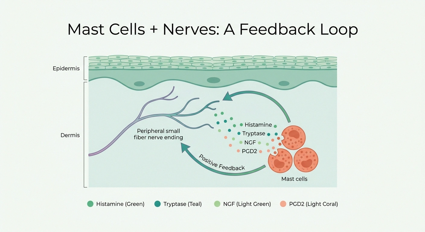
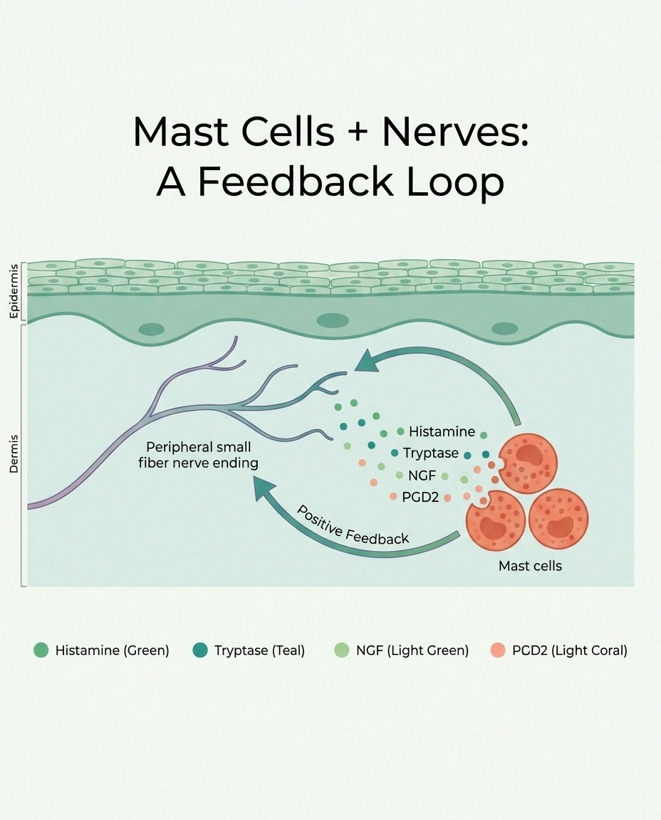
Here’s the piece that reframes everything. Mast cells and peripheral nerves are not strangers. In the skin, in the gut, around the dorsal root ganglia (the clusters of sensory nerve cell bodies just outside the spinal cord), mast cells physically cluster next to nerve endings. When they degranulate, the mediators they release don’t have to travel far — they bathe the nociceptor directly.
also have POTS
for MCAS
simultaneously
Based on 2025 review data in the hEDS patient population
Mast cells and pain nerves are physical neighbors. Laboratory studies show that mediators released during mast cell degranulation directly sensitize nociceptors:
- Histamine lowers the firing threshold of TRPV1 (the heat/capsaicin channel)
- Tryptase activates PAR2 on nerves, driving hyperexcitability
- Nerve growth factor (NGF) from mast cells sensitizes pain-carrying neurons
- Activated nerves release neuropeptides that feed back on mast cells — a positive feedback loop
Lab studies over the last decade have mapped out how this crosstalk sensitizes nerves. Histamine lowers the firing threshold of TRPV1, the heat-and-capsaicin channel pain fibers use, so warmth that used to feel neutral now feels burning. Tryptase activates a receptor on nerves called PAR2, driving hyperexcitability and CGRP release. Nerve growth factor from mast cells sensitizes pain-carrying neurons to heat. Prostaglandin D2, TNF, and IL-6 add to the inflammatory soup. Once nerves are activated, they release neuropeptides that go back to the mast cells and provoke more degranulation. A positive feedback loop takes hold.
What clinical researchers have been asking is whether, over time, this constant sensitization tips over into actual small fiber loss. Work by Novak, Doherty, and colleagues at Beth Israel Deaconess Medical Center, published in Annals of Allergy, Asthma & Immunology, reported that patients with hereditary alpha tryptasemia and idiopathic MCAS presenting with unexplained neurologic symptoms often showed objective small fiber loss on skin biopsy, autonomic testing abnormalities, and reduced cerebral blood flow during tilt-table testing. The suggestion isn’t that MCAS “causes” SFN in a simple linear way, but that a subset of patients carry both, and treating one may relieve the other.
This overlap sits inside what patients and clinicians have started calling “the trifecta”: mast cell activation syndrome, postural orthostatic tachycardia syndrome (POTS, a form of autonomic dysfunction), and hypermobile Ehlers-Danlos syndrome (hEDS). Review data from 2025 suggests that among women with hEDS, roughly a quarter carry all three; more than half have POTS; about a third meet criteria for MCAS. Small fiber neuropathy shows up as a common feature in this population and may be the mechanistic bridge tying the three together — a framework that researchers have started calling “autonomic neuro-immune axis dysfunction.”
The Symptom Picture Patients Actually Live With
People I’ve spoken with in the community describe an oddly consistent picture, even though on paper their charts read like unrelated organ systems.

There’s the neuropathy layer: burning feet, especially at night, that keeps them walking on cold tile at 2 a.m. just to feel something else. Electric-shock sensations up the legs. Numbness that isn’t quite numbness — more like the skin is thicker than it should be. Itching without a rash. Sweating that’s either too much or too little.
There’s the mast cell layer: flushing when they get warm, drink wine, eat leftovers, or take a hot shower. Hives that appear and disappear. Stomach pain and diarrhea a few hours after some meals but not others. Brain fog. Heart racing when they stand up. Anxiety that seems to come from nowhere and pass within an hour — a chemical anxiety, they’ll say, not the kind that comes from thoughts.
And there’s the flare pattern: symptoms that cluster and worsen after known triggers. Heat, emotional stress, menstrual cycles, illness, certain foods, alcohol (especially red wine), aged cheese, cured meats, leftovers, sun, exercise recovery. The pattern is idiosyncratic to each person but usually reproducible once they know to look.
Because the picture is so wide and so weird, most patients pick up diagnoses along the way that don’t quite fit: fibromyalgia, chronic fatigue, IBS, generalized anxiety, panic disorder, seasonal allergies, “atypical migraine.” A great many are told at some point that it’s psychosomatic. If you’ve been on that road, please read our piece on the mental health toll of being disbelieved — the medical gaslighting is its own injury on top of the physical one.
Getting Diagnosed — What a Real Workup Looks Like
Two separate workups have to happen, ideally coordinated. One targets MCAS, one targets SFN. Very few generalists know how to do both. This is a case where finding the right specialty center matters more than the specific test.

- Baseline serum tryptase (symptom-free day)
- Flare tryptase within 4 hours of an event (20% + 2 formula)
- 24-hr urine: N-methylhistamine, PGD2 metabolite, LTE4 (with drug washouts)
- Optional: chromogranin A, plasma heparin, HαT genetic test, gut biopsy
- 3-mm distal calf punch biopsy for IENFD
- QSART + tilt table + heart-rate variability
- Rule out diabetes, B12, thyroid, celiac, Sjögren’s, HIV, hep C
- Small fiber autoantibody panel (TS-HDS, FGFR3, plexin-D1)
The MCAS Workup
The starting labs are a baseline serum tryptase (drawn on a symptom-free day) and, if possible, a flare tryptase drawn within four hours of a documented symptomatic episode. The 20-plus-2 formula compares the two: if the flare value is at least 20% above baseline plus 2 ng/mL, that’s a mast cell activation event by Consensus-1 criteria. Most people never manage to get a lab drawn during a flare, and that’s part of why many MCAS patients have “normal” tryptase records.
Because tryptase misses so much, the workup usually adds a 24-hour urine mast cell mediator panel: N-methylhistamine (the metabolite of histamine), 2,3-dinor 11β-prostaglandin F2α (a mast-cell-specific PGD2 metabolite), and leukotriene E4 (LTE4). Ideally collected close to a flare and compared to a baseline. Certain medications interfere and need to be paused first: aspirin for two weeks and NSAIDs for 72 hours (they lower PGD2 metabolite), MAO inhibitors and aminoguanidine for three weeks (they raise histamine metabolite), and zileuton for 48 hours. Your clinician should walk you through the washout.
Supporting labs may include chromogranin A, plasma heparin, IgE, CBC with differential, and — for select patients — genetic testing for hereditary alpha tryptasemia (an extra copy of the TPSAB1 gene). Endoscopy or colonoscopy with mast cell staining of gut biopsies is sometimes added when GI symptoms dominate.
The SFN Workup
The definitive test is a 3-mm skin punch biopsy at the distal calf (10 cm above the lateral malleolus), sent to a lab that specializes in PGP 9.5 staining and IENFD counting. Some centers also biopsy the thigh for a proximal comparison. Results come back as fibers per millimeter with a normative comparison. Alongside the biopsy, an autonomic testing battery — QSART (quantitative sudomotor axon reflex test), tilt table, heart-rate variability, Valsalva — evaluates the autonomic small fibers. Our detailed guide on how neuropathy is diagnosed walks through what each of these tests actually feels like and what the results mean.
A parallel workup rules out or identifies other SFN causes: diabetes and prediabetes (fasting glucose, HbA1c, oral glucose tolerance test), B12 and folate, thyroid, celiac serology, HIV and hepatitis C, Sjögren’s antibodies (SSA/SSB), ANA, and — increasingly — small fiber autoantibody panels including TS-HDS, FGFR3, and plexin-D1. When every cause comes back negative and the biopsy still shows fiber loss, the SFN gets labeled idiopathic — but with an MCAS diagnosis in the same chart, “idiopathic” often becomes “presumed dysimmune.”
Treatment — Two Problems, Two Layers of Management
Here’s the mindset shift that helps most: this is not one disease with one drug. It’s two overlapping problems that both need attention, on separate tracks that inform each other. When the mast cell layer calms down, some patients see nerve pain improve too. When it doesn’t, nerve pain gets treated on its own terms.

SFN pain (duloxetine, gabapentin, pregabalin) is treated on a separate track.
MCAS diagnosis and treatment belong to allergy/immunology specialists (ideally MCAS-literate). SFN workup and neuropathic pain management belong to a neurologist with autonomic and small-fiber subspecialty training.
Do not start or stop antihistamines, cromolyn, ketotifen, montelukast, low-dose naltrexone, duloxetine, or gabapentinoids on your own. These medications interact with each other, with your existing prescriptions, and with the workup labs themselves. Bring this article to your appointment — don’t use it in place of one.
The Mast Cell Layer
The standard first step is combined H1 and H2 antihistamines, often at doses higher than the over-the-counter labels suggest. Non-sedating H1 blockers (cetirizine, loratadine, fexofenadine) are the daytime backbone. Sedating H1 options (hydroxyzine, diphenhydramine) can be added at night. H2 blockers (famotidine, cimetidine) sit on mast cells themselves and add stabilization — they’re not just for stomach acid. Both together beats either alone in most patients.
If antihistamines aren’t enough, the next step is a mast cell stabilizer. Cromolyn sodium is poorly absorbed, which is actually useful — it concentrates in the GI tract where many symptoms live. Dosing is usually 200 mg four times a day, 30 minutes before meals and at bedtime, and it takes four to eight weeks to reach full effect. Ketotifen is a hybrid — stabilizer plus H1 blocker — and unlike cromolyn it’s absorbed systemically, so it can quiet mast cells throughout the body.
The leukotriene inhibitor montelukast is often added to cover the leukotriene arm of mast cell mediators. Low-dose naltrexone (LDN), typically 1.5 to 4.5 mg at bedtime, is being explored by MCAS-literate clinicians as an immune modulator that appears to reduce mast cell reactivity in some patients — the evidence base is early but growing.
Non-drug measures matter. A low-histamine trial for four to six weeks (aged cheese, cured meats, fermented foods, leftovers, alcohol, and known personal triggers) is often diagnostic in itself. Long-term restriction should run under a dietitian to avoid nutritional shortfalls. A written trigger and symptom diary is worth more than most people expect — patterns show up on paper that don’t in memory.
Certain medications are known mast cell degranulators and should be avoided or used with pre-medication: many opioids (especially morphine and codeine), some NSAIDs, fluoroquinolone antibiotics, rapidly infused vancomycin, and iodinated radiocontrast. Always tell any new prescriber you have suspected MCAS.
The Nerve Pain Layer
The SFN pain layer is treated separately with neuropathic pain drugs. Duloxetine, an SNRI, is often first-line and has the added benefit of helping mood and sleep. Gabapentin and pregabalin are the anticonvulsant options — sedating, but effective for the electric and burning quality of pain. Topical lidocaine 5% patches and the specialty-clinic capsaicin 8% patch are useful for patchy or focal pain. Alpha-lipoic acid and other supplements are sometimes tried, though the evidence for SFN specifically is thinner than for diabetic neuropathy.
The Autoimmune Question and What Research Is Testing Now
One of the most important shifts in the SFN world right now is the recognition that a meaningful minority of “idiopathic” cases are actually dysimmune — the immune system is targeting the small fibers, through autoantibodies against nerve-surface proteins or inflammatory cells around nerves. The best-known autoantibodies now on specialty panels are TS-HDS (trisulfated heparin disaccharide), FGFR3, and plexin-D1. Not every SFN patient has them, but for those who do, the diagnosis moves from “unexplained” to “immune-mediated” — which opens different treatment doors.

For select autoantibody-positive patients at specialty centers, intravenous immunoglobulin (IVIG) and other immunomodulators are being trialed. This is not standard of care — IVIG is expensive, has real side effects, and access usually depends on documented autoantibodies plus a specialist committed to the workup. The relevant point for MCAS-SFN patients is that this is a live research frontier, and clinicians are actively studying whether calming the mast cell layer, treating an underlying autoantibody, or both produces the best outcomes.
There’s also the genetic side. Hereditary alpha tryptasemia (HαT) — an extra copy of the TPSAB1 gene — is recognized as a genetic driver in a subset of MCAS-spectrum patients and correlates with elevated baseline tryptase. Testing can identify it, and changes how tryptase results are read. If MCAS runs in your family, mention it. If your baseline tryptase is unusually high without signs of systemic mastocytosis, ask specifically about HαT.
Living Well With MCAS-SFN
Even with the best specialists and the right medications, this is a condition you learn to live around, not one you make disappear. The patients I know who are doing well share a few habits.

They keep a trigger diary and know their personal high-risk foods and stressors well enough to plan around them. They pace: a good day is not the day to do everything they’ve missed, because the crash is real. They advocate: they carry a printed list of their diagnoses and mast-cell-degranulating drugs to avoid, so an ER visit doesn’t turn into a flare. They find a community — patient groups for MCAS, POTS, hEDS, and SFN are unusually well-informed because these are diseases where patients have often out-researched their doctors.
And they protect their mental health. Being told for years that it’s anxiety when it isn’t is a wound in its own right, one that deserves attention from a therapist who understands chronic illness. If your workup so far has come back “normal” and you’re still symptomatic, the diagnosis may just not be found yet. Read our piece on idiopathic neuropathy — sometimes “idiopathic” is a placeholder that gets rewritten later once someone asks the right questions.
Frequently Asked Questions
Can MCAS-related small fiber neuropathy be reversed?
Research is still evolving. Some patients report significant improvement in nerve symptoms when the mast cell layer is well controlled, which fits the model that ongoing mediator release keeps nerves sensitized. Others have measurable fiber loss that doesn’t fully rebuild even with excellent MCAS management. The safest framing is that symptom improvement is realistic for many patients, particularly early on; full structural reversal is not a promise anyone can make honestly. Working with a neurologist and an MCAS-literate specialist together gives you the best shot.
How long does diagnosis usually take?
In community surveys, patients often report five to ten years or more between symptom onset and formal MCAS diagnosis. SFN diagnosis is faster once someone thinks to order a skin biopsy, but many patients never get referred because their EMG was normal and the neurologist stopped there. The fastest path is usually a referral to an academic autonomic clinic, a mast cell specialty center, or an allergist and immunologist who explicitly treats MCAS.
My tryptase is normal — does that rule out MCAS?
No. Elevated tryptase is present in only a small minority of MCAS patients, roughly 15 percent in some published estimates. Under the broader Consensus-2 criteria championed by Molderings, Afrin, and others, a normal tryptase with typical mediator symptoms, positive urinary metabolites, and a good response to mast-cell-directed therapy is enough. If a clinician tells you your tryptase is normal and therefore you don’t have MCAS, that reflects one interpretation of the diagnostic literature. It’s reasonable to seek a second opinion from an MCAS-literate specialist.
Do I have to follow a low-histamine diet forever?
Usually not. A four to six week trial can be very useful diagnostically and can bring symptoms down enough that other treatments work better. But indefinite tight restriction can cause nutritional shortfalls and social stress that outweigh the benefit. Most experienced clinicians re-introduce foods gradually once medication is optimized, keeping only the personal triggers that reliably cause flares. This is a good area to work with a registered dietitian who has MCAS experience.
Which specialist do I actually need to see?
The best case is a coordinated team: an allergist or immunologist who lists MCAS explicitly in their scope (not every allergist does), plus a neurologist with a small fiber and autonomic subspecialty at an academic center with a punch biopsy program. If you’re near a POTS or dysautonomia center, that’s often the best single starting point because those clinics tend to be fluent in the whole overlap.
Is this the same as fibromyalgia?
No, though the symptom overlap is real and many patients carry a fibromyalgia label at some point. Fibromyalgia is a clinical syndrome defined by widespread pain and tender points without a specific pathologic finding. MCAS-SFN is a distinct entity with objective abnormalities — low IENFD on biopsy, elevated urinary mediators, autonomic testing changes. Some researchers now suspect a portion of what has been called fibromyalgia is actually undiagnosed small fiber neuropathy, sometimes with a mast-cell component. If your fibromyalgia diagnosis has never been re-examined with a skin biopsy or mediator panel, it may be worth asking.
Can supplements alone manage this?
Supplements can play a supportive role but they are not a substitute for a proper workup and medical management. Quercetin, vitamin C, luteolin, and DAO enzyme (diamine oxidase) are commonly discussed in MCAS communities as mast cell support. Alpha-lipoic acid has some evidence for diabetic neuropathy and is often tried in SFN. None of these replace antihistamines, stabilizers, or neuropathic pain medication in a patient with real disease activity, and none should be started without checking for interactions with your existing medications.
What if my workup keeps coming back negative but I feel worse?
You are not making it up. Negative early workups are frustratingly common because these tests are sensitive to timing, drug washouts, and where the sample is drawn. If everything so far has come back clean, the next moves worth asking about are a properly timed flare-day mediator panel with washouts, a skin punch biopsy for IENFD (if you’ve only had EMG so far), autoantibody panels for dysimmune SFN, and a referral to an MCAS-literate specialist rather than a generalist. Protect your mental health through this — being disbelieved is its own injury and it deserves care.
About the Author