
Most neuropathy arrives as a gradient. It starts at the toes because those nerve fibers are the longest, it spreads upward slowly and evenly, and by the time it reaches the ankles it has usually done the same on both sides. The pattern is so consistent that neurologists call it length-dependent, and it fits diabetes, chemotherapy, alcohol, B12 deficiency, and most of the other common causes.
Mononeuritis multiplex does not do that. It arrives as a list.
One wrist stops working over a weekend. Three weeks later a foot drops. A month after that, a patch of numbness appears on the opposite thigh that has nothing to do with either. Each event is separate, each involves a different named nerve, and each tends to hurt considerably on the way in.
That sequence is not a strange variant of ordinary neuropathy. It is a different mechanism producing a different disease, and recognising the pattern is most of the work. It also happens to be one of the very few neuropathy presentations where speed genuinely changes the outcome.
The Pattern Is the Diagnosis
The formal definition is damage in two or more separate, named peripheral nerve distributions. Not two toes. Not both feet. Two distinct nerves, in unrelated parts of the body, each with its own territory of skin and its own set of muscles.
Three questions a neurologist is really asking
01
Did it come on over hours, or over years?
Hours to days points at something closing off a blood supply. Years points at a metabolic cause acting slowly on every nerve at once.
02
Is it in one nerve's territory, or a gradient?
A crisp border that follows a named nerve is different from numbness that fades gradually as it climbs the leg.
03
Has this happened before, somewhere unrelated?
This is the question people answer badly, because the earlier event was filed away as a separate minor problem and never mentioned.
Question three is the one worth preparing for. Go back through the last two years for anything that was dismissed at the time: a numb patch that resolved, a weak grip for a fortnight, a foot that scuffed. Bring the dates.
You will see it written both as mononeuritis multiplex and as mononeuropathy multiplex. Both names are in current use and mean the same thing.
The classic behaviour is stepwise. One nerve fails, then after an interval of days or weeks another fails somewhere else, then possibly another. Between events things can be stable, which is part of why people wait. A single sudden wrist drop looks like a pinched nerve from sleeping awkwardly. It is only when the second event happens that the shape becomes visible.
There is an important complication in that. If enough nerves are involved over enough time, the individual patches merge and the picture starts to look symmetric. A late presentation can be mistaken for ordinary polyneuropathy, and the history becomes the only thing that reveals what actually happened. This is one of the reasons it helps to understand the distinction between polyneuropathy and mononeuropathy, because the words describe patterns rather than severity, and the pattern is doing the diagnostic work.
Why It Arrives So Suddenly
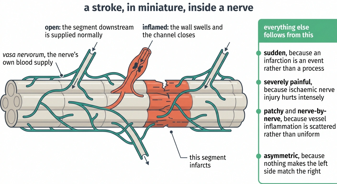
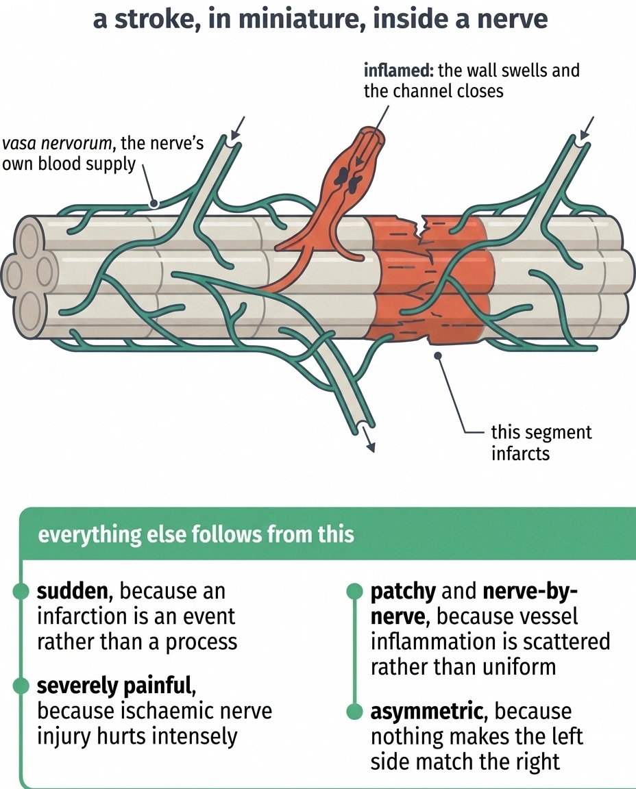
Nerves need a blood supply, and they have their own dedicated one. Running along and within every peripheral nerve trunk is a network of very small arteries called the vasa nervorum, literally the vessels of the nerves. They are tiny, and each one supplies a limited stretch of a single nerve.
When those vessels become inflamed, the walls thicken and swell and the channel through them closes. The segment of nerve downstream loses its blood supply and infarcts. This is a stroke, in miniature, in a nerve.
Every characteristic feature follows from that one event.
The suddenness follows, because an infarction is an event rather than a process. The severe pain follows, because ischemic nerve injury is intensely painful. The patchy nerve-by-nerve distribution follows, because which nerve is affected depends entirely on which small vessel happened to close, and inflammation in blood vessels is scattered rather than uniform. And the asymmetry follows for the same reason: there is no mechanism making the left side match the right.
Contrast that with diabetic neuropathy, where a metabolic insult acts on all nerves at once and the longest fibers simply fail first. One is a gradient. The other is a series of separate accidents.
The Causes, and Which One Leads
Vasculitis is the most common cause, and this shapes everything about how the condition is investigated.
What each test on the panel is actually hunting for
The blood panel looks scattershot on the request form. It is not. Each item maps to a specific candidate, and knowing which is which makes the results conversation far shorter.
ANCA
Granulomatosis with polyangiitis, eosinophilic granulomatosis with polyangiitis
ANA, rheumatoid factor
Lupus, rheumatoid vasculitis, Sjogren's syndrome
Cryoglobulins
Cryoglobulinemic vasculitis, often alongside hepatitis C
Hepatitis B and C, HIV
Infection-driven vasculitis and direct viral nerve involvement
ESR and CRP
Whether an inflammatory process is running at all, and how hot
HbA1c
Diabetes, as a contributor or a coexisting condition
Serum protein electrophoresis
Amyloid deposition and paraprotein-driven disease
Chest imaging
Sarcoidosis, and lung involvement in systemic vasculitis
A normal inflammatory marker does not clear vasculitis. Nonsystemic vasculitic neuropathy is confined to nerve and can run with an entirely unremarkable ESR and CRP.
The vasculitis can be part of a systemic disease. Polyarteritis nodosa, eosinophilic granulomatosis with polyangiitis (which older sources call Churg-Strauss), granulomatosis with polyangiitis, rheumatoid vasculitis, lupus, Sjögren's syndrome, and cryoglobulinemia can all produce it. It can also occur as nonsystemic vasculitic neuropathy, where the inflammation is confined to nerve and nothing else in the body is affected. Our overview of vasculitic neuropathy covers that family in more depth.
The other causes matter too, and the list is worth knowing because it determines which blood tests get ordered:
Diabetes. Usually through entrapment, but occasionally through genuine nerve infarction from occlusion of the vasa nervorum. Diabetes is common enough that it can coexist with something else, which occasionally delays the right diagnosis.
Sarcoidosis. Granulomatous inflammation can involve nerve in a patchy multifocal way. Sarcoid neuropathy takes several forms and this is one of them.
Infections. Leprosy remains the leading infectious cause worldwide. Hepatitis C, hepatitis B, HIV and Lyme disease all appear in the literature.
Infiltration. Amyloid deposits and lymphoma cells can both produce a multifocal pattern.
The hereditary mimic. Hereditary neuropathy with liability to pressure palsies produces repeated focal nerve failures at pressure points, often after trivial compression, in a person with a family history of the same. It looks superficially similar and is treated completely differently.
The motor mimic. Multifocal motor neuropathy produces multifocal weakness with no sensory loss, and it responds to immunoglobulin rather than immunosuppression.
The Carpal Tunnel Trap
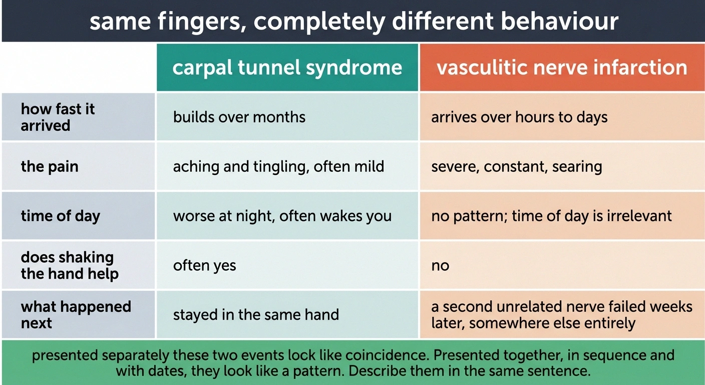
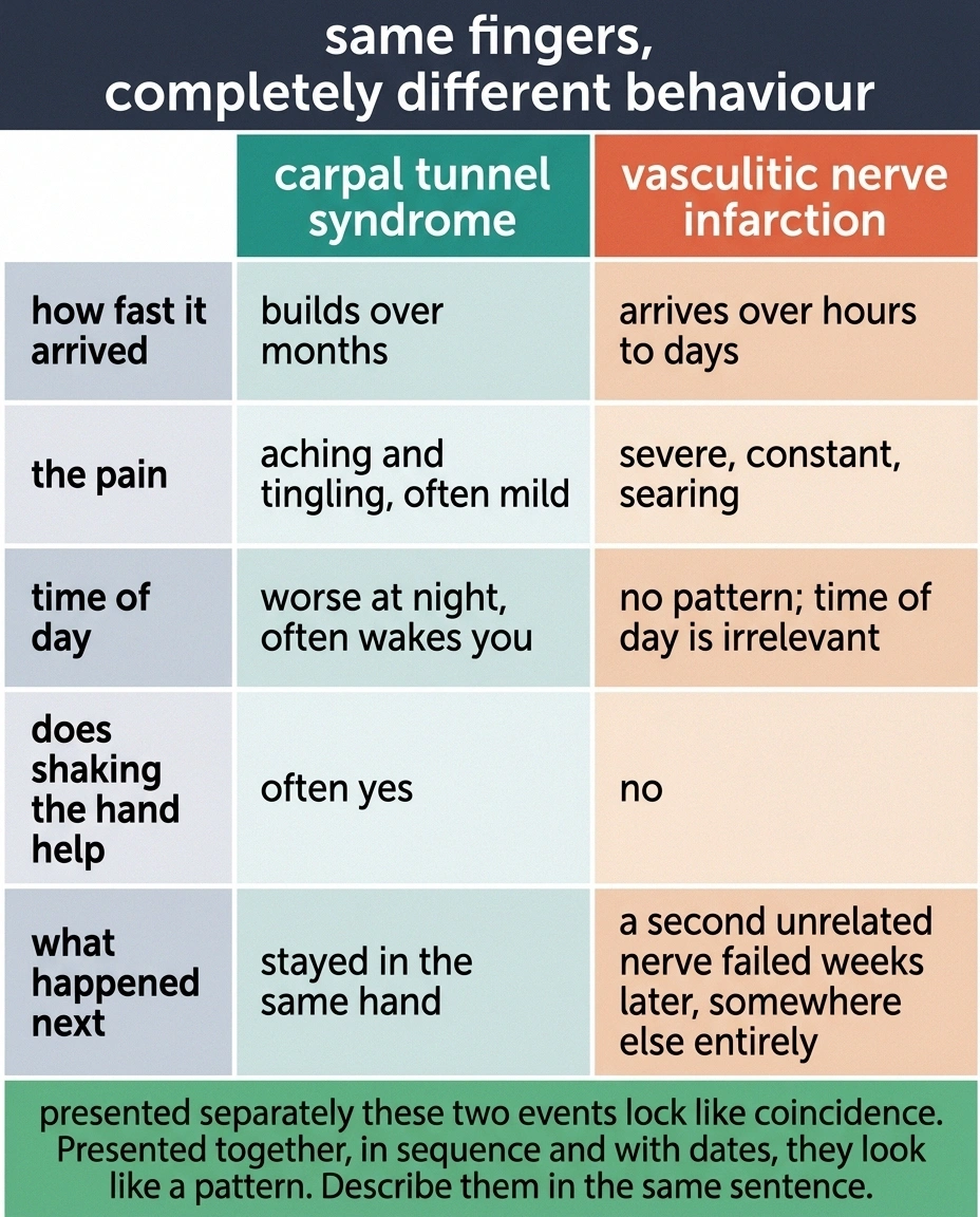
There is a documented pattern of vasculitic mononeuritis multiplex being misdiagnosed as carpal tunnel syndrome, and understanding why is genuinely useful.
The median nerve at the wrist is a common site. When it fails first, the symptoms land in the thumb, index and middle fingers, which is exactly the carpal tunnel distribution. Carpal tunnel syndrome is enormously common. Vasculitic neuropathy is not. The reasonable first guess is the common thing.
What separates them is behaviour rather than location. Carpal tunnel builds over months, is worse at night, is often relieved by shaking the hand, and does not usually hurt in a searing way. Vasculitic nerve infarction arrives over hours to days, hurts severely and constantly, and does not care what time it is.
And then a second nerve goes, somewhere with no anatomical relationship to the first. If you have been told you have carpal tunnel and something has since happened in a foot or the other arm, those two events belong in the same sentence when you describe them. Presented separately they look like coincidence. Presented together they look like a pattern, and our guide to what else causes tingling in the hands covers the wider set of conditions that get confused with each other here.
How It Gets Confirmed

Three layers of investigation, usually in this order.
Electrodiagnostic testing. Nerve conduction studies and EMG map which nerves are affected and how. The expected finding is an asymmetric axonal pattern: reduced amplitudes of the motor and sensory responses, with conduction velocities that are normal or only mildly slowed. That combination points at the axon dying rather than the insulation being stripped, which is what infarction produces. Testing multiple nerves in multiple limbs is what reveals the multifocal distribution, so a study confined to one arm can miss the whole point.
Timing catches people out here. After a nerve infarcts, the fibers below the injury need roughly seven to ten days to degenerate far enough for nerve conduction studies to register the loss. A study run within the first few days of a sudden foot drop can look much milder than the injury plainly is, occasionally close to normal. If your test happened very early and the result did not match how bad things clearly were, that is a known limitation of the timing rather than reassurance, and a repeat study a few weeks later is the standard answer.
Blood work. The panel is aimed at finding the cause: ANCA, ANA, rheumatoid factor, cryoglobulins, hepatitis B and C serologies, HIV, ESR and CRP, HbA1c, and serum protein electrophoresis. It is a broad net, deliberately.
Tissue. Nerve biopsy, usually of the sural nerve at the ankle and often combined with a sample of adjacent muscle, is the confirmatory test.
Why a Biopsy Is Worth It Here
Nerve biopsy is rarely indicated in ordinary neuropathy. It is a real procedure, it leaves a permanent patch of numbness on the outer foot, and in most cases it does not change what happens next.
Four findings, four different treatment paths
This is what makes the procedure worth its permanent patch of numbness on the outer foot. Each result sends treatment somewhere different, and guessing between them is not a safe alternative.
- Inflammation and destruction in the vessel walls
- Confirms vasculitic neuropathy. Leads to corticosteroids, often with cyclophosphamide or rituximab.
- Amyloid deposits
- Sends the investigation toward the protein producing it, and toward an entirely different set of therapies. Immunosuppression would not have helped.
- Granulomas
- Points at sarcoidosis and prompts a search for involvement elsewhere, particularly the lungs.
- Malignant cells within the nerve
- Indicates infiltration rather than inflammation, which is treated as cancer in the nerve and not as an autoimmune process.
Ask whether muscle is being sampled alongside the nerve. The same process is frequently easier to catch in muscle tissue, and taking both in one procedure raises the chance of an answer.
Here the calculation is different, and the reason is worth stating plainly: the findings it looks for are treatable ones. Inflammation in the walls of the vasa nervorum, evidence of vessel destruction, abnormal deposits such as amyloid, granulomas suggesting sarcoidosis. Each of those points at a specific treatment. Combining nerve with muscle raises the yield, because the same inflammatory process is often easier to catch in muscle tissue.
The other half of the calculation is that the treatments involved carry real risks of their own. Cyclophosphamide and long-course corticosteroids are not things to start on a hunch. Tissue confirmation is what justifies them.
Treatment Moves Quickly
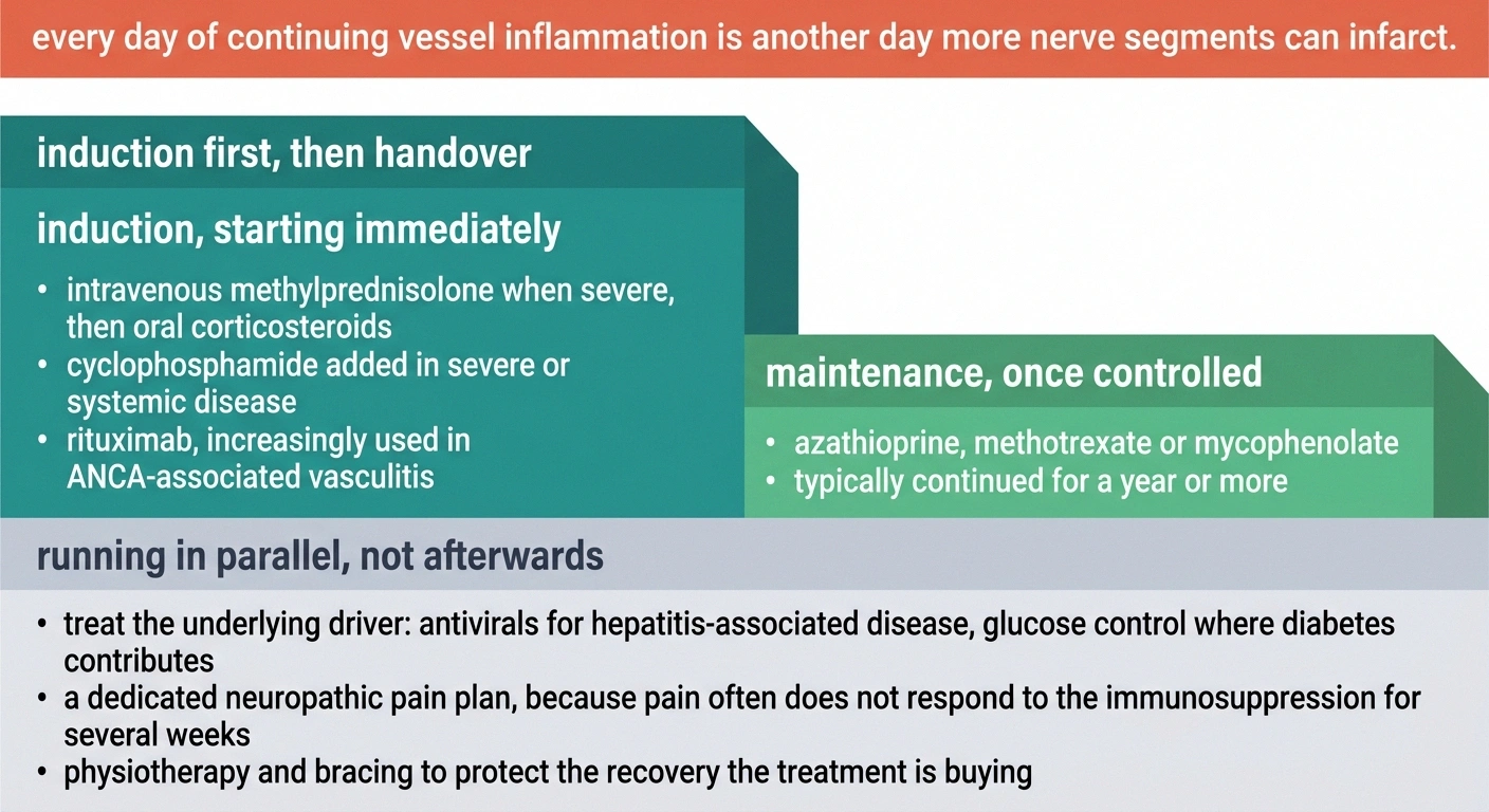
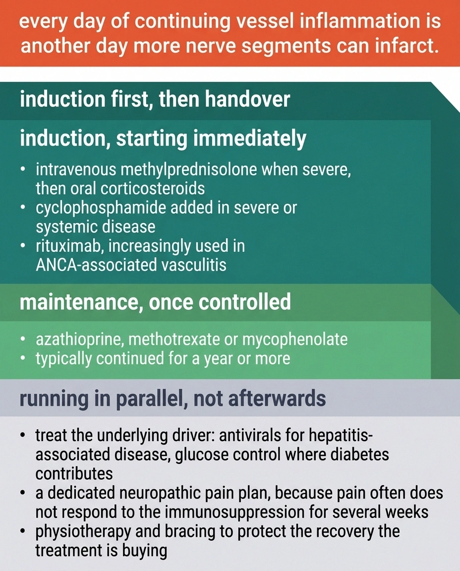
Once vasculitis is established, treatment is immunosuppression and it usually starts before every last result is back.
Corticosteroids are first-line, given intravenously as methylprednisolone when the presentation is severe, then orally. Cyclophosphamide is added as part of first-line therapy in severe or systemic disease. Rituximab is increasingly used, particularly in ANCA-associated vasculitis. Once things are controlled, maintenance shifts to a gentler agent such as azathioprine, methotrexate or mycophenolate, often for a year or more.
Where an underlying driver exists, treating it is the actual therapy. Antiviral treatment for hepatitis-associated disease, glucose control where diabetes is contributing, and so on.
Two practical things about this phase are worth expecting in advance. The steroid doses used at the start are high, and the side effects that come with them are substantial: sleep disruption, appetite and mood changes, raised blood sugar, and a flushed puffiness in the face. All of that is temporary and it is the price of stopping the vessel inflammation quickly. Separately, the nerve pain often does not respond to the immunosuppression for several weeks, because controlling inflammation and repairing an already infarcted nerve are different jobs. A dedicated pain plan running alongside the disease treatment is not an afterthought, and neuropathic pain generally responds better to nerve-specific agents than to ordinary painkillers.
The urgency is not administrative. Every day that inflamed vessels stay inflamed is another day more nerve segments can infarct, and infarcted nerve does not come back quickly or completely.
Recovery Runs on Nerve Time
The prognosis is genuinely good when the cause is found and treated. Full recovery happens. The published case follow-ups describe meaningful gains in strength and mobility at six months on appropriate treatment.
The timescale is the hard part, and it is set by biology rather than by effort.
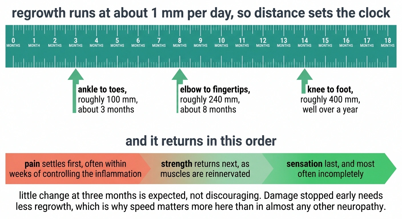
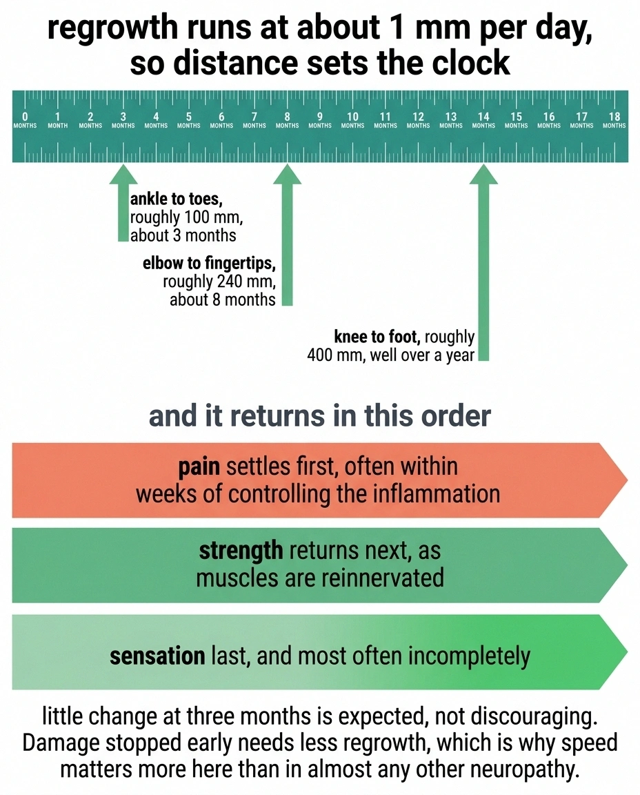
Regenerating axons grow at roughly one millimetre per day. That number sounds abstract until you apply it. A nerve infarcted near the knee has something like 400 millimetres to cover before it reaches the foot, which is well over a year of continuous regrowth. A nerve damaged at the elbow reaches the fingertips in perhaps eight months.
Two consequences follow. The first is that the absence of improvement at three months means very little, and people are often told to expect progress on a timescale the biology cannot deliver. The second is that damage stopped early requires less regrowth, which is the concrete reason speed matters more here than in almost any other neuropathy.
Recovery also tends to arrive in a particular order. Pain settles first, often within weeks of controlling the inflammation. Strength returns next as muscles are reinnervated. Sensation is usually last and frequently the most incomplete.
Physical therapy through that period is not optional filler. A muscle that has been denervated for a year has to be kept from contracting into a fixed position while it waits, and a foot drop needs bracing so you can walk safely in the meantime. That work protects the recovery the treatment is buying.
The One Section to Act On

Most neuropathy content ends with a gentle suggestion to raise it at your next appointment. This condition is the exception, so here it is directly.
Where to direct the call, and what each route can do today
A nerve that fails suddenly, painfully, and on its own, followed by a second unrelated nerve failing days or weeks later, is a presentation that warrants urgent medical assessment rather than a routine booking. That is especially true alongside any of the following: unexplained weight loss, fever, night sweats, a rash of small raised purple spots, joint swelling, blood in the urine, or new breathlessness. Those suggest systemic vasculitis, which can affect kidneys, lungs and gut as well as nerve, and it is treated as an emergency.
What to say when you call matters more than most people realise. “My hand went numb” is a low-priority complaint in any triage system. “I lost the use of my left wrist three weeks ago and now my right foot is dropping, and it was very painful both times” describes a multifocal, stepwise, painful nerve presentation, and it moves. Give the sequence, the dates, and the pain. If you are unsure whether the pattern fits, our guide on when neuropathy needs a neurologist sets out the wider criteria.
Frequently Asked Questions
What is mononeuritis multiplex?
It is damage to two or more separate, named peripheral nerves in unrelated parts of the body, usually arriving in a stepwise sequence with days or weeks between events. It is also called mononeuropathy multiplex. The mechanism is typically loss of blood supply to segments of nerve, most often because inflammation has closed the small vessels that feed them.
How is it different from regular peripheral neuropathy?
Ordinary peripheral neuropathy is length-dependent and symmetric. It starts in both feet because the longest fibers fail first, and it progresses gradually and evenly. Mononeuritis multiplex is asymmetric, patchy, sudden, and usually painful, affecting individual named nerves rather than a gradient. Over a long period the patches can merge and start to resemble a symmetric neuropathy, so the history of how it began is often the clearest evidence.
Is mononeuritis multiplex an emergency?
It warrants urgent assessment rather than a routine appointment. The most common cause is vasculitis, which can involve kidneys, lungs and gut alongside nerve, and ongoing vessel inflammation continues to infarct nerve segments while it goes untreated. Sudden painful loss of function in one nerve followed by another elsewhere, particularly with fever, weight loss, rash, joint swelling or blood in the urine, should be assessed promptly.
What causes it?
Vasculitis is the leading cause, either as part of systemic disease such as polyarteritis nodosa, eosinophilic granulomatosis with polyangiitis, granulomatosis with polyangiitis, lupus, rheumatoid vasculitis, Sjögren's syndrome or cryoglobulinemia, or as nonsystemic vasculitis confined to nerve. Other causes include diabetes, sarcoidosis, leprosy, hepatitis B and C, HIV, Lyme disease, amyloidosis and lymphoma. Hereditary neuropathy with liability to pressure palsies and multifocal motor neuropathy are important mimics.
What tests diagnose it?
Nerve conduction studies and EMG show an asymmetric axonal pattern, meaning reduced response amplitudes with normal or only mildly slowed conduction velocities, sampled across several nerves in more than one limb. Blood work looks for the cause and typically includes ANCA, ANA, rheumatoid factor, cryoglobulins, hepatitis serologies, HIV, inflammatory markers, HbA1c and serum protein electrophoresis. Nerve biopsy, often combined with muscle, provides confirmation.
Do I really need a nerve biopsy?
Often yes, which is unusual for neuropathy. Biopsy looks for findings that are directly treatable, including inflammation in the small vessels supplying nerve, amyloid deposits and granulomas. It also justifies treatments such as cyclophosphamide and prolonged corticosteroids, which carry meaningful risks of their own and are not started lightly. Sampling nerve together with adjacent muscle improves the diagnostic yield.
Can it be reversed?
Substantially, in many cases, provided the underlying cause is identified and treated. Published follow-up describes significant gains in strength and mobility by six months on appropriate immunosuppression. The extent of recovery depends heavily on how much nerve was lost before treatment began.
How long does recovery take?
Months to years. Regenerating nerve fibers grow at roughly one millimetre per day, so a nerve damaged near the knee needs well over a year to reach the foot. Pain usually settles first, strength returns as muscles are reinnervated, and sensation recovers last and least completely. Little change at three months is expected rather than discouraging.
Can diabetes cause mononeuritis multiplex?
Yes. Diabetic mononeuropathies more often result from entrapment, but genuine nerve infarction from occlusion of the small vessels supplying nerve does occur. Because diabetes is common, it can also coexist with vasculitis, so a multifocal stepwise presentation in someone with diabetes still warrants a search for another cause rather than being attributed to the diabetes by default.
About the Author