A reader named Priya wrote to me last month with a question I’ve heard some version of at least fifty times: “They can MRI my knee. Why can’t they just MRI my nerves and see what’s wrong?”
The honest answer is that they can. There’s a scan for exactly that, it’s called MR neurography, and it has been quietly getting better for twenty-five years.
The less satisfying half of the answer is that for most people reading this, it wouldn’t help. Not because the technology is weak — it’s built to answer a completely different question than the one most of us are actually asking.
So let’s sort out which question is yours. That’s the part nobody explains, and it’s the difference between a scan that finally cracks your case open and a scan that costs you a deductible and tells you nothing.
What MR Neurography Actually Is
A standard MRI of your lower back is designed to show you bones, discs, and the spaces between them. Nerves are in there somewhere, sure, but they’re not the point of the picture — think of it as a group photo taken to capture the building behind everyone.
MR neurography flips that. It uses specialized sequences that suppress the signal from fat and flowing blood so the nerve itself becomes the brightest, clearest thing on the image. A skilled radiologist can then look at an individual nerve trunk and evaluate its caliber, its internal fascicular pattern, and whether its signal is abnormally bright, which usually signals inflammation, injury, or compression.
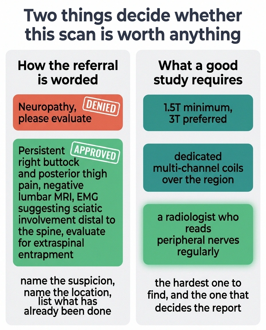
The technical requirements are real. You need a scanner of at least 1.5 Tesla, and 3T is strongly preferred, with dedicated multi-channel coils positioned over the body part in question. You also need a radiologist who reads peripheral nerves regularly. That second requirement is the harder one to find, and it’s the reason a scan done at the wrong place can come back as an unhelpful shrug even when the equipment was fine.
The Sorting Question: Is Your Problem One Nerve or All of Them?
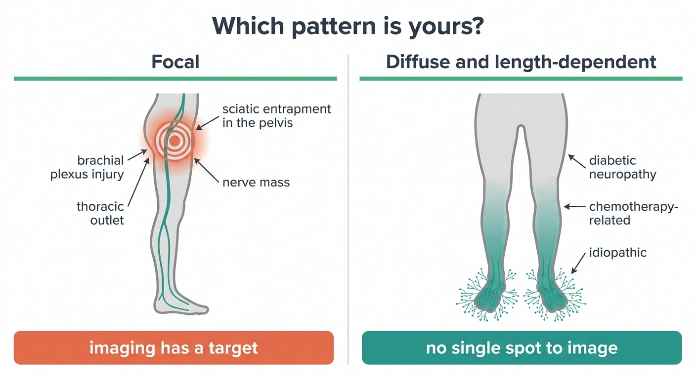
Here’s the distinction that determines everything else — and I wish someone had drawn it for me years ago.
Finish this out loud: “My worst symptoms are in ______, and they started in ______.”
If both blanks name one side or one limb, and especially if there was an injury, a surgery, or a sudden onset, that is a focal pattern. Imaging has somewhere to look.
If both blanks name both feet, and it crept upward over months or years, that is a diffuse pattern. Imaging has no target.
Mixed answers happen and they are worth flagging out loud, because a person with long-standing diabetic neuropathy can also develop a separate focal entrapment on one side. The second problem gets missed precisely because the first one explains everything.
Some nerve problems are focal. One nerve, or one bundle of nerves, is being squeezed, stretched, cut, invaded, or inflamed in one identifiable spot. Sciatic pain from something pressing on the nerve deep in the pelvis. A brachial plexus injury after a fall or a difficult surgery. Thoracic outlet syndrome. Numbness in a specific patch of thigh from the lateral femoral cutaneous nerve getting pinched.
Other nerve problems are diffuse. Thousands of the smallest, longest nerve fibers in your body are struggling at once, which is why symptoms start in both feet, spread symmetrically, and creep upward over years. That’s length-dependent polyneuropathy, and it describes the large majority of people living with diabetic neuropathy, chemotherapy-related damage, or idiopathic neuropathy.
MR neurography is a focal-problem tool. It shines a spotlight on one nerve at one location. If your problem is that millions of microscopic fiber endings are failing simultaneously across both feet, there is no single spot for the spotlight to land on. The nerves would very likely look normal, because at the scale MRI can resolve, they are normal. The damage is happening below the resolution of the picture.
That’s not a failure of the scan. It’s the wrong instrument for the question — like weighing yourself to find out your blood pressure.
Who MR Neurography Genuinely Helps
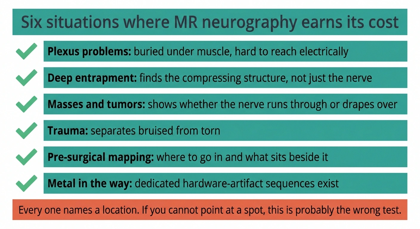
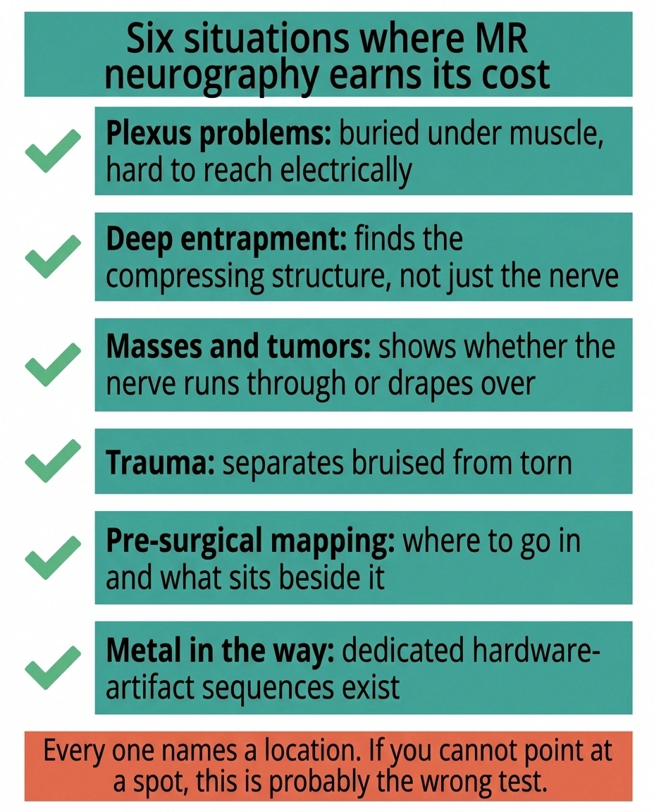
The published literature and the practice patterns at major nerve centers point at a fairly consistent list.
Plexus problems. The brachial plexus in the shoulder and the lumbosacral plexus in the pelvis are dense tangles of nerve tissue buried under muscle. They’re hard to reach with a needle, hard to interpret electrically, and easy to under-image — which is exactly the gap MRN fills. MRN is genuinely the best available look at them.
Suspected entrapment in a place your doctor can’t feel. Carpal tunnel gets diagnosed clinically because the wrist is right there under the skin. A nerve pinched deep in the pelvis by a fibrous band, a vein, or an unusual muscle is a different matter entirely. MRN can find the compressing structure, not just the compressed nerve.
Masses and tumors. Schwannomas, neurofibromas, and other growths on or beside a nerve show clearly, along with whether the nerve runs through the mass or is draped over it. Surgeons need that distinction before they open anything.
Trauma with an unclear ending. After an injury, MRN helps sort a nerve that is bruised but intact from one that has been torn. Those two situations have opposite treatment plans — and opposite timelines.
Pre-surgical mapping. When surgery is genuinely on the table, MRN answers where to go in and what’s sitting next to the target.
Cases where the metal is in the way. Radiologists have developed sequences specifically for imaging nerves near orthopedic hardware, which used to be a dead end.
Notice what runs through all six. Every one of them involves a suspected problem at a knowable location. If you and your neurologist can point at a spot on your body and say “we think something is happening right about there,” MRN becomes worth discussing. If you’re pointing at both feet and saying “everywhere below the ankle,” it usually isn’t.
The Limitation Nobody Puts in the Brochure
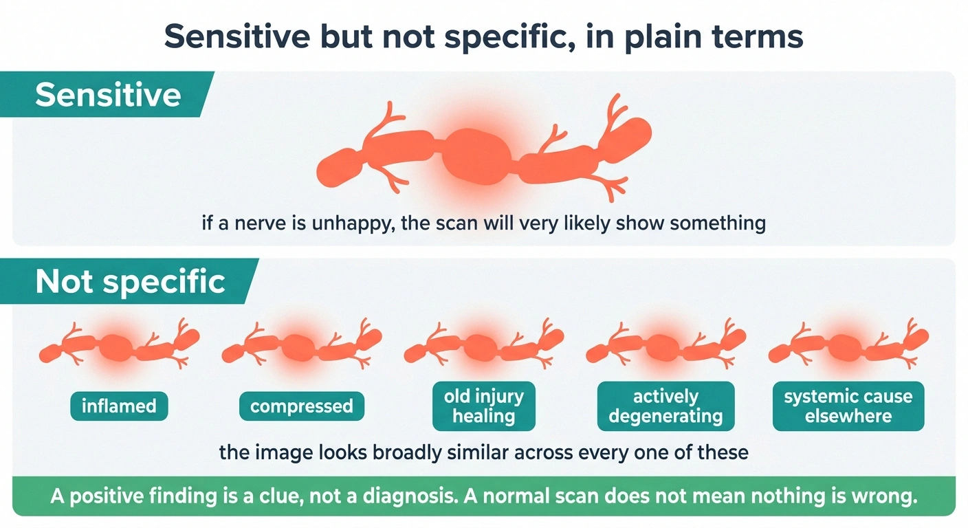
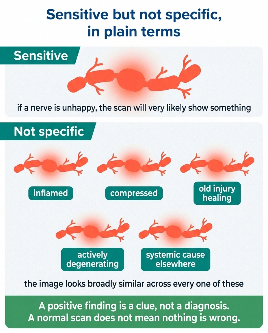
MR neurography is sensitive but it is not specific. That phrase gets used constantly in radiology papers and almost never gets translated, so let me translate it.
- T2 hyperintensity
- The nerve looks brighter than expected on one type of image sequence. It generally means extra fluid in or around the nerve, which accompanies inflammation, compression, and active degeneration alike. Bright is not a diagnosis.
- Fascicular architecture
- Nerves are bundles of smaller bundles. A normal nerve shows an orderly internal pattern in cross-section. Loss of that pattern, or swelling of individual bundles, is a more specific abnormality than brightness alone.
- Caliber change
- The nerve narrows or widens along its course. A narrowing with swelling just upstream of it is the classic look of something squeezing the nerve at that point.
- Denervation change
- Signal change in the muscles a nerve supplies, appearing as increased brightness early and as fatty replacement later. This is indirect evidence, and it can be the most useful line in the whole report, because it tells you the nerve problem has been going on long enough to affect the muscle.
Sensitive means it picks up abnormalities readily. If a nerve is unhappy, the scan will very likely show something.
Not specific means the abnormality it shows doesn’t tell you why. A bright, swollen-looking nerve on MRN can be inflamed, compressed, recovering from an old injury, actively degenerating, or caught up in something systemic that has nothing to do with that location — and the image looks broadly similar across every one of those.
This matters for one very practical reason. A positive MRN finding is not a diagnosis. It’s a clue that has to be lined up against your exam, your symptoms, and usually your EMG and nerve conduction results before it means anything. Radiologists writing about MRN say some version of this in nearly every paper: the imaging must be supported by clinical and electrodiagnostic testing.
The flip side matters just as much. A normal MRN does not mean your symptoms are imaginary. It means whatever is happening is either too small to see, too diffuse to localize, or occurring in a nerve territory the scan didn’t cover. Small fiber damage in particular is invisible here, which is why small fiber neuropathy is diagnosed with skin biopsy and specialized sensory testing rather than imaging.
MRN, Nerve Ultrasound, and EMG: Three Tools, Three Jobs
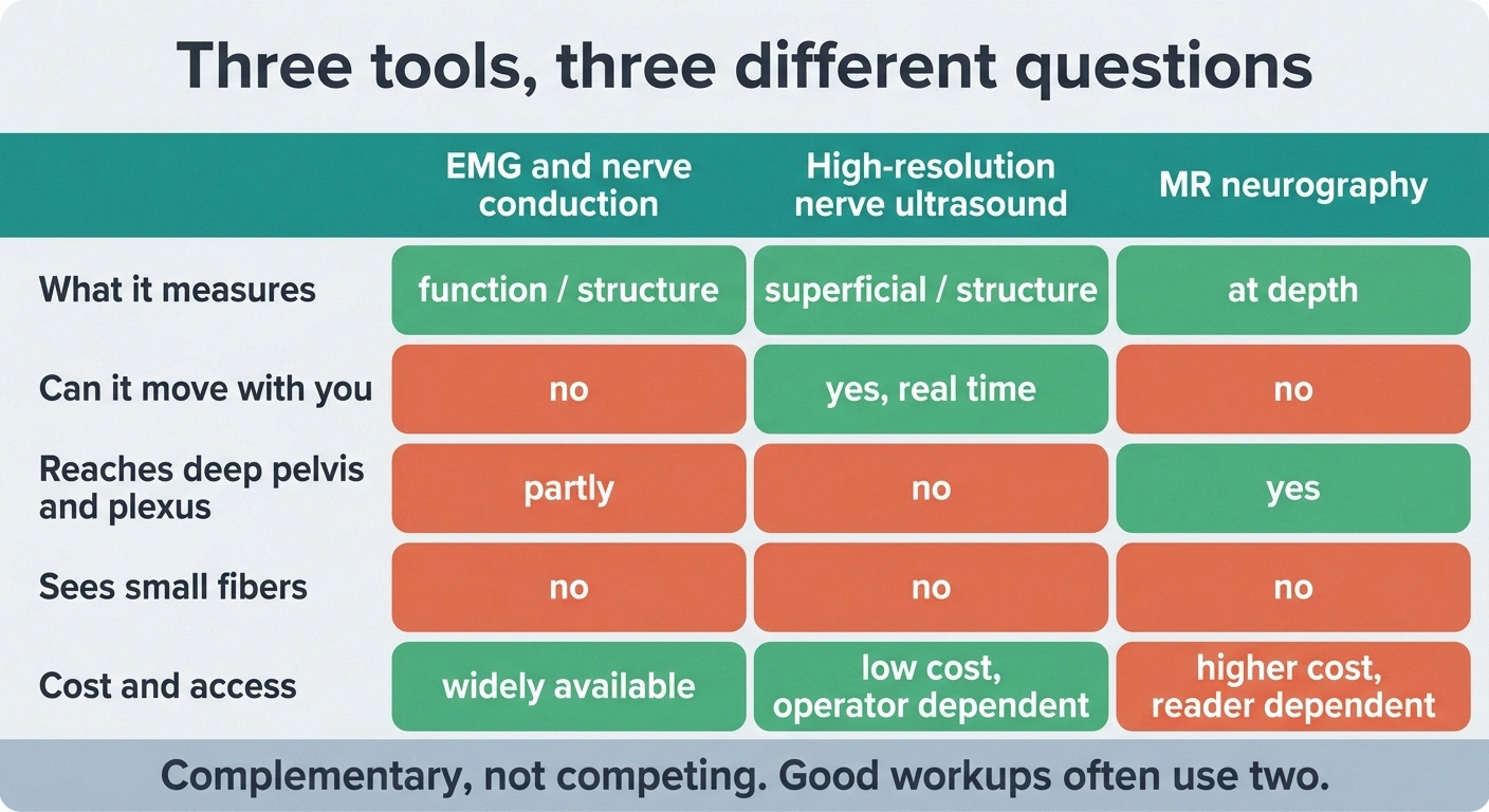
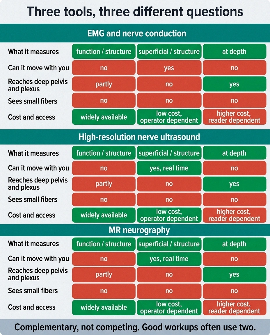
These get compared as though you’re choosing one. In practice they answer different questions and a good workup often uses two of them.
| Test | In the room | Result timing | Typical wait to book |
|---|---|---|---|
| Nerve conduction and EMG | 45 to 90 minutes | Often discussed same visit | Weeks |
| Nerve ultrasound | 20 to 40 minutes | Often same visit | Weeks, where offered at all |
| MR neurography | 45 to 75 minutes, still | Days to weeks for the read | Weeks to months, plus authorization |
The authorization step is the one that surprises people. Build in an extra two to four weeks for it, and ask the ordering office to start it the same day rather than after you call to chase the appointment.
EMG and nerve conduction studies measure function. They tell you whether signals are traveling, how fast, and how strongly, and they distinguish damage to the insulating myelin from damage to the axon itself. They cannot show you the physical structure that’s causing the trouble. If you’ve had one and the report left you baffled, it’s worth walking through what those numbers actually mean.
High-resolution nerve ultrasound shows structure, in real time, cheaply, and without a tube. It’s excellent for nerves close to the skin and it has one ability MRI simply lacks — the operator can move your limb while watching, which catches nerves that only pinch in certain positions. Its ceiling is depth. Deep pelvic nerves and plexus tissue are largely out of reach.
MR neurography shows structure at depth, covers a large field, and images the surrounding anatomy that might be causing the problem. It costs more, takes longer, and depends heavily on the reader.
Ultrasound and MRN turn out to be complementary rather than competing. Ultrasound for the superficial and the dynamic. MRN for the deep, the complex, and the pre-surgical — and plenty of good workups use both.
What the Scan Is Actually Like
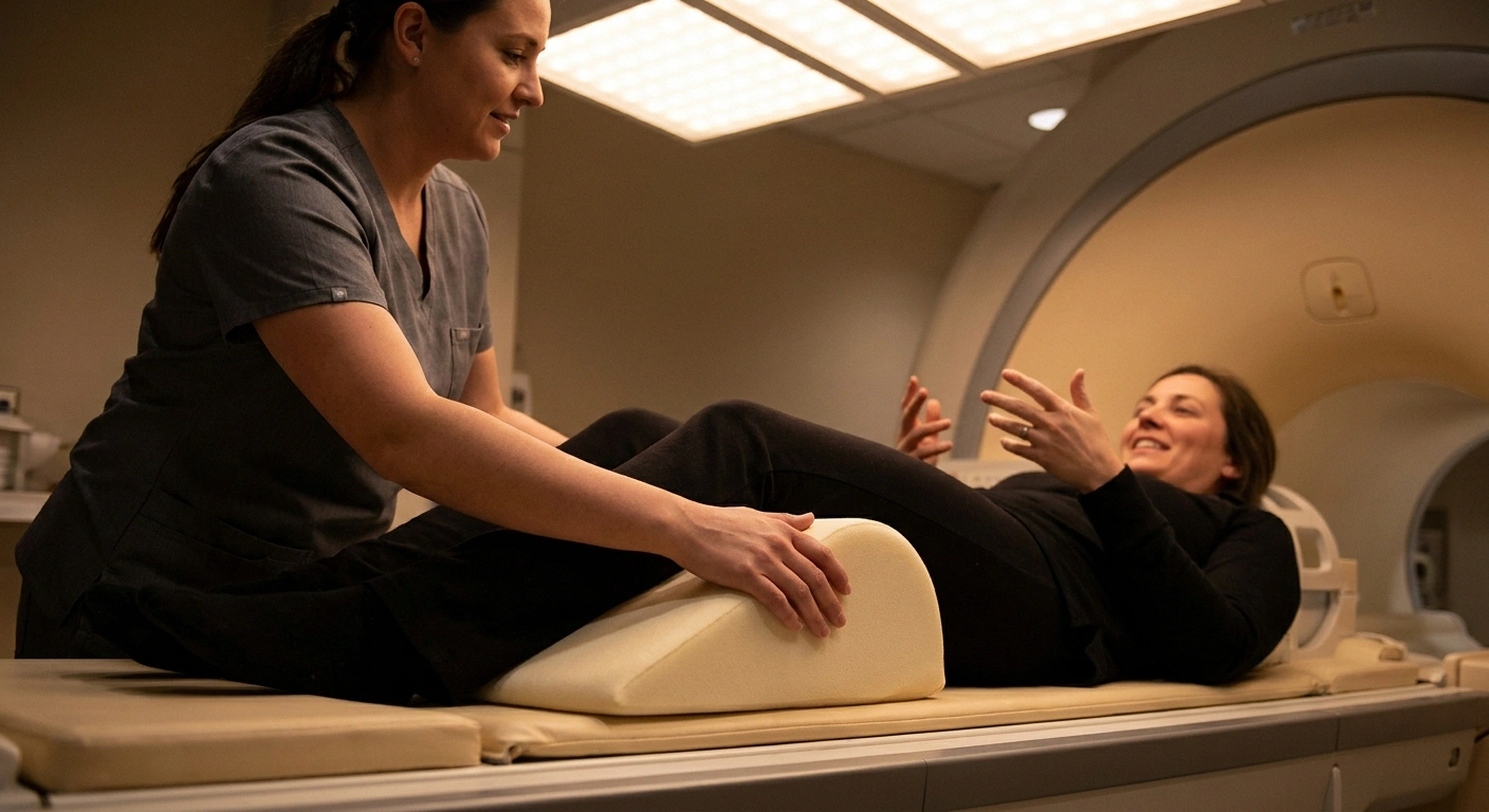
Practically speaking, it’s an MRI. If you’ve had one before, you know the drill, and our notes on getting through an MRI with neuropathy apply directly here.
A few specifics worth knowing. MRN studies tend to run longer than a routine scan, often forty-five to seventy-five minutes, because high resolution over a large field takes time. Contrast is sometimes used and sometimes not, depending on what’s being investigated. You’ll be positioned carefully with the coil over the region of interest, and staying still genuinely matters more than usual, since motion blur destroys the fine detail the whole scan exists to capture.
That last point is where neuropathy creates a real problem. Lying still for an hour with burning feet is difficult — and nobody warns you. Ask ahead about timing your pain medication so a dose lands about an hour before the scan, ask for a bolster under your knees, and ask whether the technologist can pad any pressure point that tends to flare. These are ordinary accommodations, and staff are usually glad to make them if you ask before you’re already on the table.
Getting It Covered, and Getting It Read Well
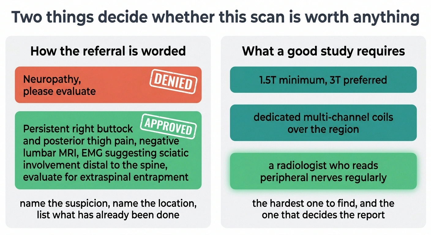
Coverage exists, but it’s conditional. Major insurers publish specific policy bulletins on MR neurography, and they generally treat it as medically necessary for defined situations, most often suspected plexus injury, suspected entrapment where standard workup has been inconclusive, evaluation of a nerve mass, and pre-surgical planning. Broad requests without that framing get denied — routinely.
- Does the radiologist reading this have a peripheral nerve focus? The single highest-value question on this list, and the one most likely to change where you go.
- Is this a 3T scanner, and will dedicated coils be used for the region? A 1.5T study can be adequate; knowing which you are getting sets expectations for the detail level.
- Has prior authorization been submitted, and on what date? Ask for the date rather than a yes. A yes that means “we will get to it” is how six weeks disappear.
- Will contrast be used, and do I need labs first? If contrast is planned, kidney function is usually checked beforehand, which is an extra appointment if nobody mentions it until you arrive.
Ask these of the imaging center’s scheduling desk, not your neurologist’s front office. The scheduler knows the equipment and the authorization status; the referring office usually knows neither.
The practical translation: the referral needs to state what is suspected and where, and it needs to document what has already been done. “Persistent right buttock and posterior thigh pain, negative lumbar MRI, EMG suggesting sciatic involvement distal to the spine, evaluate for extraspinal entrapment” is the shape of a request that gets approved. “Neuropathy, please evaluate” is the shape that doesn’t. Our rundown on insurance coverage for neuropathy testing covers the appeal process if a first request comes back denied.
Then there’s the part that isn’t about money at all. MRN is unusually reader-dependent. The same images can produce a detailed, actionable report at a center that reads peripheral nerves weekly and a two-line non-answer somewhere that reads one a year. Before you schedule, it’s fair to ask the ordering office one direct question: does the radiologist who will read this have a peripheral nerve focus? If the answer is uncertain, an academic center or a dedicated peripheral nerve program is worth the drive — even a long one.
How to Raise It Without It Landing Wrong
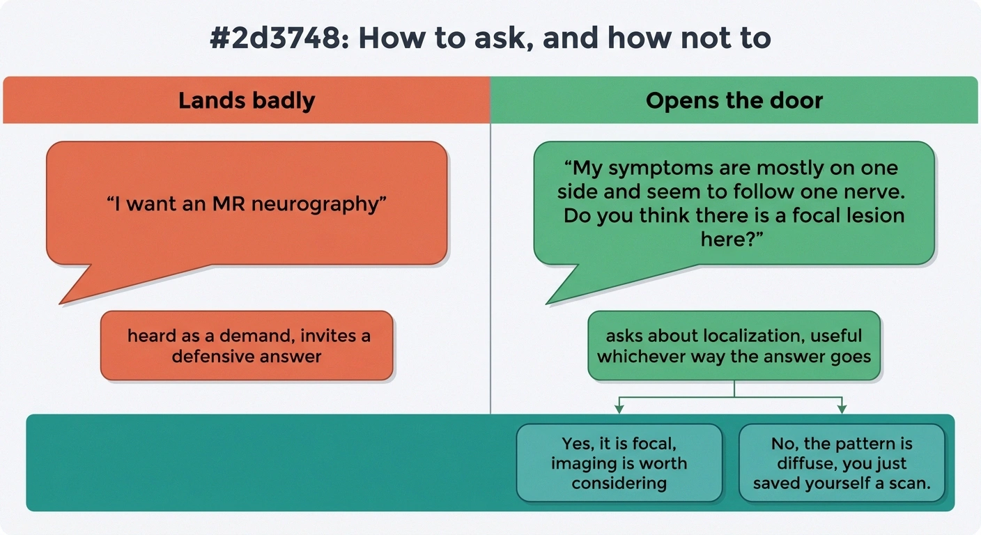
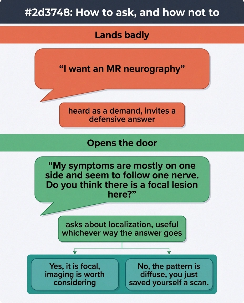
There’s a version of this conversation that goes badly, and it’s usually the version where a patient walks in asking for a specific scan by name. Doctors hear that as a demand and get defensive, which helps nobody.
The version that works reframes it as a question about localization. Something close to: “My symptoms are mostly on one side and seem to follow one nerve. Do you think there’s a focal lesion here, and if so is imaging the nerve worth considering?”
That question does something useful regardless of the answer. If your doctor says the pattern is diffuse and symmetric, you’ve learned something real about your own case, and you’ve saved yourself a scan that would have come back unremarkable. If they pause and say the one-sided pattern has been bothering them too, you’ve opened exactly the right door. Either way you’ve moved forward — which is more than most appointments manage.
If you’ve been stuck in an unexplained-symptoms loop for a long time and nobody has clearly told you whether your pattern is focal or diffuse, that’s a reasonable moment to think about getting a second opinion, ideally from someone who specializes in peripheral nerve. And if you’ve never had a formal workup at all, the standard diagnostic sequence comes well before any advanced imaging.
Where This Leaves You
I want to be careful here, because there’s a specific way this article could be misread. I’m not saying MRN is overhyped. When it’s aimed at the right problem, it can end years of guessing in a single afternoon. I’ve heard from readers whose entrapment was found on an MRN after three normal scans of the wrong body part, and one of them had surgery and got most of her function back.
What I’m saying is narrower. It’s a precision instrument for focal nerve problems, and the most common form of neuropathy isn’t a focal nerve problem. Knowing which category you’re in is the whole decision — and you can usually settle it in one honest conversation.
Priya, for what it’s worth, turned out to have a symmetric small fiber pattern. No scan was going to show it. What eventually helped her was a skin biopsy and a serious look at the causes nobody had checked yet, which is a less dramatic answer than an MRI — but a considerably more useful one.
Frequently Asked Questions
Is MR neurography the same as a regular MRI?
No. Both use the same machine, but MR neurography uses different imaging sequences designed to suppress signal from fat and flowing blood so that nerve tissue stands out clearly. It also requires higher field strength, ideally 3 Tesla, dedicated coils placed over the region being studied, and a radiologist experienced in reading peripheral nerves. A routine MRI of the spine or a joint may include nerves in the field of view, but it is not optimized to evaluate them and frequently misses nerve abnormalities that a dedicated neurography study would show.
Can an MRI show nerve damage in my feet?
Usually not, if the cause is a symmetric length-dependent polyneuropathy. That type of damage affects enormous numbers of very small nerve fibers across both feet rather than producing a single visible lesion, and the affected fibers are far below the resolution of any MRI scanner. Imaging becomes useful when symptoms are one-sided, follow the territory of a single named nerve, or began after an injury or surgery, because those patterns suggest a focal problem that a scan can actually locate.
Does insurance cover MR neurography?
Often yes, but with conditions. Insurers publish specific medical policy bulletins for MR neurography and typically approve it for suspected brachial or lumbosacral plexus injury, suspected nerve entrapment when standard evaluation has been inconclusive, assessment of a suspected nerve tumor, and pre-surgical planning. Approval depends heavily on how the referral is documented, so the request should name the suspected problem and its location and list what testing has already been completed. Denials can be appealed, and a letter from the ordering physician explaining why standard imaging was insufficient often succeeds.
How is MR neurography different from nerve ultrasound?
Ultrasound is faster, cheaper, and can image a nerve while the limb moves, which reveals compression that only occurs in certain positions. It works best on nerves close to the surface. MR neurography reaches deep structures that ultrasound cannot see, including the pelvic and plexus regions, covers a wider field, and shows surrounding anatomy that may be causing compression. The two are frequently complementary rather than alternatives, and some centers use ultrasound first and reserve neurography for deep or surgical questions.
Does a normal MR neurography result mean nothing is wrong?
No. A normal result means no visible structural abnormality was found in the nerves that were imaged. Small fiber damage, metabolic nerve injury, and diffuse polyneuropathy can all be present with entirely normal imaging, because those processes occur at a scale MRI cannot resolve. A normal scan is genuinely useful information since it argues against a mass, an entrapment, or a structural lesion in that area, but it does not rule out neuropathy and it does not mean symptoms lack a physical basis.
How long does the scan take and will I need contrast?
MR neurography studies commonly run forty-five to seventy-five minutes, longer than a routine MRI, because achieving high resolution across a large area takes additional imaging time. Contrast is used in some studies and not others, depending on whether a tumor, inflammation, or infection is suspected. Because staying still is essential for image quality, it is worth planning ahead if lying flat is painful, including timing pain medication to take effect before the scan and requesting positioning support such as a bolster under the knees.
Why did my doctor say imaging would not help my neuropathy?
Most likely because your symptom pattern is symmetric and length-dependent, meaning it started in both feet and progressed upward. That pattern indicates a diffuse process affecting many nerves at once rather than a single localized lesion, and imaging has no target in that situation. It is a reasonable clinical judgment rather than a dismissal. If your symptoms are notably asymmetric, follow one nerve distribution, or started abruptly after an injury, those features are worth raising directly, because they change the reasoning.
Where can I get MR neurography done?
Availability is uneven. The scan requires a 1.5T or preferably 3T scanner with appropriate coils, which many imaging centers have, and a radiologist who reads peripheral nerve studies regularly, which is considerably rarer. Academic medical centers, larger orthopedic and neurosurgical hospitals, and dedicated peripheral nerve programs are the most reliable places to find both. Before scheduling, ask whether the interpreting radiologist has a peripheral nerve focus, since interpretation quality varies more for this study than for most routine imaging.
About the Author