Before I say anything else, I need you to hear this: your symptoms are real, and the research has finally caught up. If you got COVID once — mild, moderate, or severe — and weeks or months later your feet started burning, your hands started tingling, your heart started racing when you stood up, or your body stopped tolerating heat and cold the way it used to, you have not lost your mind. You have not become a hypochondriac. You are not “just anxious,” no matter how many appointments have ended with a shrug and a suggestion to try meditation. Since 2022, neurologists at Harvard, at the Mayo Clinic, and at academic centers around the world have biopsied the skin of long COVID patients with these exact symptoms and found real, measurable, quantifiable nerve damage. Small fiber neuropathy after COVID-19 is a documented, biopsy-provable condition. And if this article does one thing for you, I want it to be this: give you the language, the research, and the specific requests you need to walk into your next appointment and be taken seriously.
I write here as a patient advocate, not a doctor. I have my own neuropathy story — different from yours, but overlapping enough that I know how heavy it feels to have a body that isn’t working the way it did a year ago and a medical system that keeps telling you the tests came back fine. In the last four years, a rigorous body of research has emerged on post-COVID SFN. It’s evolving fast, treatment is symptomatic, and no honest article can promise reversal. What I can promise: what’s known, what’s suspected, and what you can reasonably ask for right now.
What Small Fiber Neuropathy Is — And Why It’s Different
Your peripheral nerves are not a single kind of wire. They’re bundles of very different fibers doing very different jobs, and understanding the difference is the whole key to understanding what may have happened to you after COVID.
Large fibers are the thick, well-insulated ones. They carry vibration, joint position sense, discriminative touch, and motor commands to your muscles. If you can feel a tuning fork buzzing on your ankle, wiggle your toes with your eyes closed, and grip a coffee mug without dropping it, that’s large-fiber territory. These are the fibers standard nerve conduction studies and needle EMG were built to measure.
Small fibers are the opposite. Thin, either completely unmyelinated (the C-fibers) or only lightly wrapped (the A-delta fibers), and slow. They carry pain, temperature, itch, and the autonomic signals that regulate sweating, blood flow, blood pressure, heart rate, and digestion. Small fibers make up roughly 70 to 80 percent of your peripheral nerves by fiber count — the vast majority of your peripheral nervous system.
Here is the piece almost no one explains: standard EMG and nerve conduction studies cannot see small fibers. There is no way to send a meaningful electrical pulse down a fiber that thin and unmyelinated. So when a long-COVID patient with burning feet and racing heart shows up in a neurology office, the EMG comes back completely, misleadingly normal — because it was measuring the fibers that are still working. The damaged fibers, the ones causing every one of your symptoms, are invisible to that test. My earlier piece on small fiber neuropathy walks through the full anatomy and testing gap.
In classic small fiber neuropathy, large fibers are typically preserved. Your reflexes look fine. Your strength is intact. You can still stand on one foot with your eyes open. And your test result says “normal.” Meanwhile, your feet feel like they’ve been dipped in gasoline every night.
The COVID-19 Connection: What the Research Actually Shows
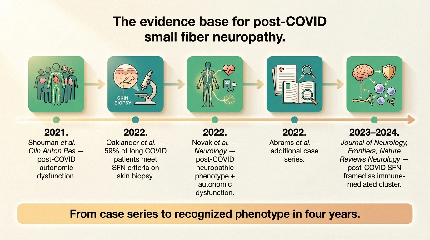
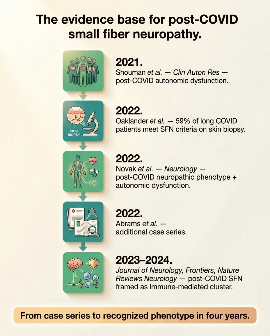
The turning point came in March 2022, when Dr. Anne Louise Oaklander and her team at Massachusetts General Hospital published a case series in Neurology: Neuroimmunology & Neuroinflammation. They studied 17 patients with persistent neuropathic symptoms after COVID — most previously healthy, most with mild acute infections — and did formal small fiber testing. Ten of those 17 patients, 59 percent, met the diagnostic criteria for small fiber neuropathy on skin biopsy. Their intraepidermal nerve fiber density was reduced. The damage was measurable, quantifiable, and real.
That paper broke the door open. Later in 2022, Peter Novak’s group at Brigham and Women’s Hospital published a companion cohort in Neurology documenting a post-COVID neuropathic phenotype combining small fiber loss on biopsy with autonomic dysfunction on tilt table testing. Rebecca Abrams and colleagues at Mount Sinai reported a similar case series. Kamal Shouman at the Mayo Clinic had already described the autonomic side — orthostatic intolerance, POTS-like tachycardia — in COVID survivors as early as late 2021.
By 2023 and 2024, reviews stopped calling this a curiosity and started framing post-COVID SFN as an emerging, likely immune-mediated cluster within long COVID. Papers in the Journal of Neurology, Frontiers in Neurology, and Nature Reviews Neurology now describe it as a recognizable phenotype worth screening for in any long-COVID patient with burning, tingling, or dysautonomic symptoms.
None of this makes the diagnosis universally accepted, and I want to be honest about that. Not every neurologist has read these papers. Not every clinic offers the skin biopsy. But the evidence base moved from anecdote to peer-reviewed cohort in only four years, and it’s more than enough to justify asking for the test.
How Small Fiber Neuropathy Feels After COVID

Post-COVID small fiber neuropathy doesn’t always look like the neuropathy people picture. Most readers, if they’ve heard of neuropathy at all, are thinking of a person in their 70s with diabetes describing numb feet. Long-COVID SFN tends to look different — the audience is younger, often previously healthy, often in their 20s to 50s, and the symptom pattern is frequently more chaotic than a classic diabetic presentation.
- Burning feet or hands
- Patchy or stocking-glove tingling
- Allodynia (bedsheet feels painful)
- Thermal reversal (cold feels burning)
- Pinprick or temperature loss
- POTS-like tachycardia on standing
- Orthostatic lightheadedness
- GI dysmotility, early satiety
- Hyperhidrosis or anhidrosis
- Exercise intolerance, temperature dysregulation
Burning. The most common symptom. Sometimes stocking-and-glove — hands and feet symmetrically — and sometimes patchy, showing up on a forearm one week, an inner thigh the next. The patchy, non-length-dependent pattern is actually MORE common in immune-mediated SFN, which is one reason clinicians increasingly consider a post-viral autoimmune trigger.
Pins and needles. Not the passing kind from sitting cross-legged. The kind that shows up at rest, at night, radiating up the calves or across the palms for hours.
Allodynia. Light touch that should not hurt, hurts. The bedsheet against your feet feels like something between a scratch and a burn. A sock seam scrapes. A shower massage is intolerable.
Thermal dysesthesia. This one is uncanny. Cold water in the shower feels burning hot. A warm compress feels icy. Your temperature-sensing nerves are misfiring, sending the wrong signal for the actual stimulus. Many post-COVID SFN patients report this and think they are losing their minds — you are not. If you’re looking for the fuller sensory map, burning feet syndrome covers it in detail.
Autonomic symptoms. This is the layer that trips up doctors and patients alike. When the small autonomic fibers are damaged, the symptoms don’t feel like “neuropathy” at all. Racing heart when you stand up. Lightheadedness that gets worse in the shower. Digestive slowness or unpredictability. Sweating too much on one side of the body and not enough on the other. Exercise intolerance where your heart rate spikes into the 150s during light activity. These are not psychiatric symptoms. They are your autonomic nerves misfiring.
The Autonomic Layer — Why POTS-Like Symptoms Show Up
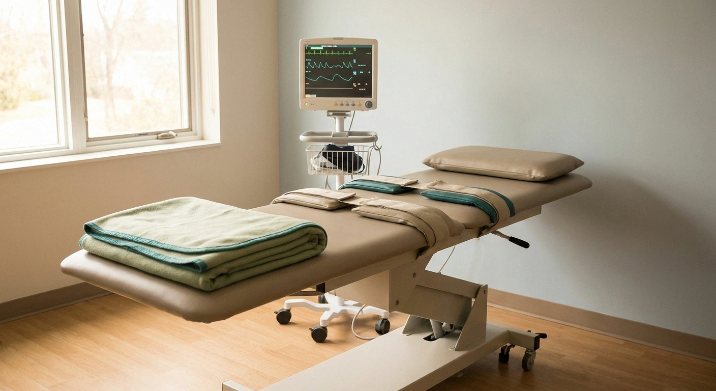
Postural orthostatic tachycardia syndrome, or POTS, is one of the most commonly recognized forms of long COVID. It’s also one of the most common companions to post-COVID small fiber neuropathy — and the overlap is not a coincidence. The autonomic small fibers that regulate the vessels in your legs to constrict when you stand up (so blood stays in your head) are the same category of fibers damaged in SFN. Damage them, and standing becomes a physiologic event your body cannot manage. Your heart races to try to compensate. You feel dizzy, breathless, foggy.
The pattern to watch for: within 10 minutes of standing, your heart rate climbs by 30 beats per minute or more (40 for teenagers) without a corresponding drop in blood pressure. Standing is exhausting. Showers are hard because heat pools blood in your skin. Meals sometimes make you dizzy because blood goes to your gut. Compression, salt, fluid, and lying flat help.
Hold onto this: the constellation of POTS symptoms plus burning feet plus normal EMG is now one of the clearest fingerprints of post-COVID neuropathic-autonomic injury. If both are present, you have a strong case for asking your doctor to work up both the sensory and autonomic small fiber systems. My deeper piece on autonomic neuropathy lays out the full autonomic map.
Getting a Diagnosis — Skin Biopsy, QSART, and What Standard Tests Miss
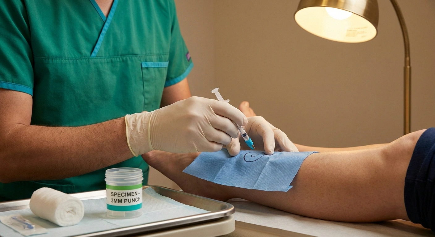
Here is the piece that changes everything if you can get it: the skin punch biopsy for intraepidermal nerve fiber density, or IENFD. This is the test that would have found what a normal EMG missed. It’s same-day, in-office, done with a 3-millimeter punch tool at the distal calf, usually with a single stitch. The tissue is sent to a specialty pathology lab where the small nerve fibers running up into the epidermis are counted per millimeter. Small fiber neuropathy shows a reduced count. The report comes back in two to four weeks with a hard number. This is the single test that turns a “we don’t know what’s wrong” appointment into a diagnosis.
QSART, the Quantitative Sudomotor Axon Reflex Test, is the autonomic companion. It measures how well tiny sweat-controlling nerve endings respond to a gentle chemical stimulus. Non-invasive, specifically probing the autonomic small fibers. Combined with skin biopsy, QSART builds a rounded picture of both the sensory and autonomic small fiber systems.
A full autonomic workup adds the tilt table test, heart rate variability testing with deep breathing and Valsalva, and the thermoregulatory sweat test. Not every clinic has all of them, but a dedicated autonomic center will.
The frustration comes back to the same issue: standard EMG and nerve conduction studies were never designed to see any of this. In isolated SFN, EMG comes back normal every time. A normal EMG in a long-COVID patient with burning feet and orthostatic tachycardia is not reassurance. It’s a signal to escalate to small fiber testing. If you want the full breakdown, neuropathy diagnosis: tests your doctor may order walks through the whole workup.
What We Know About Why This Happens
I want to be careful here, because this section is where honest science and viral misinformation both tend to overreach. Nobody has fully proven the mechanism of post-COVID small fiber neuropathy. What we have are several leading hypotheses that the research is actively probing.
Immune-mediated / dysimmune injury. The strongest current theory. In this model, the immune system’s response to SARS-CoV-2 produces antibodies that cross-react with components of small nerve fibers — a post-viral autoimmune phenomenon well-documented for other pathogens (Guillain-Barré syndrome after Campylobacter, for example). Multiple long-COVID small fiber cohorts show inflammatory findings consistent with this picture, and some patients test positive on emerging small-fiber autoantibody panels (TS-HDS, FGFR3, plexin D1).
Direct viral neurotropism. The idea that the virus itself enters peripheral neurons, perhaps via ACE2 receptors, and damages them directly. There is evidence for viral RNA in various tissues after infection, and some support for direct neural involvement, but the direct-damage mechanism is less well-established than the immune-mediated one for the small fiber picture specifically.
Microvascular injury. COVID clearly damages blood vessel endothelium and produces micro-clots in acute severe disease. The tiny blood vessels that feed peripheral nerves — the vasa nervorum — may be compromised, starving the small fibers of their blood supply.
Persistent inflammation. Chronic low-grade cytokine elevation in long-COVID patients may drive small fiber die-back over time — a slower, cumulative mechanism.
Most researchers now suspect the true story is a combination, with the mix varying from patient to patient. What matters clinically is that the leading theory is dysimmune, because that framing opens the door to considering immunomodulating therapy in refractory cases — more on that below, with important caveats.
Treatment Options: Managing Symptoms While the Research Evolves
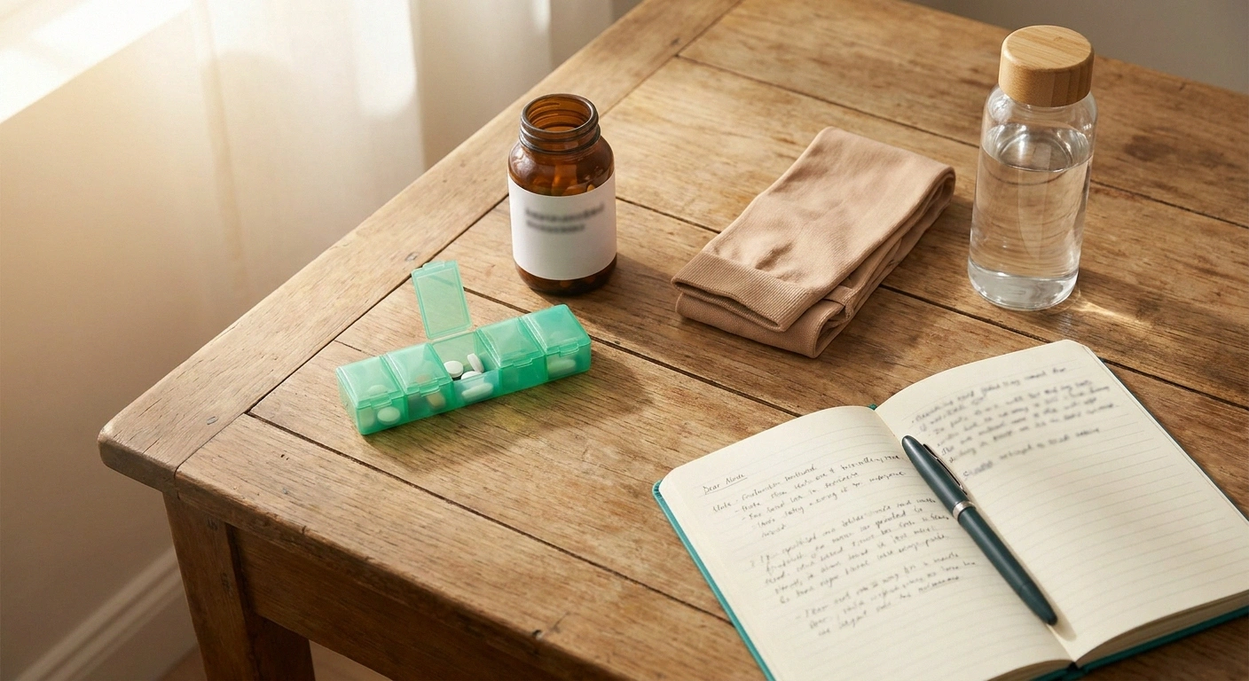
Here is where I have to be most honest with you. There is no small-fiber-neuropathy-specific cure, and there is no COVID-specific nerve fix. Treatment right now is symptomatic — managing pain, managing autonomic dysfunction, and supporting the body while it either heals or stabilizes.
Neuropathic pain medications. The first-line drugs are the same as for other small fiber neuropathies. Duloxetine, an SNRI, is often the top choice because it treats pain and mood simultaneously. Gabapentin and pregabalin are calcium-channel modulators that reduce nerve pain, and both have decent evidence in small fiber conditions. Tricyclic antidepressants at low doses (nortriptyline, amitriptyline) are another option, particularly for burning that disrupts sleep.
Topical treatments. Lidocaine patches (5%) can help with focal burning or allodynia in defined areas. Compounded capsaicin cream, and in select cases the prescription 8% capsaicin patch, can reduce burning in a localized region — the 8% patch is a specialist procedure. Menthol creams help some patients through simple cooling.
Autonomic support (especially with POTS overlap). Waist-high compression stockings (30–40 mmHg). Salt loading — often 8 to 10 grams of sodium per day. Fluid loading — 2 to 3 liters per day. Beta blockers or ivabradine for the tachycardia. Midodrine or fludrocortisone for orthostatic hypotension. A long-COVID or POTS specialist can tailor this stack.
Rehab and pacing. Graded, cautious reintroduction of activity — never pushing through into post-exertional malaise, which can worsen the whole picture for weeks. Works best with a therapist experienced in long COVID or ME/CFS.
Lifestyle basics. Anti-inflammatory eating, sleep protection, alcohol moderation, blood sugar stability. Not the whole story, but they matter more than most patients are told.
IVIG — an investigational option, with a real caveat. Intravenous immunoglobulin has shown promise in a handful of small studies for autoimmune-mediated SFN, including a subset of post-COVID cases (Dr. Oaklander’s group has published on this specifically). But IVIG is not standard of care for post-COVID SFN. It’s expensive, insurance approval is often difficult, side effects can be significant, and it should only be pursued at an academic neuromuscular clinic experienced in autoimmune neuropathies — typically after other options are exhausted and after autoantibody or biopsy evidence supports an immune-mediated picture. Plasma exchange and rituximab are similarly investigational.
What Recovery Looks Like
Here is where I have to hold two things at once. Some long-COVID small fiber patients do improve — often gradually, sometimes with clear turning points, usually over a period of 6 to 24 months. Some plateau at a lower baseline than before COVID but stable enough to build a life around. And some, unfortunately, persist with symptoms years out.
We do not yet have the long-term outcome data to say which pattern is most common, because the pandemic itself is only a few years in the rearview and research cohorts have not been followed long enough. What the field is watching for: whether early treatment of the immune component (in appropriate cases) changes the long-term trajectory; whether specific autoantibody profiles predict response; whether the microvascular contribution reverses over time. None of these questions has a settled answer yet.
What I will not do is tell you that your nerves will fully heal. I do not know, and neither does anyone else with certainty. What I can say is that the biology of small fibers is more plastic than the biology of large fibers — small fibers can regenerate in ways that motor nerves largely cannot, especially when the underlying insult is removed or damped. That biology is the honest source of hope.
Advocating for Yourself in the Doctor’s Office
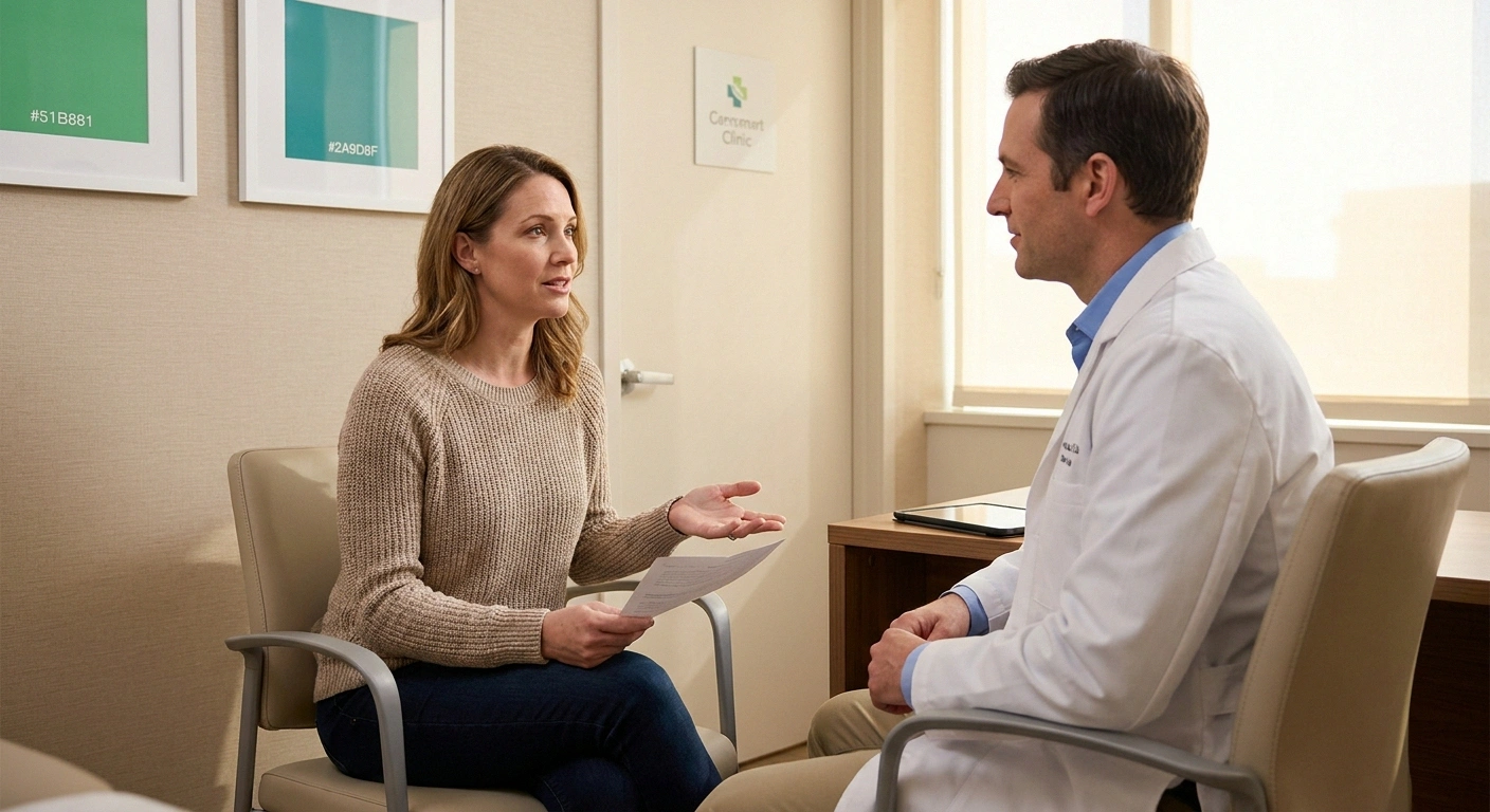
This is the section I most want you to actually use. Bring the language. Bring the specific requests. The three sentences that most reliably move things forward:
“My symptoms fit the picture of small fiber neuropathy after COVID — burning, temperature sensitivity, and orthostatic symptoms that started weeks to months after my acute infection. Would a skin punch biopsy for intraepidermal nerve fiber density be reasonable?” This names the condition, names the test, and gives your doctor an easy path to yes.
“Along with the burning, I’ve been having racing heart when I stand up and lightheadedness. Could we discuss an autonomic workup — QSART, tilt table, autonomic function testing?” This asks specifically for the autonomic layer.
“Would it be reasonable to refer me to a long COVID clinic or a neuromuscular clinic that specializes in small fiber neuropathy?” If your local practice doesn’t have the equipment or expertise, escalation is the answer. Academic medical centers — Mayo, Hopkins, Cleveland Clinic, Mass General, UCLA, and many university hospitals — have long COVID programs and neuromuscular clinics that do skin biopsy and autonomic testing in-house.
A note on tone: you don’t need to be combative. You do need to be specific. Long COVID patients who go in naming the condition, naming the test, and asking for the referral often get taken seriously the first appointment. If the response is dismissive despite the specific request, that is information about the fit with that practice, and a second opinion is your right.
One more thing. The emotional weight of a body that isn’t working the way it did, plus a medical system that has often dismissed long COVID patients specifically, is real. If the exhaustion and frustration are affecting your mood, please read neuropathy and mental health. Supporting the emotional layer is part of the workup, not a sign of weakness.
Frequently Asked Questions
Can long COVID really cause small fiber neuropathy?
Yes. Since 2022, multiple peer-reviewed studies have confirmed post-COVID small fiber neuropathy on skin biopsy. The most-cited paper is Anne Louise Oaklander’s 2022 case series in Neurology: Neuroimmunology & Neuroinflammation, which found that 59 percent of long COVID patients with neuropathic symptoms met formal small fiber neuropathy criteria on intraepidermal nerve fiber density testing. Additional cohorts by Novak, Abrams, and Shouman documented similar findings, often combined with autonomic dysfunction. Post-COVID small fiber neuropathy is now recognized as a biopsy-provable cluster within long COVID, most likely driven by immune-mediated nerve injury after the acute infection.
How soon after COVID does small fiber neuropathy typically start?
Symptoms usually start weeks to a few months after the acute COVID infection, not during the acute illness itself. Some patients trace their first burning or tingling to two to four weeks post-infection; others describe a gradual onset over several months. The pattern has been reported after mild initial infections as well as more severe acute disease — you do not need to have been hospitalized. If new burning, tingling, temperature sensitivity, or orthostatic symptoms appeared in the weeks or months after a COVID infection, the timing is consistent with post-COVID nerve involvement.
Why did my EMG come back normal if I have long COVID neuropathy?
Standard EMG and nerve conduction studies only measure large, myelinated nerve fibers — the ones that carry vibration, position sense, and motor signals. Small fibers, which carry pain, temperature, and autonomic function, are too thin and unmyelinated to produce a signal that standard equipment can detect. Post-COVID small fiber neuropathy typically leaves large fibers intact, so the EMG comes back normal even when there is real, biopsy-provable damage to the small fiber system. A normal EMG in a long-COVID patient with burning feet or orthostatic tachycardia does not rule out neuropathy — it points toward the need for skin biopsy and autonomic testing.
What is the gold standard test for post-COVID small fiber neuropathy?
The gold standard is a skin punch biopsy that measures intraepidermal nerve fiber density, often abbreviated IENFD. A 3-millimeter skin sample is taken from the distal calf, sent to a specialty pathology lab, and the small nerve fibers running up into the epidermis are counted per millimeter. A reduced density confirms small fiber neuropathy. Additional useful tests include QSART for autonomic small fiber function, tilt table testing, heart rate variability with deep breathing, thermoregulatory sweat testing, and increasingly corneal confocal microscopy at academic centers. Emerging autoantibody panels, including TS-HDS, FGFR3, and plexin D1, are used at specialty labs to explore an immune-mediated mechanism.
Does long COVID small fiber neuropathy go away?
The honest answer is that it varies. Some patients see gradual improvement over 6 to 24 months. Some stabilize at a new baseline that is lower than before COVID but manageable. And some patients continue to experience symptoms years after the initial infection. Long-term outcome data is still limited because the pandemic itself is only a few years old and research cohorts have not been followed long enough. Small nerve fibers do have some regenerative capacity, especially when the underlying immune or inflammatory driver is controlled, so improvement is biologically possible. But no honest doctor or article can promise reversal at this stage of the research.
Is IVIG used to treat post-COVID small fiber neuropathy?
IVIG (intravenous immunoglobulin) is being used investigationally for select cases of suspected autoimmune-mediated small fiber neuropathy, including some post-COVID cases. Small studies, notably from Dr. Oaklander’s group, have shown improvement in refractory patients. However, IVIG is not standard of care for post-COVID small fiber neuropathy. It is expensive, insurance approval is often difficult, side effects can be significant, and it should only be pursued at an academic neuromuscular clinic with experience treating autoimmune neuropathies — typically after other treatment options have been exhausted and after autoantibody or biopsy findings support an immune-mediated mechanism. Plasma exchange and rituximab are similarly investigational.
Do I need to have had severe COVID to develop small fiber neuropathy?
No. Post-COVID small fiber neuropathy has been reported in patients whose initial infection was mild — many never hospitalized, some who tested positive without being significantly ill. Severity of the acute infection does not appear to predict who develops post-COVID nerve involvement. This is one of the harder parts of long COVID to accept, both for patients and for family members who assume a mild acute infection means no lasting consequences. The immune and inflammatory processes that appear to drive post-COVID SFN can be triggered by relatively mild infections in previously healthy people.
A note as I close. This is a young research field. Treatment guidelines will keep evolving, new autoantibody markers will be validated, and the picture will sharpen. What is no longer up for debate is whether post-COVID small fiber neuropathy exists. It exists, it is measurable, and it deserves a real workup. Take this article — or the specific sentences from it — to your next appointment. Ask for the skin biopsy. Ask for the autonomic testing. Ask for the long COVID clinic referral. Trust your body. You have the language now. You deserve to be heard.
About the Author