
Here’s a sentence I’ve heard some version of a dozen times in support group: “They told me it was anxiety for four years.”
Usually the story goes like this. Something happens over a few weeks — a flu, a surgery, sometimes nothing identifiable at all. Then standing up becomes an event. The room grays out. Food sits in the stomach like a stone. The mouth goes dry, the eyes go dry, sweating stops entirely, and the bladder either won’t empty or won’t wait.
Every one of those symptoms, taken alone, has a boring explanation. Taken together, they describe something specific: the whole autonomic nervous system failing at once, at a single choke point.
That condition is autoimmune autonomic ganglionopathy, usually shortened to AAG. It’s rare, it’s frequently missed, and unlike most neuropathies I write about, it is sometimes genuinely treatable — because the nerves aren’t destroyed. They’re blocked.
That distinction is the entire article. Let me show you why it matters.
What a Ganglion Actually Is
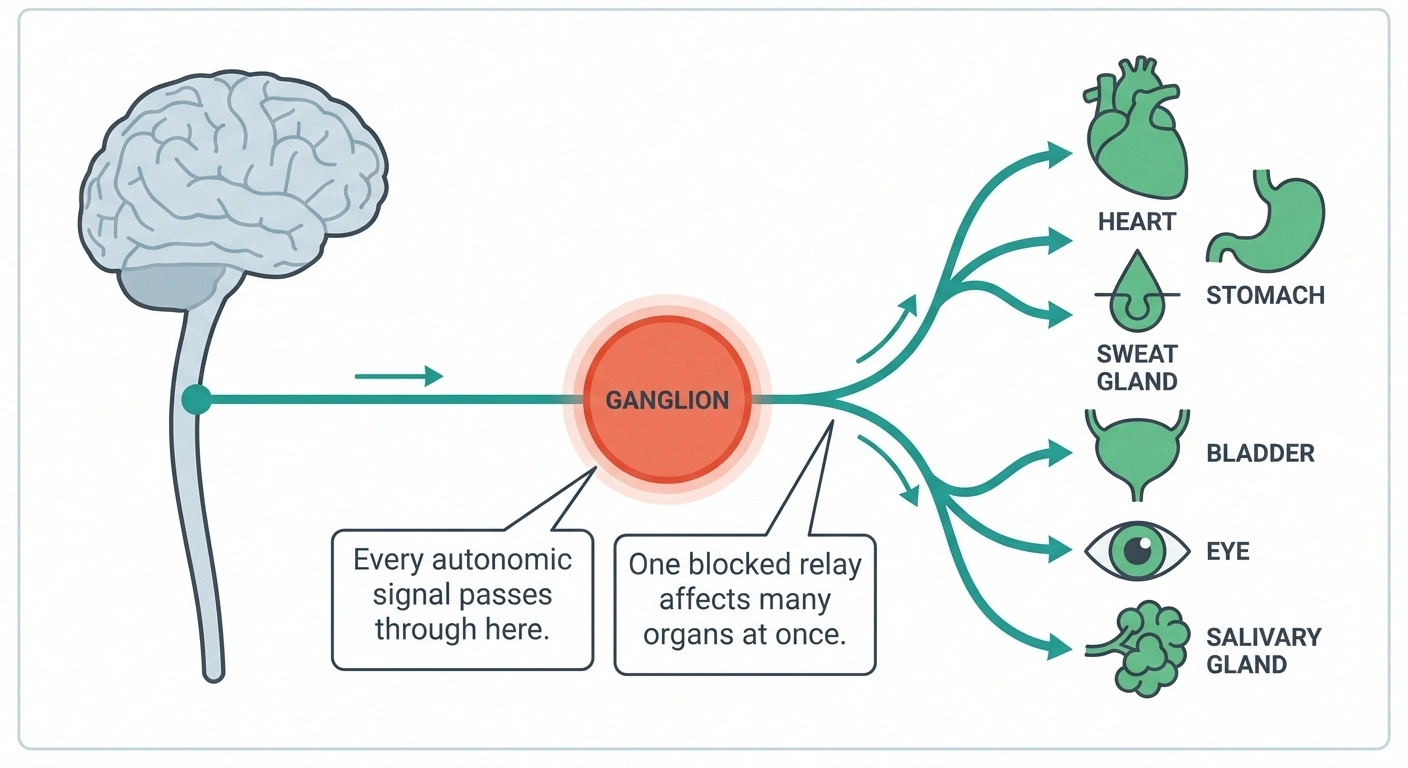
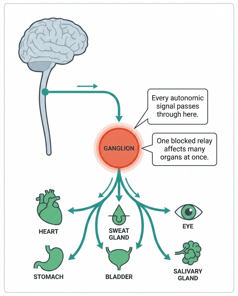
The word “ganglionopathy” scares people off, and it shouldn’t, because a ganglion is a simple idea with an intimidating name.
Your autonomic nervous system runs everything you don’t consciously control: heart rate, blood pressure, digestion, sweating, pupils, bladder, tear and saliva production. It’s the background staff of your body.
The signals don’t travel from your brain to your organs on a single unbroken wire. They travel on two nerves in sequence, and the place where the first hands off to the second is a relay station called a ganglion. Clusters of these relay stations sit alongside your spine, scattered near your organs, and woven directly into the wall of your gut.
Every autonomic instruction your body sends passes through one of these relays. That’s an efficient design and also a vulnerable one, because it means a single point of failure can take down output to many organs simultaneously.
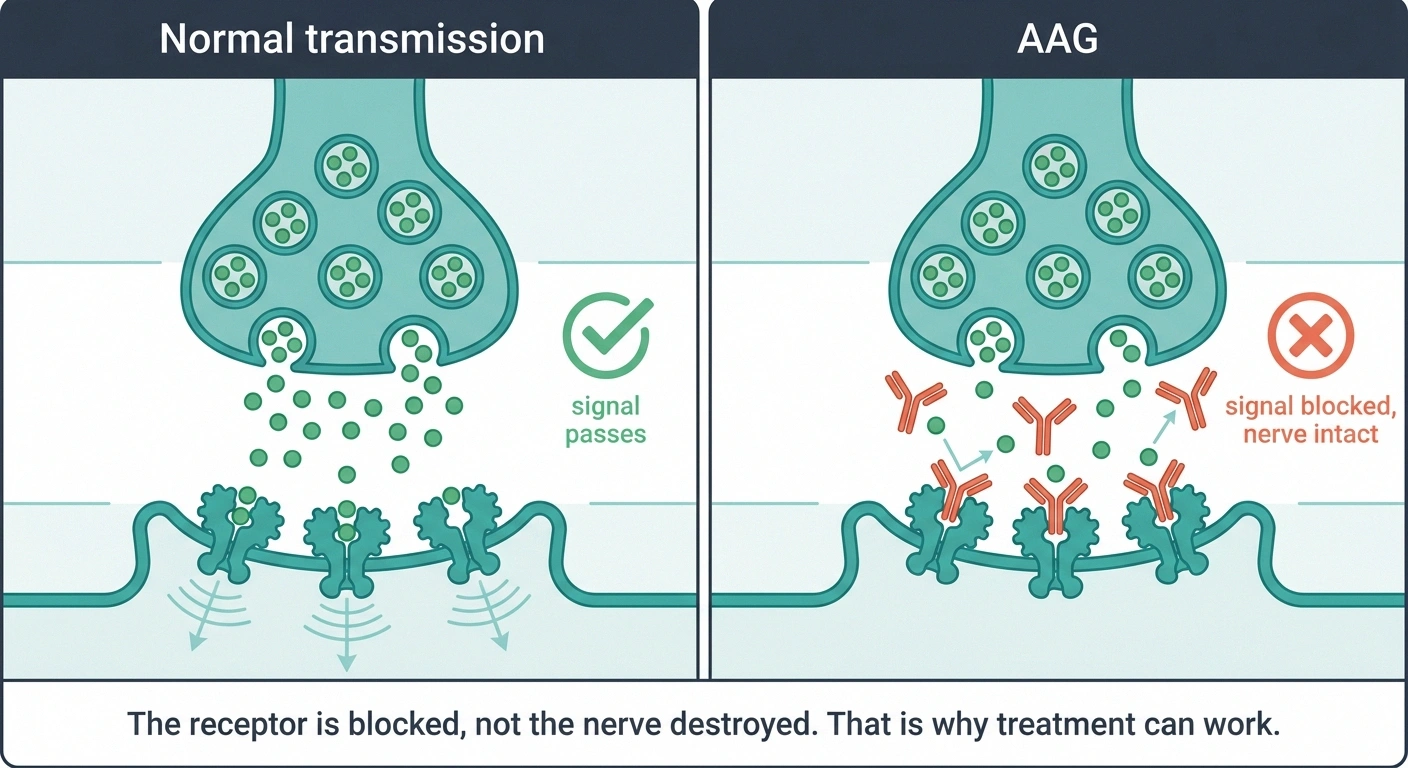
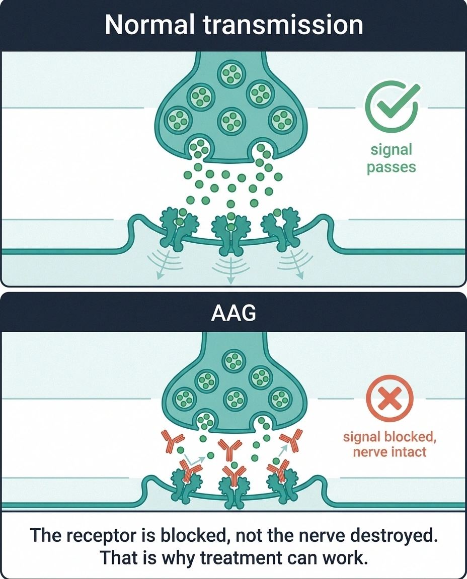
The handoff at the relay happens chemically. The incoming nerve releases acetylcholine, and the outgoing nerve catches it with a receiver called the ganglionic acetylcholine receptor. Message received, signal continues.
In AAG, the immune system produces antibodies against that receiver.
The Antibody, and What It Does to the Signal
Here’s the part that changes how you should think about this condition.
In most neuropathies the fiber degenerates. Regrowth runs at roughly a millimeter a day at best, and often does not happen at all. In AAG the fiber is intact and the receptor is occupied, which is a reversible state.
Two findings support that reading. Antibodies from patients directly inhibit ganglionic receptor currents in the laboratory. And antibody titer tracks with severity, falling in the patients who improve clinically.
What that buys you: a treatment target. Removing or suppressing the antibody is a plausible route to recovery in a way that removing a degenerative process is not.
The antibodies in AAG don’t kill the nerve. They sit on the ganglionic receptor and block it, jamming transmission at the relay. Laboratory work has shown directly that antibodies from AAG patients inhibit ganglionic receptor currents and impair transmission through the ganglia.
Compare that to a typical small fiber neuropathy, where the nerve fibers themselves degenerate. Dead fibers regrow slowly and incompletely, if at all. A blocked receptor is a different situation entirely. Clear the blockade and the relay works again.
There’s a second piece of evidence that ties this together neatly. The level of ganglionic receptor antibody in the blood correlates with how severe the autonomic failure is — higher titer, sicker patient — and when patients improve clinically, their antibody levels fall.
That relationship is why immunotherapy is even on the table here. In most neuropathy, we treat symptoms because the damage is done. In AAG, there’s a real argument for treating the cause.
The Symptom Picture: A Whole System Going Quiet
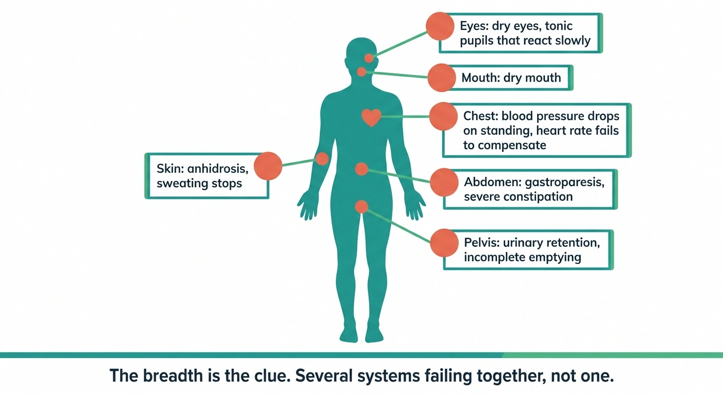
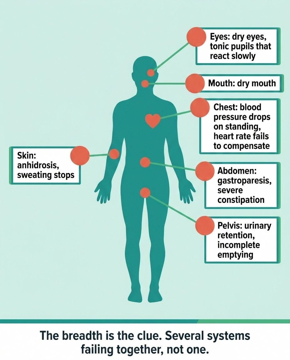
The classic presentation is a full house of autonomic failure. Not everyone has all of it, but the breadth is the tell.
Orthostatic hypotension. Blood pressure drops on standing because the vessels can’t be told to constrict. This is often the most disabling piece — some people cannot stand for more than a minute or two. Our page on autonomic neuropathy generally covers the mechanics of blood-pressure control in more depth.
Gastrointestinal shutdown. Gastroparesis, severe constipation, early fullness, nausea. The enteric nervous system in the gut wall is full of these same ganglia, which is why the gut takes such a direct hit. Some cases have presented with constipation as the leading complaint and nothing else obvious. Our guide to gastroparesis from nerve damage covers the management side.
Anhidrosis. Sweating stops, sometimes completely. This is dangerous in hot weather and it’s also diagnostically valuable, since the pattern of lost sweating can be mapped precisely in a lab.
Bladder dysfunction. Retention, incomplete emptying, or urgency.
Sicca symptoms. Dry eyes and dry mouth, because tear and saliva glands take their orders through the same relays. This one sends a lot of people down a Sjögren’s evaluation first, which is reasonable and often the right first move.
Tonic pupils. Pupils that react poorly and slowly to light. An ophthalmologist may spot this before anyone else connects the dots.
Now, the thing that makes AAG distinctive: in its purer form there’s no somatic neuropathy. Sensation in the feet is normal. Strength is normal. Reflexes are intact. Everything that a standard neurological exam checks comes back fine, while the autonomic system is in freefall.
You can probably see how that plays out in an exam room. A patient reports feeling catastrophically unwell, the neurological exam is unremarkable, and the encounter drifts toward a psychological explanation.
Two Ways It Arrives
AAG shows up in two patterns, and they’re different enough that the slower one is much more likely to be missed.
| Abrupt | Insidious | |
|---|---|---|
| Timeline | Days to weeks | Months to years |
| Trigger | Often a preceding viral illness | Usually none identifiable |
| Typical labels collected first | Few, the change is too dramatic to dismiss | Chronic fatigue, IBS, anxiety, unexplained dysautonomia |
| Diagnostic risk | Usually worked up properly | Frequently missed for years |
Abrupt onset. Days to weeks, often following a viral illness. Someone goes from healthy to profoundly autonomically impaired over the span of a month. Because the timeline is dramatic and the onset is clear, this pattern tends to get worked up properly.
Insidious onset. Months to years of gradual decline. This is the version that collects other labels along the way — chronic fatigue, IBS, anxiety, dysautonomia of unclear cause — because no single visit shows enough change to alarm anyone.
The post-viral trigger is worth pausing on, since it’s become a much bigger conversation in the last few years. Plenty of people developed serious autonomic symptoms after COVID infections, and most of those cases are not AAG. But “most are not” isn’t “none are,” and the overlap is real enough that anyone with severe, persistent post-viral autonomic failure deserves to have this considered rather than assumed away. We’ve written separately about small fiber neuropathy after COVID, which is the more common finding in that group.
Why It Gets Called Something Else First
Four conditions sit close enough to AAG to absorb it, and knowing the differences helps you ask better questions.
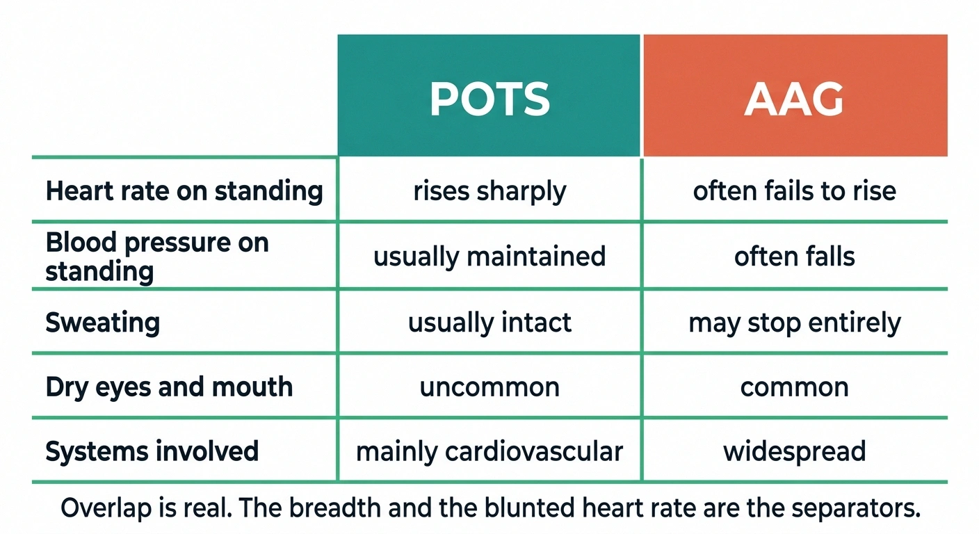
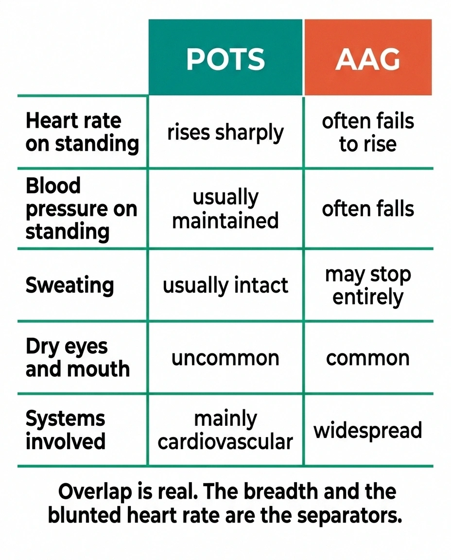
POTS. The overlap here is substantial and the distinction genuinely matters. POTS involves a large heart-rate rise on standing, usually with blood pressure maintained. AAG classically involves blood pressure falling, often with a heart rate that fails to compensate — because the signal telling it to speed up is blocked too. That blunted heart-rate response to a falling blood pressure is a meaningful clue. Our comparison of POTS and small fiber neuropathy works through the related overlap.
Sjögren’s syndrome. Dry eyes and dry mouth send people to rheumatology, and Sjögren’s can cause its own autonomic neuropathy. Ganglionic antibodies have also been detected in various autoimmune rheumatic diseases, so the two aren’t strangers.
Anxiety and panic disorder. Racing heart, dizziness, nausea, feeling faint. The symptom overlap is nearly complete on the surface. What separates them is that anxiety doesn’t cause anhidrosis, tonic pupils, urinary retention, or a measurable pressure drop on a tilt table.
Functional neurological disorder. This one requires care in both directions. Anti-ganglionic antibodies have been reported in people diagnosed with functional neurological symptom disorder, which means a low-positive antibody result is not by itself a diagnosis of AAG. A number on a lab slip doesn’t override the clinical picture — it has to fit with it.
The Antibody Test, and What a Borderline Result Means
The serum test looks for antibodies to the ganglionic acetylcholine receptor, the alpha-3 subunit specifically. Major reference laboratories run it, usually as part of an autonomic or paraneoplastic panel.
Negative: does not rule this out. Only about half of clinically diagnosed patients carry high-titer antibodies, and seronegative AAG is diagnosed on clinical grounds plus autonomic testing.
Low positive: one data point, not a diagnosis. These antibodies have turned up in other autoimmune conditions and in people diagnosed with functional neurological symptom disorder.
High titer: correlates with the classic severe presentation, and is the result most likely to change management.
Expect imaging either way if it is positive. AAG can be paraneoplastic, so a positive result routinely triggers a search for an occult tumor. Knowing that is coming makes it far less frightening when it arrives.
Two things about interpreting it.
First: only about half of clinically diagnosed AAG patients have high-titer antibodies. Seronegative AAG exists and is diagnosed on clinical grounds plus autonomic testing. A negative antibody test does not close the case, and it shouldn’t be treated as if it does.
Second: titer height carries real information. High titers correlate with the classic severe presentation. Low-positive results are murkier, showing up in other conditions and in some people without clear autonomic disease at all. If your result comes back barely positive, the honest reading is “this is one data point among several,” not “confirmed.”
There’s one more thing the panel is doing that nobody explains to patients: screening for cancer. AAG can occur as a paraneoplastic syndrome, meaning an immune response provoked by a tumor — small cell lung cancer and thymoma being the usual suspects. A positive ganglionic antibody typically prompts imaging to rule that out. It’s an unnerving thing to have happen without warning, so it’s better to know it’s coming and why.
The Tests That Build the Rest of the Case

Antibodies alone don’t make the diagnosis. Autonomic function testing does the heavy lifting, and these tests are the reason a referral to a center with an autonomic lab is worth the drive.
- Tilt table
- Blood pressure and heart rate against gravity. Documents the drop objectively instead of relying on your description of it.
- QSART
- Sweat output from small skin sites after chemical stimulation. Detects reduced sweating and localizes it.
- Thermoregulatory sweat test
- Whole-body sweat mapping using a powder that changes color. The pattern of loss indicates where the lesion sits.
- Deep breathing and Valsalva
- Parasympathetic function via heart rate variability. Takes minutes, involves no needles, and is often the first thing done.
These labs live at academic medical centers. Documentation from them frequently determines insurance approval for immunotherapy, so the drive is usually worth it.
Tilt table testing measures blood pressure and heart rate as you’re tilted upright, documenting the drop objectively rather than relying on symptom description.
QSART measures sweat output from small skin sites after a stimulus, showing whether the sweat response is reduced and where.
Thermoregulatory sweat testing maps sweating across the whole body surface. The pattern of loss says a great deal about where the lesion sits.
Heart rate variability with deep breathing and Valsalva tests the parasympathetic pathways in a few minutes with no needles.
Gastric emptying study if the gut symptoms lead.
Together these establish that autonomic failure is objectively present and how widespread it is. That documentation matters for treatment decisions and, practically speaking, for insurance approval of the treatments that follow.
Treatment: Holding the Line vs. Changing the Course
Treatment splits into two categories that do different jobs, and conflating them causes disappointment.
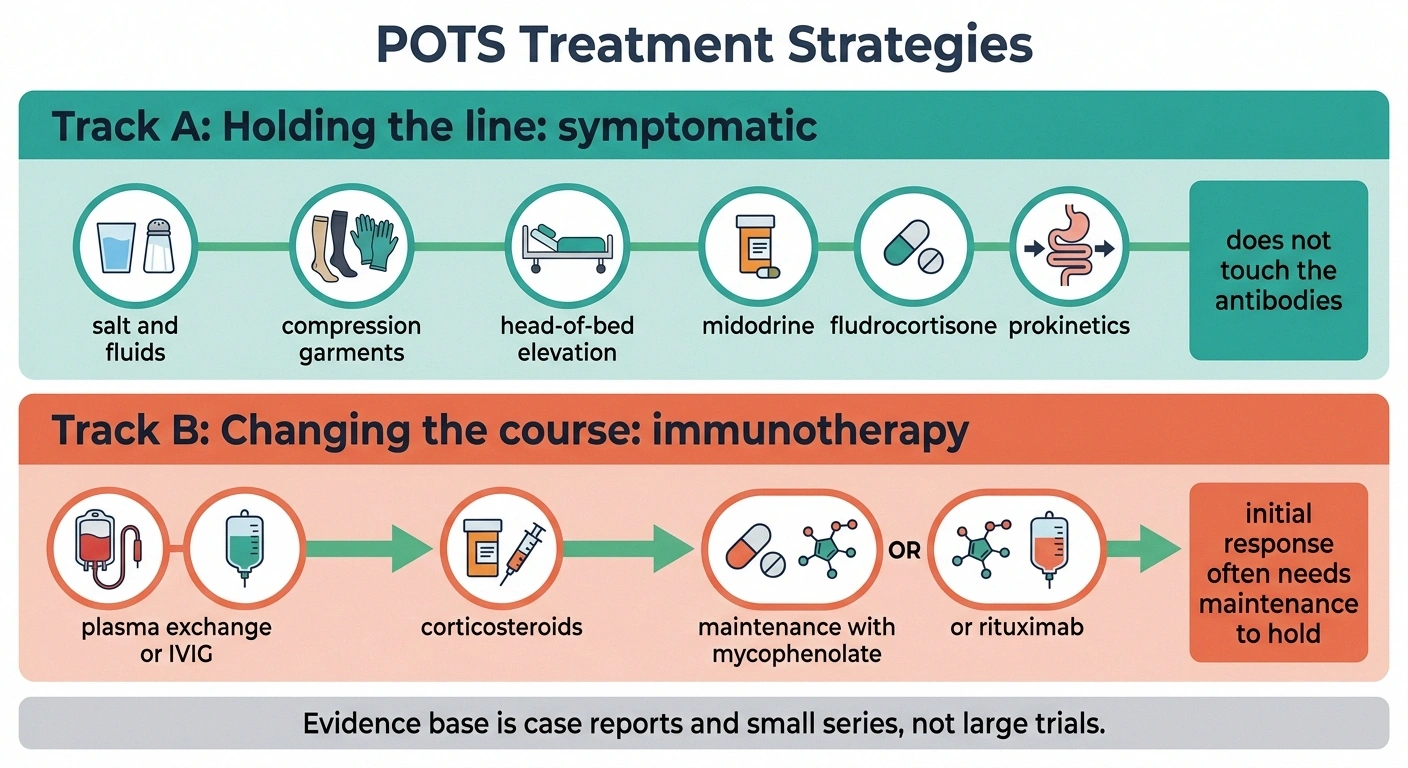
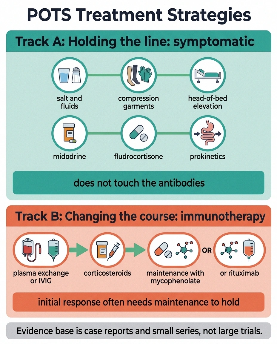
Symptomatic treatment holds the line. For orthostatic hypotension: increased salt and fluid, compression garments, sleeping with the head of the bed raised, and medications such as midodrine or fludrocortisone. For the gut: prokinetics and dietary modification. For the bladder: catheterization schedules where needed. None of this touches the antibodies. It makes daily life livable while the underlying question is worked out, and for milder cases supportive care may be all that’s needed.
One caution that comes with the territory: aggressive treatment of low standing blood pressure can produce very high pressures lying down. Supine hypertension is a real and common consequence, and it’s why the head-of-bed elevation advice keeps coming up.
Immunotherapy aims to change the course. This is where AAG diverges from most neuropathy.
Plasma exchange physically removes antibodies from the circulation and can produce rapid improvement. IVIG is used similarly. Corticosteroids reduce antibody production more broadly.
But here’s the pattern reported repeatedly in the literature, and it’s worth knowing in advance: patients often respond to IVIG or plasma exchange initially, then need an ongoing immunosuppressant to hold or extend the gains. The first treatment clears the antibodies already made. It doesn’t stop the immune system from making more.
For that maintenance role, mycophenolate mofetil and rituximab both appear in the published experience. Rituximab depletes the B cells that produce the antibodies, and long-term B-cell depletion has been described as producing suppressed antibody production alongside sustained clinical improvement. One small series reported that steroids plus mycophenolate plus plasma exchange did better than any of those alone.
This is a small evidence base — case reports and small series rather than large randomized trials, which is what happens with rare diseases. It’s enough to justify trying, and honest to say it isn’t enough to predict who responds.
What Recovery Actually Looks Like
I want to be careful here, because “treatable” gets heard as “curable” and those aren’t the same word.
What the literature supports: meaningful improvement is possible, sometimes substantial. Antibody levels drop in patients who improve. Some people go from housebound to functional.
What it also supports: many require ongoing immunosuppression to maintain gains, relapses happen, and response varies widely between individuals. Some improve dramatically. Some improve partially. Some don’t respond.
Timing appears to matter, as it does across autoimmune neurology — treating during the blockade phase makes more sense than treating after prolonged failure has led to secondary degeneration. That’s an argument for pursuing diagnosis promptly rather than waiting to see how things go.
If You Think This Might Be You

Let me be straight about the odds: AAG is rare, and most people with dizziness and digestive trouble don’t have it. If you’re reading this and recognizing yourself, the most likely explanation is still something more common.
But the profile that genuinely warrants asking is narrower than “some autonomic symptoms,” and here it is.
Multiple autonomic systems failing together, not one. A subacute onset over weeks to months, especially after an illness. Anhidrosis — actual loss of sweating, which is uncommon and specific. Severe orthostatic hypotension with a heart rate that doesn’t rise to meet it. A normal neurological exam despite feeling profoundly unwell. And dryness of eyes and mouth alongside all of it.
If that’s a fair description of your situation, the request to make is a referral to a neurologist with autonomic expertise, and the two words to bring are “autonomic testing.” Academic medical centers are where those labs live. A ganglionic acetylcholine receptor antibody panel is a reasonable ask alongside it.
Go in with your symptoms written down, organized by system rather than by when they bothered you most. Blood pressure and standing. Digestion. Sweating. Bladder. Eyes and mouth. Pupils. A list arranged that way tells a story that a list of complaints in order of annoyance never quite manages, and it makes the pattern visible to someone who might otherwise treat each item as its own small problem.
That’s what four years of “it’s anxiety” is usually made of — six real findings, never once looked at in the same frame.
Frequently Asked Questions
What is autoimmune autonomic ganglionopathy?
It is an acquired disorder in which the immune system produces antibodies against the ganglionic acetylcholine receptor, the receiver that allows signals to pass through the relay stations of the autonomic nervous system. These relays sit in sympathetic, parasympathetic and enteric ganglia, so a blockade there disrupts blood pressure control, digestion, sweating, bladder function, and tear and saliva production simultaneously. The defining feature is widespread autonomic failure without accompanying damage to sensation or strength.
What is the difference between AAG and POTS?
POTS is characterized by an excessive heart rate rise on standing, typically with blood pressure maintained, and most people with POTS do not have widespread failure of other autonomic functions. AAG classically involves blood pressure falling on standing, often accompanied by a heart rate that fails to compensate because that signal is also blocked, along with anhidrosis, gastrointestinal dysmotility, bladder dysfunction and dry eyes and mouth. The breadth of systems involved and the blunted heart-rate response are the main distinguishing features.
Does a negative antibody test rule out AAG?
No. High-titer ganglionic acetylcholine receptor antibodies are found in roughly half of clinically diagnosed patients, meaning seronegative AAG is common and is diagnosed on clinical grounds combined with objective autonomic function testing. A negative result reduces the likelihood but does not exclude the diagnosis. Conversely, a low-positive result is not confirmation on its own, since these antibodies have been detected in other conditions including functional neurological symptom disorder.
Can autoimmune autonomic ganglionopathy be cured?
Meaningful improvement is achievable in many patients, but cure is not a reliable expectation. The published experience shows plasma exchange, IVIG and corticosteroids producing improvement, with antibody levels falling in those who respond. However, many patients require ongoing immunosuppressive treatment such as mycophenolate mofetil or rituximab to sustain those gains, and relapse can occur. Response varies considerably between individuals, and the evidence base consists largely of case reports and small series rather than large trials.
Can a virus trigger AAG?
A preceding viral illness is commonly reported before the abrupt-onset form, which develops over days to weeks. The proposed mechanism is that the immune response to an infection generates antibodies that cross-react with the ganglionic receptor. Not all cases follow an identifiable infection, and the insidious form develops gradually over months or years without a clear trigger. Severe autonomic symptoms after a viral illness have other more common explanations, so a viral trigger alone does not indicate AAG.
Why would a doctor order cancer imaging after a positive antibody result?
Because AAG can occur as a paraneoplastic syndrome, meaning the antibody production is provoked by an underlying tumor rather than arising on its own. Small cell lung cancer and thymoma are the malignancies most often associated. A positive ganglionic antibody therefore usually prompts imaging to look for an occult tumor, since finding and treating the cancer is essential and can improve the autonomic syndrome. The screening is routine practice rather than an indication that cancer has been found.
What tests confirm autonomic failure?
Tilt table testing documents blood pressure and heart rate responses to upright posture. QSART measures sweat output from small skin sites after stimulation, and thermoregulatory sweat testing maps sweating across the whole body to show the pattern of loss. Heart rate variability during deep breathing and Valsalva maneuvers assesses parasympathetic pathways. A gastric emptying study is added when digestive symptoms predominate. These tests establish objectively that autonomic failure is present and how extensive it is, which is necessary for both diagnosis and treatment decisions.
Is anhidrosis dangerous?
Yes, and it deserves specific planning. Sweating is the body’s primary cooling mechanism, so losing it substantially raises the risk of heat exhaustion and heat stroke during hot weather, exertion or hot baths. People with significant anhidrosis need active external cooling strategies, including limiting time in heat, using cooling vests or wet cloths, and staying in air conditioning during hot spells. It is also one of the more diagnostically useful findings, since it is uncommon and can be measured precisely in an autonomic laboratory.
About the Author