Roll your sock down and look at your shin.
Not your toes. The stretch of skin from the ankle bone up to about mid-calf. Is the hair there thinner than it used to be, or gone altogether? Does the skin have a faint gloss to it, the way a balloon does, like it’s been pulled slightly too tight over whatever is underneath?
If you’re nodding, you’ve just found something worth writing down. Not something to panic about at eleven at night. But a real finding, and one that gets missed constantly, because the person best positioned to notice it is you, in the shower, filing it under getting older.
Sometimes it is just getting older. And sometimes those two changes together — smooth shiny skin, plus hair that quietly stopped growing below the knee — are your legs reporting on your nerves, your circulation, or both at once. The both-at-once version is the one that matters most, and it’s the one almost nobody writes about.
What You Are Actually Looking At
Clinicians have a word for this cluster of changes. They call them trophic changes, and the word is less intimidating than it sounds. Trophic just means related to nourishment. Trophic changes are what tissue looks like when its supply line, whether that’s blood or nerve signaling, has been degraded a long while.
Do this in good light, both legs bare to the knee, comparing left against right. Comparison is what makes it informative, since almost nobody remembers what their own shin looked like five years ago.
- Hair. Note the highest point on each leg where hair still grows. A difference of more than a couple of inches between legs is worth reporting.
- Shine. Tilt the leg toward a window. Skin that reflects a soft highlight along the shin the way a balloon would is the finding.
- Temperature. Back of your hand on one foot, then the other, then each calf. You are looking for one foot cooler than its partner, not for cold feet in general.
- Heels. Any crack deep enough to catch a fingernail counts, whether or not it hurts.
- Color on the move. Lie down, raise both legs for 60 seconds, then hang them over the edge. Marked pallor on raising or deep red on lowering is a circulation finding.
Photograph anything you find, with a ruler or a coin in frame for scale. Skin changes are slow, and a dated photo beats a memory at your next appointment.
On the lower legs and feet, the full list runs like this:
Skin that looks glossy or waxy. Thinned, tight, a little translucent. The shine isn’t oil. It’s the loss of the fine surface texture normal skin has, which is the thing that scatters light and keeps skin looking matte.
Hair loss in a stocking pattern. Starting at the toes and the top of the foot, working up the shin. It tends to be symmetrical when nerves are driving it, and it can be lopsided when circulation is.
Nails that grow slowly, thicken, or yellow. Nails are among the first things to change — and among the last things anybody connects to anything.
Skin dry enough to flake or crack, especially at the heel, often in someone whose skin everywhere else is perfectly ordinary.
Color that’s off. Pale, dusky, reddish when the leg hangs down, blanching when you raise it.
You won’t have all of these. Most people have two or three, and have had them long enough to have stopped seeing them.
Why Nerves Change Skin
This is the part that surprises people: your skin is under nerve control, and not only for sensation.
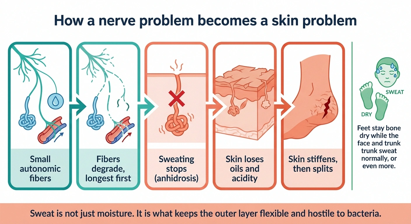
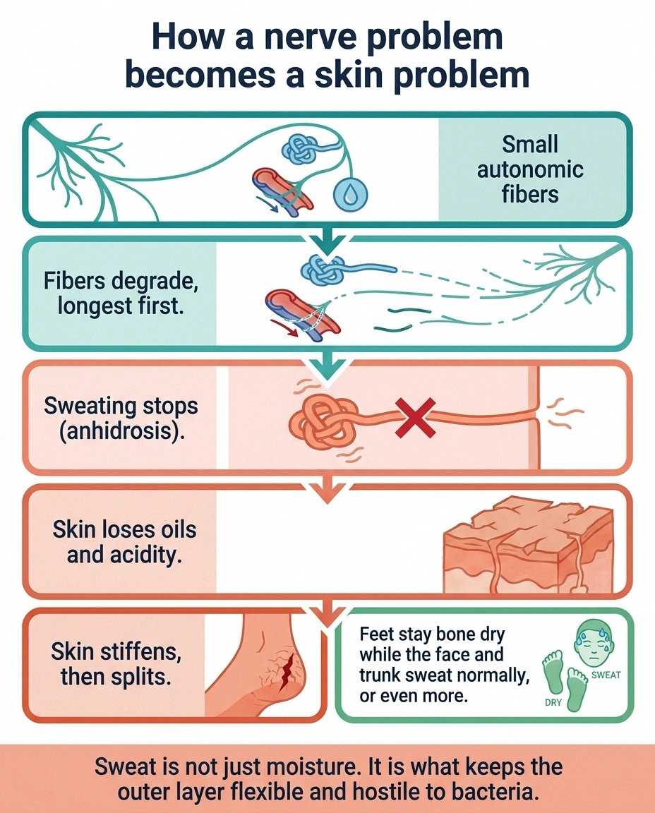
A separate set of small, unmyelinated fibers runs to your sweat glands and to the tiny blood vessels in your skin. These are autonomic fibers — the ones handling all the business you never think about. They set how much you sweat and how much blood moves through the skin’s smallest vessels. They’re also, unfortunately, among the first fibers damaged in many neuropathies, and they’re precisely the fibers that small fiber neuropathy is named for.
When the sweat-gland fibers degrade, the feet stop sweating. The clinical word is anhidrosis, and it produces a very specific and very common picture: feet that are bone dry and cracking while the rest of the body sweats normally, or even sweats more, because the body compensates elsewhere. If you’ve ever wondered why your face and trunk pour while your feet stay parched, that’s the mechanism, and our piece on why feet stay dry when everything else is soaked goes further into that trade-off.
Sweat isn’t only moisture. It carries the oils and the mild acidity that keep the outer layer of skin flexible and unfriendly to bacteria. Take it away and the skin stiffens, then splits. Those splits, particularly at the heel, aren’t a cosmetic problem. They’re an opening in the only barrier you’ve got — and it doesn’t grow back on its own.
The hair loss works through a related route. Follicles need consistent blood supply and normal signaling, and in a length-dependent neuropathy the longest nerves fail first — which is why every bit of this starts at the toes and climbs. That distal-first pattern is the signature of nerve damage rather than something local, and it’s why the symmetry is worth noticing.
None of this is rare. Sudomotor dysfunction in the feet has been linked to autonomic involvement of the heart in people with type 2 diabetes, which is a good reminder that autonomic neuropathy rarely stays in one organ. Dry feet can be the visible end of something with a longer reach.
The Other Cause, and It Is the Urgent One
Shiny skin and hair loss on the lower legs is also a textbook sign of peripheral artery disease, where narrowed arteries cut blood flow to the limb.
- Trophic changes
- Changes in skin, hair or nails caused by a long-term shortfall in blood supply or nerve signaling. A description, not a diagnosis.
- Anhidrosis
- Loss of sweating. In the feet it is caused by damage to the small autonomic fibers supplying sweat glands.
- Claudication
- Muscle pain brought on by walking a predictable distance and relieved by rest. A circulation symptom, and one that numbness can hide.
- Ankle-brachial index (ABI)
- Ankle blood pressure divided by arm blood pressure. Under 0.90 indicates artery disease. Above roughly 1.30 suggests vessels too stiff to compress, which makes the number unreliable rather than good.
- Length-dependent
- Affecting the longest nerves first. It is why findings start at the toes and climb, and why they are usually symmetrical.
The mechanism there is starvation rather than miscommunication. Skin that chronically gets too little oxygen turns thin, brittle and smooth, and takes on that same glossy look. Hair follicles, which are metabolically expensive, shut down. Nails slow to a crawl. Wounds that should close in a week stay open for a month.
Artery disease brings its own tells, and they’re worth knowing cold:
Pain that comes on with walking and stops with rest. Calf, thigh or buttock, reliably at about the same distance, gone a few minutes after you stand still. It’s called claudication and it’s close to diagnostic.
A foot that’s cool compared with the other one, or compared with your own calf.
Color that shifts with position. Pale when you elevate the leg, dusky red when you hang it off the edge of the bed.
Weak or missing pulses at the ankle and the top of the foot.
A cut or blister that just won’t close.
Artery disease matters more urgently than the cosmetic questions that probably brought you here, for two reasons. It’s a marker of atherosclerosis elsewhere, including the arteries feeding your heart and brain. And in a foot that also has neuropathy, poor blood supply turns a small unnoticed injury into a wound that can’t heal. That combination is the road to diabetic foot ulcers, and it’s why this article doesn’t end with a moisturizer recommendation.
Telling Them Apart at Home
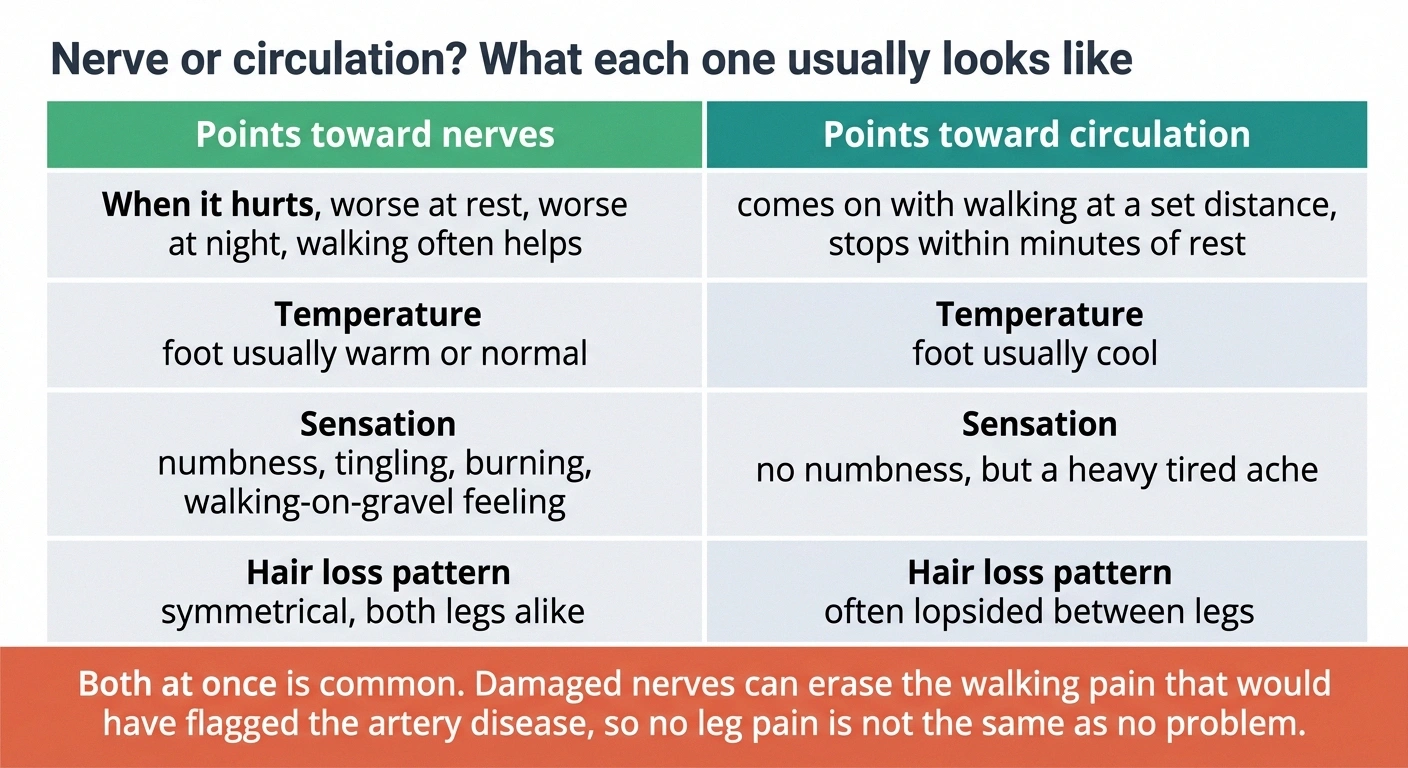
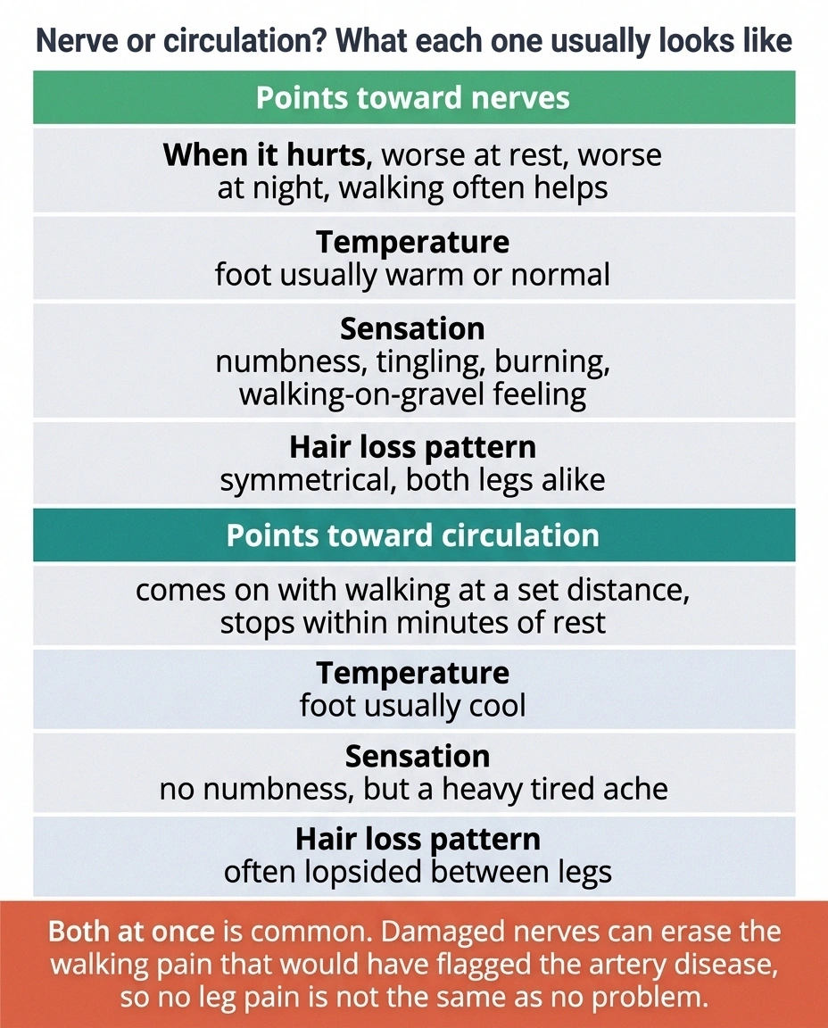
You can’t settle this yourself, and I want to be straight about that before I hand you a way to think about it. What follows will help you describe the problem accurately and ask for the right test. It won’t give you an answer.
The single most useful question is when it hurts.
Nerve pain is typically worse at rest and worse at night, and walking often takes the edge off for a while. Circulation pain is the mirror image — fine at rest, brought on by exertion, relieved by stopping. If you tell me your legs hurt most at two in the morning while you’re lying perfectly still, that points one direction. If you tell me you can walk exactly two blocks before your calf seizes, that points the other.
Temperature is the second question. A neuropathic foot is usually warm or normal. An ischemic foot is usually cool.
Sensation is the third. Neuropathy brings numbness, tingling, burning, or that sense of walking on gravel. Plain artery disease doesn’t cause numbness, though it can absolutely cause a heavy, tired ache.
Now the complication, and it’s the reason this page exists in the shape it does. In long-standing diabetes both conditions are common, they share risk factors, and they routinely show up together. Worse, neuropathy can erase the warning sign of the other one. Claudication is a pain signal. If the nerves carrying that signal are damaged, you can have significant arterial narrowing and never feel the walking pain that would’ve sent you to a doctor years earlier.
So a person with numb feet, no leg pain at all, shiny skin and no hair from the knee down isn’t reassuring. That person may simply have lost the alarm. Our comparison of numbness in the feet across neuropathy, artery disease and sciatica lays the overlap out in more detail.
The Tests That Settle It
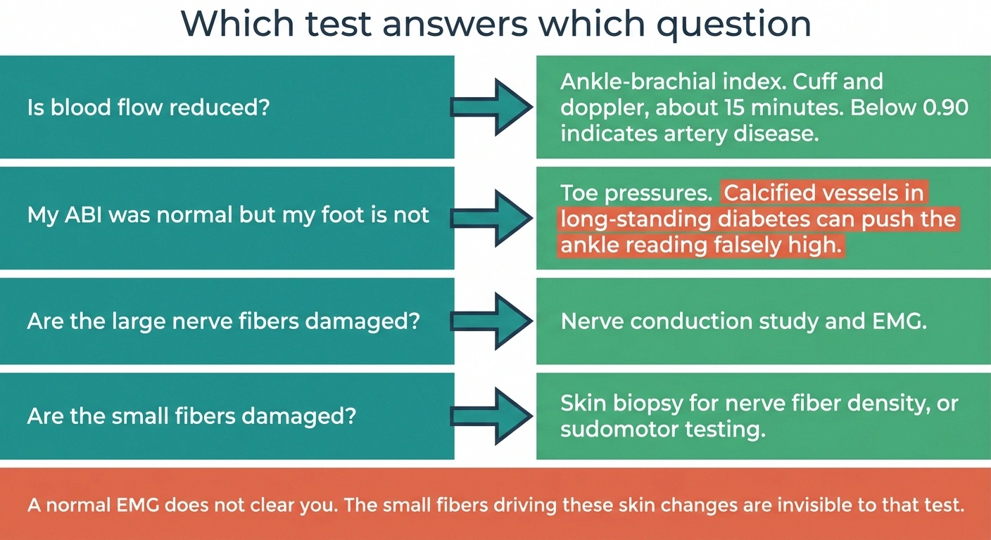
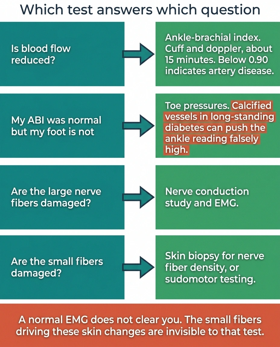
The good news: separating these two is neither expensive nor slow. The tests exist, they’re routine, and most of them happen in one visit.
Vague reports produce vague responses. These three sentences are specific enough to act on, and each one names the finding rather than the fear.
To open: “The hair on my lower legs has stopped growing and the skin looks shiny. I would like an ankle-brachial index to check circulation.”
If you have numbness and no walking pain: “I know I do not get leg pain when I walk, but my feet are numb, so I am not sure I would feel it. Does that change how much the absence of claudication tells us?”
If the ABI comes back normal: “I have had diabetes a long time. Would toe pressures be more reliable than the ankle reading in my case?”
Bring the dated photographs. A visible change over six months moves a conversation faster than any adjective you can supply.
For circulation, the workhorse is the ankle-brachial index. A cuff and a handheld doppler compare the blood pressure at your ankle with the blood pressure in your arm. A ratio under 0.90 indicates peripheral artery disease. It takes about fifteen minutes, there are no needles and no dye — and it’s the test to ask for by name.
One caveat matters a great deal for people with long-standing diabetes: stiff, calcified arteries can resist the cuff and push the reading falsely high, so a normal ankle-brachial index in a diabetic leg isn’t automatically a clean result. Toe pressures get around this, because the small vessels in the toes calcify far less. If your result comes back fine and your foot still looks and behaves wrong, that’s a reasonable and very specific thing to raise.
For nerves, the panel depends on which fibers are suspect. Standard nerve conduction studies and EMG measure the large fibers, and here’s the catch worth understanding: they’re frequently normal in pure small fiber neuropathy, because the small fibers driving your skin changes are invisible to that test. A normal EMG doesn’t clear you. When small fiber disease is the question, a skin biopsy measuring nerve fiber density in the epidermis, or sudomotor testing that measures the sweat response directly, is the tool that actually answers it.
In practice the ordering is simpler than it sounds. A primary care doctor can order the ankle-brachial index, vascular reads it, and neurology handles the nerve side.
Caring for Skin That Cannot Sweat

Whatever the underlying answer turns out to be, the skin in front of you needs looking after now. Dry, cracking skin on a foot with reduced sensation is the most preventable step on the road to a serious wound.
| What the skin looks like | What to reach for | Why |
|---|---|---|
| Mildly dry, no thickening | A plain thick ointment or cream, urea 5 to 10 percent | Holds water in. Nothing stronger is needed yet. |
| Thickened, scaly, rough heels | Urea 10 to 25 percent, or ammonium lactate | Breaks down the hardened outer layer so it flexes rather than splits. |
| Deep fissures, any bleeding | Cover it and get it looked at first | This is an open wound on an insensate foot, not a moisturizing problem. |
| Soggy or white between the toes | Nothing. Dry the area and keep it dry. | Trapped moisture macerates skin and invites fungal infection. |
Two practical notes. Urea can sting on cracked skin, which is a signal to treat the crack before continuing rather than to abandon the product. And a tub lasts far longer than a tube at the amount this actually takes, which for two feet is roughly a teaspoon a day.
A short, unglamorous routine does most of the work.
Moisturize daily, and pick the right product. Ordinary lotion is mostly water and won’t do much here. What works on thickened, fissured skin is a keratolytic — urea at 10 to 25 percent, or ammonium lactate. These don’t just sit on top of the skin. They soften and break down the hardened outer layer so it can flex instead of splitting.
Put it on within a few minutes of bathing, onto skin that’s still slightly damp, which traps the water already there.
Never between the toes. This is the exception that catches everybody. Moisture trapped in that airless space macerates the skin and invites fungal infection. Everywhere else on the foot, yes. Between the toes, dry gently and leave them be.
Keep the water lukewarm and the soak short. Hot water strips what little oil the skin has left, and with reduced temperature sensation you may not register that the water’s hot enough to burn you.
Look every day. Tops, soles, heels, and between every toe, using a mirror or a phone camera for the parts you can’t see. The two-minute daily foot check exists precisely because skin that can’t sweat and can’t feel won’t tell you when something’s gone wrong.
About the hair: it usually doesn’t come back, and I’d rather say so plainly than leave you hoping. Hair loss from long-standing nerve or vessel damage is generally permanent. It’s also, in the ranking of things happening to your legs, near the bottom of the list of things that matter. The hair is a signal, not the problem. The intact skin barrier is the problem worth protecting.
The Findings That Mean Call Today
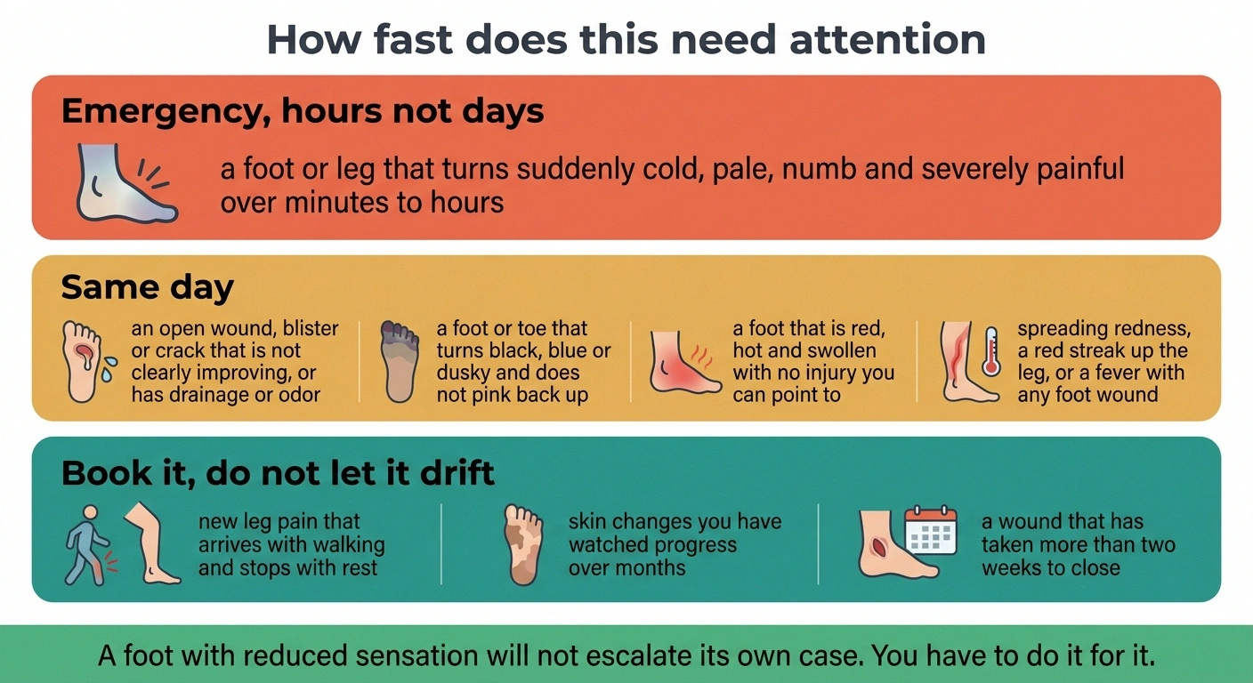
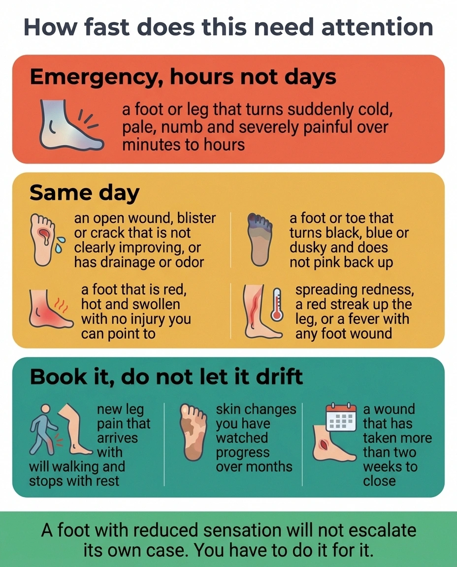
Most of what’s on this page can wait for a scheduled appointment. A few things can’t, and the distinction is worth keeping straight, because a foot with reduced sensation won’t escalate its own case.
Get seen the same day for any of these: an open wound, blister or crack that isn’t clearly improving, or that has drainage, odor or surrounding redness. A foot or toe that turns black, blue or dusky and doesn’t pink back up. A foot that’s red, hot and swollen with no injury you can point to. Spreading redness, a red streak traveling up the leg, or a fever alongside any foot wound.
Treat as an emergency: a foot or leg that goes suddenly cold, pale, numb and severely painful over minutes to hours. That combination suggests an abruptly blocked artery, and it’s measured in hours, not days.
And book it, without particular urgency but without letting it drift: new leg pain that reliably arrives with walking and stops with rest, skin changes you’ve watched progress over months, or a wound that’s taken more than two weeks to close. Any of those earns an ankle-brachial index. Knowing when to bring in a podiatrist is most of the battle, and the honest answer is that it’s earlier than most people think.
One last thought. The reason to look at your shin tonight isn’t that shiny skin is dangerous. It’s that shiny skin is information, arriving from a part of your body that has largely stopped sending you any. When sensation goes quiet, appearance becomes the channel that’s still open. Use it.
Frequently Asked Questions
Does neuropathy cause hair loss on the legs?
Yes, it can. Damage to the small autonomic nerve fibers that regulate blood flow and sweating in the skin can reduce follicle function, producing hair loss that typically begins at the toes and moves up the shin. It is described in small fiber neuropathy and in nutritional neuropathies. That said, the same pattern is a classic sign of peripheral artery disease, so leg hair loss should not be attributed to nerves without checking circulation as well.
Why is the skin on my lower legs shiny?
Shine comes from thinning and the loss of the normal fine surface texture of skin, which happens when tissue has been chronically underserved by blood supply or nerve signaling. Both reduced circulation and long-standing nerve damage produce it. Chronic swelling, certain medications and some skin conditions can also cause it, which is why the finding is worth showing to a clinician rather than interpreting on your own.
How do I know whether it is circulation or nerve damage?
The most useful clue is timing. Circulation pain tends to arrive with walking and fade with rest, while nerve pain is often worse at rest and at night. A cool foot points toward circulation; numbness, burning and tingling point toward nerves. Neither pattern is reliable enough to act on alone, and the two conditions frequently coexist. An ankle-brachial index is the usual first test for the circulation half of the question, though in long-standing diabetes a normal result can be falsely reassuring and toe pressures may be needed.
Will the hair on my legs grow back?
Usually not. Hair loss from long-standing nerve or vascular damage tends to be permanent, though hair sometimes returns partially when the underlying cause is reversible, such as a corrected nutritional deficiency or restored blood flow after a vascular procedure. The hair itself is a cosmetic matter. Its disappearance is useful mainly as a sign.
My feet are extremely dry but I sweat normally everywhere else. Why?
This is a recognized pattern in autonomic neuropathy. The small fibers supplying the sweat glands in the feet are damaged in a length-dependent way, so the feet lose sweating while the rest of the body is unaffected or compensates by sweating more. The result is dry, cracking skin on the feet in someone whose skin is otherwise normal.
Is cracked heel skin actually dangerous?
It can be, in a foot with reduced sensation. A deep fissure is a break in the skin barrier, and it can admit bacteria and progress to an infected wound, particularly if blood supply is also reduced. Because sensation is diminished, the crack may never hurt enough to prompt action. Daily moisturizing with a urea-based product and daily visual inspection are the practical defenses.
What test should I ask for first?
An ankle-brachial index. It is quick, noninvasive, widely available and often orderable in primary care, and it answers the more time-sensitive of the two questions. If you have long-standing diabetes and the result comes back normal while your foot still looks and behaves abnormally, ask whether toe pressures would be more reliable, since calcified vessels can make the ankle reading falsely reassuring.
About the Author